

Year: 2002 Vol. 68 Ed. 2 - (14º)
Artigo Original
Pages: 245 to 249
 PDF PT
 Contralateral ear in chronic otitis media: "Orloff ® Effect"
Author(s):
Ana Bárbara Scheibe 1,
Mariana M. Smith 2,
Letícia P. Schmidt 1,
Viviane B. Schmidt 3,
Cristina Dornelles 4,
Lúcia Helena S. K. Carvalhal 5,
Lisiane Kruse 6,
Sady S. da Costa 7
Keywords: chronic otitis media, contralateral ear, cholesteatoma
Abstract:
Introduction: Chronic otitis media (COM) is an entity of high prevalence and worldwide distribution. Despite being studied in depth, its pathogenesis is still a matter of discussion. One of the theories for its pathogenesis is the continuum. According to this theory, otitis media is described as a sequence of events, initiated by an insult that would lead to a cascade of events. Based on the conception of the Continuum, we studied the contralateral ear of patients with COM. Study design: Clinical prospective randomized. Material and Method: 108 patients were included in this study. All had been diagnosed as having COM (with or without cholesteatoma) and were being followed by the Research on Middle Ear Pathologies Group of the Hospital de Clínicas de Porto Alegre. Otoendoscopy with optic fiber was carried out on the affected ear just as in the contralateral ear, witch was classified as normal or abnormal. Results: 59,2% of all patients had COM with cholesteatoma and 40,8% had COM without cholesteatoma. In 46,3% of patients the contralateral ear was found to have some abnormality. In the group of patients with COM with cholesteatoma, 57% had abnormal contralateral ear, and in the other group the contralateral ear was found to be abnormal in 39%. The abnormality more frequently found on both groups was retraction of the eardrum. Discussion: The results suggest that patients with COM have grater probability of having bilateral disease, witch corroborates the idea that COM is not only an isolated event on the middle ear, but a constitutional one, and, therefore, bilateral.
![]()
INTRODUCTION
Chronic otitis media (COM) is defined as the presence of tissue abnormalities in the middle ear, of inflammatory origin and irreversible character1, 2. It has high prevalence and worldwide distribution - characteristics that added up provide it with the position of "public health issue", even in the present moment3, 4.
COM, undoubtedly represents one of the main areas of interest within modern Otorhinolaryngology, considering the great variety of research material recently published about the topic5, 6, 7, 8. The main issue concerning COM is probably its pathogenesis. What are the mechanisms that lead to central and marginal perforations? How do cholesteatomas surge and expand? Other very intriguing questions are: Why do different patients, starting from the same initial insult, sometimes present clearly different evolutions? In other words, the questions concerning the issue are still abundant and the answers are nothing more than a list of hypothesis proposed by the literature.
In our group we have followed the pathogenesis model suggested by the Minneapolis group - the so-called continuum theory4,9,10. According to the theory, otitis media seems to exist through a continuous series of events, and after the initial triggering episode, a serous or purulent otitis becomes serous-mucoid, mucoid or, if there is no resolution of the case, there would be chronic transformation. Therefore, COM does not seem to be an isolated event that affects a specifically sick ear. It seems to be, in fact, the result of a series of "constitutional" events of the subject.
According to the continuum theory, it is admitted that otitis media with effusion is an initial pathologic process that may progress to chronic transformation. Thus, the question is: considering that the presence of bilateral effusion is high11,shouldn't the prevalence of bilateral COM be equally high?
Therefore, we believe that the presence of COM in one ear has higher likelihood of causing contralateral ear abnormalities. Based on such reasoning and the few data described in the literature12, 13, the current study reports the characteristics of the contralateral ear in a series of COM patients.
MATERIAL and method
The patients selected for the present study were followed up at the ambulatory of the Study Group on Middle Ear Pathology (GEPOM) of Hospital de Clínicas, Porto Alegre (HCPA). The ambulatory serves patients with COM, cholesteatomatous or not (CCOM or NCCOM), without previous surgical treatment. In the first visit, we perform detailed history and complete otological examination, comprising acumetry, otoscopy, otoendoscopy and otomicroscopy. Whenever necessary, we proceed with careful and comprehensive external acoustic canal cleaning. Otoendoscopy with 0o and 4mm optic fiber in both ears is recorded as a sequence, followed by clear identification of the patients. The recorded images are revised and described in a specific protocol in a systematic format, always by the head of the service, in weekly meetings. Patients cared for in the ambulatory have charts of symptoms - otorrhea, vertigo, tinnitus, hearing loss, headache and others, carefully recorded every visit. They all undergo pure tone and vocal audiometry in the Service of Audiology at HCPA and data are stored in specific protocols. Complementary tests are required according to the case. Similarly, the therapeutic approach is proposed based on the identified pathology.
To conduct the present study, we analyzed all patients followed up in the ambulatory between August 2000 and July 2001, all of them classified as having NCCOM (no cholesteatoma in the otological examination) or CCOM (cholesteatoma was identified in the otological examination). Contralateral ear (CLE) was defined as the normal ear, the asymptomatic ear (when both were affected) or as the less symptomatic ear (in case both ears were symptomatic).
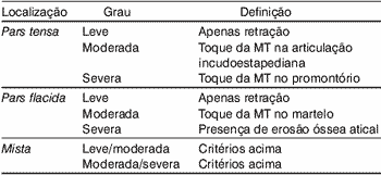
Otoendoscopy of CLE was revised and the ears were classified as normal or abnormal. Alterations were classified as effusion, retraction, perforation, cholesteatoma or presence of sequels (presence of neotympanum or tympanosclerosis). Tympanic membrane retraction was divided as belonging to the pars tensa, pars flaccida or mixed (when both portions were retracted). They were classified as mild, moderate and severe, according to the criteria shown in Table 1. Data about the CLE of each patient were recorded in the specific protocols.
All patients authorized storage of data and recording of otoendoscopy.
RESULTS
Out of 108 studied patients, 64 (59.2%) presented NCCOM and 44 (40.8%) presented CCOM. By analyzing the groups as a whole, 84 (77.8%) of them presented some of the alterations listed above detected at the otoendoscopy of the CLE.
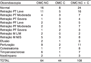
In the group of patients with NCCOM, 16 (25%) presented normal tympanic membrane (TM). The most frequent alterations in the remaining 75% were TM retractions, present in 28 patients with NCCOM, divided into 16 cases of isolated pars tensa retraction (11 mild, 4 moderate and 1 severe), 5 cases of pars flaccida retraction (2 mild, 2 moderate and 1 severe) and 7 cases of mixed retractions pars flaccida and tensa (2 mild/moderate and 5 moderate/severe). The remaining detected alterations were: perforation in 9 patients (14%), tympanosclerosis in 6 (9.37%), neotympanum in 2 (3.1%), effusion in 2 (3.1%) and cholesteatoma in 1 case (1.56%).
Among patients with CCOM, 8 (18.1%) presented the CLE without abnormalities at the otoendoscopy. The most frequent abnormalities in this group were also TM retraction, totaling 24 cases. Pars tensa alterations were described in 11 patients (5 mild, 3 moderate and 3 severe), pars flaccida in 10 patients (3 mild, 5 moderate and 2 severe), whereas mixed retraction was present in 3 patients (all of them moderate/severe). The second most common abnormality in the group of patients with CCOM was the presence of cholesteatoma on the CLE: 15.9% of the cases (7). The other abnormalities detected were: presence of fluid (1), perforation (2), neotympanum (1) and tympanosclerosis (1). Table 2 describes the abnormalities found for NCCOM and CCOM separated and together.
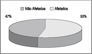
Once we excluded the presence of mild retractions, fluid in the middle ear, tympanosclerosis or neotympanum, trying to assess the ears with more significant affections, we found a total of 46.3%, being 25 (57%) of CLE of NCCOM and 25 (39%) of CLE of CCOM (Graph 1). The difference between the two groups was not statistically significant.
Upon globally analyzing the groups of retractions, 21 patients presented mild retraction, 16 moderate retractions and 15 patients had severe retractions. Out of the total number of patients, 48.1% presented some degree of tympanic membrane retraction.
Graph 1. Distribution of abnormal contralateral ear in patients with COM, ignoring mild retractions, presence of effusion, neotympanum and tympanosclerosis.
Tabela 1. Classificação das retrações da membrana timpânica. MT: membrana timpânica.
Tabela 2. Descrição das otoendoscopias das orelhas contra-laterais (OCL) estudadas OMC NC: otite média crônica não colesteatomatosa; OMC C: otite média crônica colesteatomatosa; PT: pars tensa; PF: pars flacida. M: mista. L/M: leve/moderada. M/S: moderada/severa.
DISCUSSION
COM has been classified in two main groups: cholesteatomatous chronic otitis media (CCOM) and non-cholesteatomatous chronic otitis media (NCCOM)4. There is also silent chronic otitis media in which there are irreversible tissue affections present in the auditory region, but with no contiguity to the tympanic membrane1, 6, 14. The term simple COM (sometimes used to characterized NCCOM) does not seem to be appropriate, since the absence of cholesteatoma does not guarantee that the chronic otitis media is a "simple" affection.
Although COM classifications are didactically clear, this is a topic that still generates controversy in the literature, showing the difficulty most authors have to describe in a static fashion such a dynamic pathology. According to the theory of continuum, there is no acute otitis, serous otitis media, secretory otitis media, cholesteatomatous and non-cholesteatomatous pathologic specific affections with beginning, middle and end. It is the same disease going through growing evolution stages of epithelial and subepithelial events. The transition to one stage or the other depends basically on the aggressive factor, the affected ear and the therapeutic intervention received by the patient in the course of the disease5, 10, 15.
Concomitance of middle ear chronic pathologic processes have been addressed by some studies13, 16. However, few studies have been published describing the findings of otoscopy in the CLE of patients with COM. Chalton & Stears12 published the first study in 1984 about the findings. The authors assessed CLE of 73 patients submitted to wall-down tympanomastoidectomy because of acquired cholesteatoma and found 53.4% of abnormal CLE, and pars tense retraction was the most frequent one. In 1996, Vartianen et al.16 (1996) described a series of 493 CLE of patients with otological surgery indication for COM (CCOM and NCCOM). The authors found 63% of CLE with some abnormality (defined as severe retraction, perforation or cholesteatoma), and retraction was also the most frequent finding. However, none of them defined in a direct and clear fashion the criteria adopted to classify the alterations found, similarly to the fact that there was no standardization of description.
Our series showed high prevalence of CLE alterations in patients who had COM. Upon the exclusion of mild retractions and sequels, we analyzed only patients with the most significant CLE and found prevalence of 46.3% of abnormal CLE. The findings confirmed the tendency to bilateral affection of chronic middle ear diseases.
We believe in the importance of valuing CLE findings in cases of COM, regardless of being NCCOM or CCOM, because of two key aspects: understanding of pathogenesis of COM and treatment and education of patients.
When we study the two ears of the patient, that is, when we take into account the CLE, we can understand the dynamic pathologic process that the patient shows us. There are a large number of patients that clearly show the different stages of as continuous COM pathologic process. They are patients that on the one side present attic classical cholesteatoma and on the CLE they present an attic retraction pocket, (still) cholesteatoma-free. Or cases on the one side with major central perforations of the tympanic membrane and on the CLE they present diffuse moderate perforation with clear effusion present in the cavity.
Precise and critical analysis of both ears of a specific patient, in our opinion, has a key role in the prognostic assessment of the case, since the ear established with COM may help guide us towards the likely evolution of the CLE. If a patient presents on one side aggressive cholesteatoma with significant ossicle destruction and unfavorable evolution, and on the CLE he presents clean attic retraction pocket, we should carefully follow up this patient because we believe that the "affected" ear is showing us that the retraction pocket is very likely to present an unfavorable evolution.
Another situation is the one experienced by many patients submitted to tympanotomy with bilateral ventilation tube. In many occasions, one of the tubes is extruded early and the other is maintained in situ for varied and, sometimes, prolonged periods of time. Although it is not part of our routine to remove ventilation tubes, sometimes we consider the hypothesis as a result of the patient's anxiety to restore normal life routine. In order to decide whether the ventilation tube can be removed or not, we should investigate the CLE. If it presents normal characteristics, we will be able to perform the procedure since the CLE is showing us that there are no trends towards progression of the diseases.
Such situations represent what we informally name in our ambulatory the "Orloff ® effect". Orloff® is a popular drink in Brazil whose advertisement slogan is "I am today what you will be tomorrow".
CONCLUSION
Patients with COM diagnosed in one ear are very likely to present associated CLE affections, and the most frequent one is tympanic membrane retraction.
Consequently, the CLE should always be comprehensively studied in patients with unilateral COM in order to early diagnose the alterations and, if necessary, provide timely therapeutic intervention.
REFERENCES
1. Costa SS, Paparella MM, Schachern PA et al. Histopathology of chronic otitis media with perforated and non-perforated tympanic membrane. Presented at Midwinter Meeting of the Association for Research in Otolaryngology. Clearwater; 1989.
2. Meyerhoff WL, Kim CG, Paparella MM. Pathology of chronic otitis media. Ann Otol Rhinol Laryngol 1978;87(6):749-61.
3. Brown OE, Meyerhoff WL. Complications and sequelae of chronic suppurative otitis media. Ann Rhinol Otol Laryngol ; 1988: 97(131):38-40.
4. Costa SS, Souza LCA, Piza MRT. The flexible endaural tympanoplasty. Pathology-guided, pathogenesis-oriented surgery for the middle ear. Otolaryngol Clin North Am 1999;32(3): 413-441.
5. Junh SK, Paparella MM et al. Pathogenesis of otitis media. Ann Otol Rhinol Laryngol 1977;86(4):481-93.
6. Meyerhoff WL, Giebink GS, Shea DA. Silent otitis media: an animal study. Ann Otol Rhinol Laryngol 1984;93:136-9.
7. Paparella MM. Middle ear effusions. Definitions and terminology. Ann Otol Rhinol Laryngol 1976;85(25):8-11.
8. Sade J, Ar A. Middle ear and auditory tube. Middle ear clearance, as exchange and pressure regulation. Otolaryngol Head Neck Surg 1997;116:499-524.
9. Giebink GG, Ripley MI et al. Clinical histopathological correlations in experimental otitis media: implications for silent otitis media in humans. Pediatr Res 1985;19(4):389-97.
10. Paparella MM, Hiraide F, Juhn SK, Kaneco J. Cellular events involved in middle ear fluid production. Ann Rhinol Otol Laryngol 1970;79(4):766-79.
11. Casebrant M, Brostoff LM et al. Otitis media with effusion in preschool children. Laryngoscope 1985;95:428-36.
12. Chalton R, Stearns M. The incidence of bilateral chronic otitis media. J Laryngol Otol 1984;98:337-9.
13. Vartiainen E, Kansanen M, Vartiainen J. The contralateral ear in patients with chronic otitis media. Am J Otol 1996;17:190-2.
14. Costa SS. Contribuição ao estudo da otite média crônica. [Dissertação de Mestrado]. Faculdade de Medicina de Ribeirão Preto, USP; 1991.
15. Costa SS, Ruschel C, Cruz OL, Paparella MM. Otites Médias - Aspectos Gerais. In: Cruz OL & Costa SS. Otologia clínica e cirúrgica. 1999 b. Ed Revinter.
16. Vartiainen E, Kärjä J. Bilateral Otitis Media. Arch Otorhinolaryngol 1986; 243:190-3.
1 Undergraduate, Medical School, Federal University of Rio Grande do Sul.
2 Resident physician, Service of Otorhinolaryngology, Hospital de Clínicas, Porto Alegre.
3 Undergraduate, Medical School, Lutheran University of Brazil.
4 Biologist with the Study Group on Middle Ear Pathology, Hospital de Clínicas, Porto Alegre.
5 Master studies under course, Department of Surgery, Medical School, Federal University of Rio Grande do Sul.
6 Otorhinolaryngologist.
7 Joint Professor, Department of Ophthalmology and Otorhinolaryngology, Federal University of Rio Grande do Sul.
Study Group on Middle Ear Pathology - GEPOM
Service of Otorhinolaryngology - Hospital de Clínicas de Porto Alegre - HCPA
Address correspondence to: Mariana Magnus Smith - R. Artur Rocha, 825 - Porto Alegre - RS - CEP 90459-171 - Tel: (55 51) 3332.6002 E-mail: marimagnussmith@hotmail.com
Article submitted on November 8, 2001. Article accepted on December 13, 2001.
Print: ![]()