

Year: 2002 Vol. 68 Ed. 2 - (11º)
Artigo Original
Pages: 224 to 228
 PDF PT
 Orbital complications in patients with acute sinusitis
Author(s):
Richard Louis Voegels 1,
Fábio Tadeu Moura Lorenzetti 2,
Walmir Eduardo P. A. D'Antonio 3,
Cláudio Márcio Y. Ikino 3,
Ossamu Butugan 4
Keywords: sinusitis, orbital complications, orbital abscess
Abstract:
Introduction: The orbital complications of sinusitis continue to be an important topic in otolaryngology despite the reduction in their prevalence observed after the advent of antibiotic therapy. Aim: With the objective to determine the clinical and therapeutic characteristics of cases of complicated sinusitis. Study design: Clinical prospective randomized. Material and method: We conducted a retrospective study on 128 patients admitted to the Department of Otorhinolaryngology, University of São Paulo Medical School, over the last 15 years. Most of these patients were children or young adults (82,81% were less than 30 years old) and 57,81% were males. All of the patients had a favorable outcome, with no sequels. Conclusion: An early diagnosis and the institution of adequate treatment are of fundamental importance for the prognosis and outcome of these patients. The observation of these two factors contributes to the low mortality of these conditions.
![]()
INTRODUCTION
Nasosinusal infections are highly prevalent clinical entities. However, secondary complications to these affections have clearly decreased after the advent of antibiotic therapy1, 2, 3, 4, 5. In spite of that, high morbidity and occasional mortality associated to sinusitis complications justify the careful assessment of all cases of acute or chronic sinuses pathologies and prompt investigation of any orbital, intracranial or bone complications6, 9, 10, 11, 12. Acute sinusitis seems to be the most frequent cause of orbital infections1, 4, and they are responsible for 50-75% of all intracranial abscesses9, 13.
Orbital complications of sinusitis have been known for more than 60 years 14. In 1937, Hubert made the first description of a case of acute sinusitis with orbital complications, proposing the first classification of orbital complications of sinusitis14, which was later modified by Smith & Spencer and then in 1970, by Chandler et al1,4,7. In 1983, Moloney reviewed Chandler's classification and emphasized the difference between orbital abscess and cellulitis, and finally, in 1997, Mortimer et al. proposed to exclude cavernous sinus thrombophlebitis from the classification of orbital complications of sinusitis, since it is a central affection.
The great importance of sinusitis as a source of orbital infection may reside in some of the anatomical characteristics of paranasal and orbital cavities that seem to facilitate the dissemination of an infectious process, as follows:
1. the presence of a thin bone lamina - papyraceous lamina - dehiscent in some points, that separates the anterior ethmoidal sinus mucosa from the orbital content;
2. venous drainage of the paranasal sinuses through non-valve veins, enabling free blood flow between the ethmoid, orbit and intracranial content;
3. orbital septum or palpebral fascia, which is a deflection of the periorbital region, separating the pre-septal content from the post-septal space (involves the orbit)1, 7, 8, 9.
Therefore, we observed that dissemination routes of sinusitis may be hematogenic, by contiguity and by continuity1, 4, 9, 11, 14, 15.
Orbital complications used to present mortality rates of up to 17% in pre-antibiotic age, in addition to visual loss in another 20%14. Intracranial complications today have a high mortality rate of approximately 11%6,8,9,13,16,17,18,19.
The objective of the present study was to present clinical, radiological, treatment and follow-up characteristics of patients hospitalized in the last 15 years in the Clinical Otorhinolaryngology of Hospital das Clínicas, Medical School, University of São Paulo (HC-FMUSP), with diagnosis of acute sinusitis and orbital complications.
MATERIAL AND METHOD
The authors conducted a retrospective study using medical chart analysis of 128 patients hospitalized in the Clinical Otorhinolaryngology of HC-FMUSP between December 1982 and December 1997, with diagnosis of acute sinusitis with orbital complication, including only patients who had been followed-up in the ambulatory for at least 6 months after hospital discharge. We excluded patients who had mucocele or complications of chronic sinusitis, and cavernous sinus thrombophlebitis (Chandler V), since it is considered an intracranial complication.
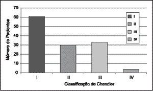
Patients were divided into 4 groups according to the diagnosed orbital complication, following the proposal set forth by Chandler et al, 1970: Chandler I- pre-septal complication; Chandler II- orbital cellulitis; Chandler III- subperiosteal abscess, and Chandler IV- orbital abscess.
We analyzed the following data: sex, age, main complaint and duration, radiology (x-ray and Computed tomography scan - CT scan), affected paranasal sinuses (by radiological assessment or surgery description), diagnosis, clinical or surgical treatment performed, and evolution.
RESULTS
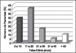
In our study, ages ranged from 10 months to 62 years of age, mean age of 17.39 years. The distribution of patients according to age range was 0-10 years - 28.91% of the patients; 11-20 years - 40.63%; 21-30 years - 16.41%; 31-40 years - 4.69% and over 40 years - 9.38% (Figure 1). Seventy-four patients (57.81%) were male subjects, amounting to a male/female ratio of 1.34:1.
We observed that 71 patients (55.47%) presented the complaint for less than 5 days. The most common symptoms were periorbital edema (118 patients - 92.19%), headache (64 cases - 50.00%) and rhinorrhea (41 patients - 32.03%).
One hundred and nineteen patients (92.97%) were submitted to simple x-ray of the paranasal sinuses, whereas 91 patients (71.09%) were submitted to CT scan (axial and coronal sections) of paranasal sinuses and/or orbit and/or skull. The 9 (7.30%) patients that did not undergo simple facial x-ray presented clinical picture typical of post-septal complication, and they were immediately submitted to CT scan of the paranasal sinuses and orbit. Thirty-seven patients (28.91%) did not undergo paranasal sinuses CT scan. They all presented picture of palpebral cellulitis with good clinical evolution during hospitalization.
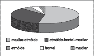
Patients were distributed according to the affected paranasal sinuses, and this distribution is presented in Figure 2.
The distribution of patients according to Chandler's classification of orbital complications is presented in Figure 3.
Intravenous antibiotic therapy was indicated in all cases. In 83 cases (64.84%) of patients (47 patients Chandler I, 24 Chandler II and 12 Chandler III) there was excellent evolution only with clinical treatment. In 45 cases (35.16%) surgical approach was required: of the total, 14 were classified as Chandler I (22.95% of the patients with this diagnosis), and 13 presented palpebral abscess and 1 was submitted to sinusectomy because of unsatisfactory clinical evolution; 6 patients with Chandler II (20%); 21 patients with Chandler III (63.63%), and 4 patients with Chandler IV (100%).
All patients progressed to significant improvement or complete remission of orbital and sinusal manifestations upon hospital discharge. Seven patients (5.47%) presented recurrence after hospital discharge, requiring new treatment intervention after which they finally had remission of the affection, without new events. There was no mortality or cases of ophthalmologic sequelae detected during ambulatory follow-up.
Figure1. Distribution of patients according to age range.
Figure 2. Distribution of patients according to affected paranasal sinus.
Figure 3. Distribution of patients according to Chandler's classification of orbital complications.
DISCUSSION
As observed in the present study, there is a high predominance of affection of subjects in the 1st and 2nd decades of life. These findings coincide with the reports made by different authors4,9,11,20, although the reason for such distribution is not clearly defined. Singh et al21 reported in the literature that the degree of pneumatization of ethmoid bone in childhood facilitates the progression of infectious processes, which justifies the higher occurrence of complications of acute sinusitis in children and young adolescents.
The duration of complaint (less than 5 days) is a variable rarely reported in the studied literature. Mortimer et al (1997) found a mean duration of complaint of 8 days.
As to initial symptoms, our findings (periorbital edema, headache, rhinorrhea and fever) are compatible with those reported by other authors20, 21. We point out to the high rates of orbital bulging in such patients (92.19% in our sample), also reported by other studies6,8,12,13,16,17,18,19,21,22.
As to complementary diagnosis, simple paranasal sinuses x-rays were performed in 92.97% of the patients, whereas CT scan were ordered in 71.09% of the patients. According to Chandler et al (1970), diagnosis of sinusitis with orbital complications may be performed with simple paranasal sinuses x-rays and careful clinical assessment. In recent studies, the authors described the need to perform paranasal sinuses CT scan as the first choice for diagnosis, classification and follow-up of these patients4,10,11,14,20,21. In our study, patients hospitalized between 1982 and 1987 with diagnosis of palpebral cellulitis and acute sinusitis with good evolution after clinical treatment were those who did not undergo paranasal sinuses CT scan. The use of magnetic resonance should be reserved for those cases in which even after investigation, there are still doubts concerning potentially severe affections, such as cavernous sinus thrombophlebitis 11, 13, 17, 18.
The paranasal sinuses most frequently affected were maxillary, ethmoid and frontal, as previously reported9,10,11,14,15,22.
Patients with orbital complications usually presented initial manifestation of palpebral cellulitis, enabling isolated clinical treatment (intravenous antibiotic therapy) in most cases that were classified as Chandler I (77.05%). Out of the patients with Chandler I submitted to surgical procedures, drainage of palpebral abscess under local anesthesia was the most widely performed treatment. Endonasal sinusectomy was necessary in only one patient, indicated after 48 hours of clinical treatment, when the evolution was not considered satisfactory. These findings were compatible with the data reported in the literature1,6,7,10,11,15,22.
Subperiosteal abscesses (Chandler III) were the second most frequent complication. They required surgical treatment in 63.63% of the patients, but 32.32% presented complete spontaneous remission after clinical treatment. The patients presented unclear clinical manifestation concerning post-septal complication, and paranasal sinuses CT scan was performed later (24 to 48 hours after hospitalization). At that time, small subperiosteal collections were observed, but maintenance of conservative treatment was adopted since the clinical evolution was satisfactory. In fact, the literature is controversial about the indication of surgery in such cases. Some authors advocate formal surgical intervention whenever subperiosteal collection is diagnosed 7, 9, 21, whereas others believe that it is possible to maintain clinical conservative treatment in some cases, followed by careful assessment of visual acuity1, 6, 10, 14, 22. Among the latter, Backer (1995) reported how difficult it is to radiologically differentiate between subperiosteal cellulitis (in which clinical treatment could be satisfactory) and the presence of small subperiosteal collections (which would require surgical intervention), and the author found in his study a 20% rate of false positive results with CT scan. According to Backer (1995), in addition to other authors1, 6, 10, 14, 22, the surgery should be indicated after 48 hours of treatment if there is no improvement of proptosis, chemosis, ocular extrinsic mobility impairment, or visual acuity compromise. In our service, we normally indicate early surgical procedure in patients classified as Chandler III.
Orbital cellulitis (Chandler II) affected 23.43% of the patients, being that 20% required surgery. Such data coincide with what was reported by previous studies6, 10, 11, 22. Orbital abscess (Chandler IV) was observed in 3.21% of the cases. In such patients, the likelihood of vascular compression and optic nerve ischemia leading to amaurosis requires emergency surgical decompression11. It justifies the indication of surgery in 100% of the cases. The abscess may be a consequence of the organization of orbital cellulitis or rupture of the periorbit in cases of subperiosteal abscess1, 4, 6, 7, 15, 18.
We did not observe cases of fatal evolution or late sequelae in our patients, despite the relatively high rate of mortality/morbidity reported by the literature5. We believe that intensive clinical treatment, associated to early surgical indication in cases of more aggressive complications and/or unsatisfactory clinical evolution were the primordial reasons for us to reach such results.
CONCLUSION
The 128 patients hospitalized in the Division of Clinical Otorhinolaryngology, HCFMUSP, were mainly in their 20's, male patients, who complained of palpebral bulging, headache and rhinorrhea, usually for less than 5 days.
All patients were treated with intravenous antibiotic therapy, requiring surgical intervention in 45 cases. Most of the patients submitted to surgery (21 cases) had subperiosteal abscess. All patients presented satisfactory evolution and there were no cases of sequelae or death.
The authors concluded that early diagnosis together with appropriate clinical therapy and precise surgical indication are essential conditions to prevent fatal evolution or irreversible sequelae in patients that present orbital complications of sinusitis.
REFERENCES
1. Chandler JR, Laugenbrunner DJ, Stevens ER. The pathogenesis of orbital complications in acute sinusitis. Laryngoscope 1970;80:14148.
2. Dale BAB, Mackenzie IJ. The complications of sphenoid sinusitis. J Laryngol Otol 1983;97:661-70.
3. Giannoni CM, Stewart MG, Alford EL. Intracranial complications of sinusitis. Laryngoscope 1997;107:863-7.
4. Ognibene RZ, Voegels RL, Bensadon RL, Butugan O. Complications of sinusitis. Rhinology 1994;8(4):175-9.
5. Pirana S, Ognibene RZ, Sitchin G, Butugan O, Silveira JAM, Miniti A. Doenças do seio esfenoidal: relato de 4 casos. Rev Bras de Otorrhinolaringologia 1996;62(1):53-60.
6. Backer AS. Role of anaerobic bacteria in sinusitis and its complications. Ann Otol Rhinol Laryngol 1995 [Suppl.] 154:17-22.
7. Stankiewicz JA, Newell DJ, Park AH. Complications of inflammatory diseases of the sinuses. Otolaryngologic Clinics of North America 1993;26(4):639-550.
8. Stoll D, Dumon TH, Adjibabi W. Sphénoidites inflammatoires isolées compliquées. Ver Laryngol Otol Rhinol 1997;118(2):87-9.
9. Wagenmann M, Naclerio RM. Complications of sinusitis. J Allergy Clin Immunol 1992;90(3):552-6.
10. Healy GB. Classics of Laryngoscope: Chandler et al. Laryngoscope 1997;107:441-6.
11. Mortimer S, Wormald PJ. The Groote Schur Hospital classification of the orbital complications of sinusitis. J Laryngol Otol 1997; 111:71923.
12. Xenos C, Rosenfeld JV, Kleid SM. Intracranial extension of sphenoid sinusitis. Head & Neck 1995;7(4):346-50.
13. Maniglia AJ, Goodwin J, Arnold JE, Ganz E. Nasal, sinus and orbital infections in adults and children. Arch Otolaryngol Head Neck Surg 1989;115:1429-9.
14. Clary RA, Cunningham MJ, Eavey RD. Orbital complications of acute sinusitis: comparison of computed tomography scan and surgical findings. Ann Otol Rhinol Laryngol 1992;101:598-600.
15. Banfield GK, Daya H. Sinusitis, the orbital and visual complications. Br J Hosp Med 1996;55:656-7.
16. Chua R, Shopiro S. A mucopiocele of the clivus: case report. Neurosurgery 1996;39(3): 589-90.
17. Jones RL, Violaris NS, Chauda SV, Pahor AL. Intracranial complications of sinusitis the need of aggressive management. J Laryngol Otol 1995;109:1061-2.
18. Kriss TC, Kriss VM, Warf BC. Cavernous sinus Thrombophlebitis: case report. Neurosurgery 1996;39(2):585-9.
19. Singh B. The management of sinogenic orbital complication. J Laryngol Otol 1995;109:300-3.
20. Sassiad I, Riding K. Orbital complications of ethmoiditis BC Children´s Hospital experience, 1982-89. J Otolaryngol 1991;20(6):400-3.
21. Singh B, Dellen JV, Ramjettan S, Maharaj TJ. Sinogenic intracranial complications. J Laryngol Otol 1995; 109:945-50.
22. Mann W, Amedee RG, Mauree J. Orbital complications of Pediatric sinusitis: treatment of periorbital abscess. Ann J Rhinol 1997;11(2):14953.
1 MD, Ph.D., Professor, Clinical Otorhinolaryngology, Hospital das Clínicas, FMUSP.
2 MD, Ph.D., Professor, Clinical Otorhinolaryngology, Hospital das Clínicas, FMUSP.
3 MD, Former residents, Clinical Otorhinolaryngology, Hospital das Clínicas, Medical School, University of São Paulo (FMUSP).
4 Associate Professor, Discipline of Otorhinolaryngology, FMUSP.
Study conducted at the Division of Clinical Otorhinolaryngology, Hospital das Clínicas, FMUSP.
Address correspondence to: Dr. Richard Louis Voegels - Rua Pedroso Alvarenga, 1255 cj. 26/27
CEP 04531-021 São Paulo, SP Brazil - E-mail: voegels@ibm.net
Article submitted on October 27, 1999. Article accepted on December 27, 2001.
Print: ![]()