

Year: 2002 Vol. 68 Ed. 2 - (10º)
Artigo Original
Pages: 219 to 222
 PDF PT
 A proposal for a practical method of sialometry
Author(s):
Daniella B. Pupo 1,
Ivo Bussoloti Fo 2,
Bianca M. Liquidato 3,
Gustavo P. Korn 4
Keywords: xerostomia, Sjögren's syndrome, sialometry
Abstract:
Introduction: Xerostomia is a subjective complaint that may or not be related to decrease in salivation. It should be always investigated because it is frequently associated to systemic diseases, including Sjögren's syndrome. Despite there is not a universal standard to identify hiposalivation, we consider that sialometry is an important tool for treatment evaluation through serial exams in the same patient. After the analysis of several sialometry techniques, we opted to adapt and to standardize the Bolwig and Rafaelsen method (1972) for being the most practical and reliable method among all. We pursue the large-scale use of this method. Aim: To evaluate the results of a systematized sialometry method. Material and method: 10 subject were evaluated, 5 assymptomatic and 5 with xerostomy. The subjects were submitted to a silalometry pre and post stimulus. The results were corroborated. Results: All individuals presented informed salivary thru measurable by the method. Conclusion: The method is easy and trustworthy to factory.
![]()
Introduction
Xerostomia is the subjective sensation of dry mouth as a consequence or not of reduction/interruption of the salivary gland function1, 2, 3, 4, 5, 6, 7. Xerostomia is more frequent among the elderly8 and female patients9. It may manifest by difficulty to masticate, swallow and speak, burning sensation in the tongue and mouth, reduction of gustatory sensation, mucositis and ulcerations in the mouth10. Oral flora affections predispose to opportunistic infections, especially by Candida albicans, and contribute to the proliferation of cariogenic microorganisms. Thus, patients with xerostomia are predisposed to decays and periodontal diseases11.
In most cases, xerostomia is part of a systemic condition and it may be associated with extra-oral symptoms, such as skin, vagina and eye drying9, 12. The most common causes of xerostomia are auto-immune diseases, especially Sjögren's syndrome, the use of drugs that reduce saliva flow and irradiation of salivary glands2, 5. There are, nevertheless, various others conditions associated to this symptom, including psychiatric affections (especially depression)2, 4, 6, 13, salivary gland obstruction and/or infection10, diabetes melitus7 and dehydration13.
The therapeutic approach of the patient with xerostomia varies according to some specific individual characteristics. The treatment aims at relieving symptoms, preventing or correcting sequelae of salivary dysfunction and the cure of the associated systemic disease, but it all depends on the amount of functioning glandular tissue.
The confirmation that at least part of the salivary gland may be stimulated, increasing its production, allows us to start treatment with topical stimulants. Systemic stimulation, represented mainly by the use of pilocarpine, is not frequently used in Brazil owing to its great number of side effects14, 15. Patients whose salivation can not be stimulated because of lack of healthy and functioning glandular tissue have the alternative of using artificial saliva3, 5. Thus, it is extremely important to measure the salivary flow before and after topical stimulation so that we can determine which treatment to use.
Sialometry may be used in two ways: to measure the total saliva produced or to measure the saliva produced by each individual gland. The salivary flow of parotid, submandibular and sublingual glands may be determined independently by using specialized collectors that are placed over the orifices of Stenson & Wharton ducts6. The secretion of minor salivary glands may be quantified with the use of cellulose stripes adhered to the internal aspect of the lips, hard palate and jugal mucosa16.
The total saliva production is easily measured; this method is enough to quantify hyposalivation. In such cases, salivary flow may be determined with or without previous stimulation. As previously exposed, both dosages are important and should be analyzed consecutively. There are different sialometry methods available. Among them, we highlight the one proposed by Sreebny & Valdini (1987)17, in which the patient expels passively, through a collecting tube, the saliva that is accumulated in the mouth every 2 minutes, for a total of 6 minutes, allowing saliva swallowing only between the 2-minute intervals. The second part of the test is performed similarly, but with previous stimulation, which may be mastication (paraffin tablets) or gustatory stimulus (citric acid). Salivary flow is stimulated and the weight of the tube before and after collection is compared, then converted in ml/min. Values that are below 0.1ml/min without stimulus or below 0.5ml/min with stimulus are considered abnormal17. This method accommodates some modifications, such as when the patients "spit" in the collecting tube the saliva accumulated in the mouth2, 3 or when the saliva is aspirated through specific collecting tubes18.
Another method of sialometry that is used by different authors but without any formal standardization consists of placing two cotton balls previously weighed on the floor of the mouth, close to the internal aspect of the gingival region, where they shall remain for some seconds before being weighed again. The difference between the weights is converted into ml/min19, 21, 21. In our opinion, this is the easiest and most reliable test and that is why we proposed to standardize it so that it can be used in large scale.
Material and method
We have randomly selected 5 patients who came to the Department of Otorhinolaryngology, Santa Casa de São Paulo, in April/2001, complaining of different otorhinolaryngological complaints that did not manifest oral signs and symptoms of any nature and without any known predisposing factors to xerostomia. Three of the patients were female and 2 were male and their ages ranged from 40 to 63 years (mean age of 54 years). We also selected the 5 first patients with Sjögren's syndrome seen in our ambulatory of Stomatology in March/2001. They were all women, aged 39 to 72 years (mean of 55 years). The 10 patients were submitted to sialometry with and without stimulation.
We prepared 6 cotton balls to each patient, arranged in groups of two. Each pair of cotton balls was placed in a plastic container (an 80ml universal collector) and the set was weighed in a scale model Acculabr V1200. The weight of the set varied from 14.5g to 15.5g. Patients were then submitted to 3 consecutive sialometry procedures. Initially, we asked patients to swallow the saliva from their mouths and then we placed two cotton balls on the floor of the mouth of the patients, close to the internal aspect of gums on both sides. The cotton remained in place for 2 minutes and the patients were asked not to swallow saliva during this period. We removed the cotton soaked in saliva and placed it again in the plastic container and the set was weighed again. Salivary flow was then stimulated with a solution that contained citric acid 2.5%, aspartame 2.5g and saturated with calcium carbonate: we placed two drops on the lingual back portion of patients who were instructed to immediately swallow after this procedure, and then they were submitted to a new sialometry following the procedure described before. The last stage of the test was the over stimulation of salivary flow, useful in cases in which the salivary gland is severely compromised and does not respond to a single stimulus. We instilled 2 drops of the solution with citric acid on the back of the tongue, patients swallowed all the saliva and then the last pair of cotton balls was placed on the floor of the mouth. During the 2 next minutes, citric acid was again instilled at regular intervals of 30s, amounting to 8 drops (0 sec, 30sec, 60 sec and 90 sec). The differences between the weights before and after sialometry in their three stages were converted into ml/min.
Results
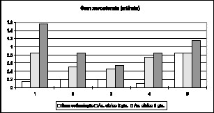
The results collected from the study are presented in the Tables that follow.
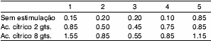
Table 1. Patients without xerostomia (ml/min).
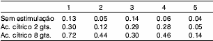
Table 2. Patients with xerostomia (ml/min).
DISCUSSION
The method of sialometry proposed here is an adaptation of the technique described by Bolwig and Rafaelsen in 197222, and previously used by different authors19, 20, 21, but with no standardization. We believe the method is practical, reliable and non-invasive. Other techniques are more frequently used, based on active or passive expectoration of saliva through collecting tubes for a pre-defined period of time3, 5, 23. We consider this kind of sialometry inappropriate for the elderly who usually have compromised oral motor coordination, impairing the validity of the results, especially when we consider that xerostomia is very prevalent in this age range. The dosage of saliva produced by each independent gland is a reliable method but difficult to perform and should be reserved for cases in which quantitative analysis of saliva is required. It should be performed considering that total saliva measurement is contaminated by food debris, epithelial desquamation and serum fluids from the gingival juice3, 24. The quantitative dosage of the salivary flow produced by one single gland also requires this type of technique but this information rarely influences diagnosis/treatment of xerostomia.
Patients who are going to undergo sialometry should avoid eating, drinking, smoking, brushing the teeth, or chewing gum two hours before the test. Saliva should be collected in a quiet room, after a period of at least 2-hour fast. Serial sialometry in the same patient should start at the same time of the day because the salivary flow varies according to the circadian rhythm6, 8, 17, 25. These cares were not strictly observed in the present study because the authors wanted only to demonstrate the proposed sialometry technique but such norms will be incorporated in future trials in order to add to data reliability.
There is no universally accepted standard of sialometry for hyposalivation or exclusion of this hypothesis6. The results obtained sometimes do not correspond to signs and symptoms manifested by the patients. Salivary flow varies a lot, including in asymptomatic people2, a fact observed in our sample, in which patient 5 presented salivary flow without stimulation nearly 6 times greater than that of patient 1. Nevertheless, we adopted the criteria by Sreebny & Valdini (1987)17, that stated that abnormal salivary flow is below 0.1 ml/min without stimulation and below 0.5ml/min with stimulation. Based on these criteria, we observed that all patients with Sjögren's syndrome presented hyposalivation. It is important to point out again that xerostomia is a subjective complaint and sometimes it is not compatible with the salivary flow presented by the patient. We decided to perform a 3-stage sialometry because the initial stimulation with only 2 drops of citric acid is sometimes not enough to increase salivary flow (example 5, Table 1), requiring additional stimulation to confirm that the gland tissue can still be stimulated.
Sialometry helps choosing the treatment to be implemented, but also enables assessment of its efficacy by means of performing serial tests in the same patient. In our opinion, the main purpose of this test is to compare pre and post-treatment outcomes. Small increases in salivation tend to benefit substantially the patients that produce little or no saliva, whereas equivalent increase in patients with higher salivary flow may not trigger any improvement26. We also advocate the performance of sialometry before and after procedures/treatments that may promote hyposalivation (for example, head and neck radiotherapy). This simple procedure will help quantify the impairment caused to saliva production.
Conclusion
The method proposed by the authors is simple and reliable provided that some standards are followed, such as previous preparation of patients (feeding, brushing the teeth, etc.). Sialometry facilitates the study of xerostomia thanks to its clinical and scientific applications.
REFERENCES
1. Anttila SS, Knuutilla MLE, Sakki TK. Depressive symptoms as an underlying factor of the sensation of dry mouth. Psychosom Med 1998;60(2):215-8,.
2. Field EA, Longman LP, Bucknall R, Kaye SB, Higham SM, Edgar WM. The establishment of a xerostomia clinic: a prospective study. Br J Oral Maxillofac Surg 1997;35:96-103.
3. Fox PC. Management of dry mouth. Dent Clin North Am 1997;41(4):863-75.
4. Longman L.P, Higham S.M, Bucknall R, Kaye S.B, Edgar W.M, Field E.A.- Signs and symptoms in patients with salivary gland hypofunction. Post. Grad. Med. J.
5. Narhi TO, Meurman JH, Ainamo A. Xerostomia and hyposalivation. Causes, consequences and treatment in the elderly. Drugs Aging 1999;15(2):103-16.
6. Sreebny LM. Recognition and treatment of salivary induced conditions. Int Dent J 1989;39:197-204.
7. Sreebny LM, Yu A, Green A, Valdini A. Xerostomia in diabetes mellitus. Diabetes Care 1992;15(7):900-4.
8. Osterberg T, Landahl S, Hedegard B. Salivary flow, saliva, pH and buffering capacity in 70-year-old men and women. J. Oral Rehab 1984;11:157-70.
9. Epstein J.B, ScullyC. The role of saliva in oral health and the causes and effects of xerostomia. J. Can. Dent. Assoc. 58(3):217-21 1992.
10. Crockett DN. Xerostomia: the missing diagnosis? Aust Dent J 1993;38(2):114-8.
11. Astor CA, Hanft KL, Ciocon JO. Xerostomia: A prevalent condition in the elderly. Ear nose throat J 1999;78(7):476-9.
12. Sreebny LM, Valdini A. Xerostomia. A neglected symptom Arch Intern Med 1987;147:1333-7.
13. Ettinger RL. Review: Xerostomia: a symptom which acts like a disease. Age Ageing 1996;25:409-12.
14. Hamlar DD, Schuller DE, Gahbauer RA, Buerki RA, Staubus AE, Hall J, Altman JS, Elzinga DJ, Martin MR. Determination of the efficacy of topical oral pilocarpine for post-irradiation xerostomia in patients with head and neck carcinoma. Laryngoscope 1996;106:972-6.
15. Van der Reijden WA, Vissink A, Veerman ECI, Amerongen AVN. Treatment of oral dryness related complaints (xerostomia) in Sjögren's syndrome. Ann Rheum Dis 1999;58(8):465-74.
16. Loesche WJ, Bromberg J, Terpenning MS, Bretz WA, Dominguez BL, Grossman NS, Langmore SE. - Xerostomia xerogenic medications and food avoidance in selected geriatric groups. J Am Geriatr Soc 1995;43:401-7.
17. Sreebny LM, Broich G. Xerostomia (dry mouth). The Salivary System. Boca Raton, Fl.: Ed. CRC Press; 1987. p.179-202.
18. Billings RJ, Proskin HM, Moss ME. Xerostomia and associated factors in a community-dwelling adult population. Community Dent Oral Epidemiol 1996;24:312-6.
19. Bagheri H, Schmitt L, Berlan M, Montastruc JL. A comparative study of the effects of yohimbine and anetholtrithione on salivary secretion in depressed patients treated with psychotropic drugs. Eur J Clin Pharmacol 1997;52:339-42.
20. Clemmesen L. Anticholinergic side-effects of antidepressants: studies of the inhibition of salivation. Acta Psychiatr Scand 1988 (Suppl) 345;78:90-3.
21. Hamada T, Nakane T, Kimura T, Arisawa K, Yoneda K, Yamamoto T, Osaki T. Treatment of xerostomia with the bile secretion-stimulating drug Anethole Trithione: a clinical trial. Am J Med Sci 1999;318(3):14651.
22. Bolwig TG, Rafaelsen OJ. Salivation in affective disorders. Physiol. Med. 2:232-8 1972.
23. Bivona PL. Xerostomia. A common problem among the elderly. NY State Dent J 1998;64(6):46-59.
24. Baum BJ. Salivary gland fluid secretion during aging. JAGS 1989;37:4538.
25. Sreebny LM, Valdini A, Yu A. Xerostomia. Part II: Relationship to nonoral symptoms drugs and diseases. Oral Surg Oral Med Oral Pathol 1989;68:419-27.
26. Johnson JT, Ferretti GA, Nethery J, Valdez IH, Fox PC, Ng D, Muscoplat CC, Gallagher SC. - Oral pilocarpine for post-irradiation xerostomia in patients with head and neck cancer. N Engl J Med 1993;329:390-5.
1 Master studied under course, Department of Otorhinolaryngology, Santa Casa de São Paulo.
2 Joint Professor, Department of Otorhinolaryngology, Santa Casa de São Paulo.
3 Doctorate studies under course, Department of Otorhinolaryngology, Santa Casa de São Paulo.
4 Resident physician, Department of Otorhinolaryngology, Santa Casa de São Paulo
Address correspondence to: Daniella Belotto Pupo - R. Itambé 367 apto. 72-A - Higienópolis São Paulo - SP - CEP 01239-001 - Tel.: (55 11) 9789-7210 - e-mail: danipupo@hotmail.com.
Article submitted on January 24, 2002. Article accepted on February 14, 2002.
Print: ![]()