

Year: 2002 Vol. 68 Ed. 2 - (8º)
Artigo Original
Pages: 203 to 207
 PDF PT
 Prototype development for CO2 laser application on human larynx at small distances
Author(s):
Plínio F. Morgado 1,
Paulo Wagner 2,
Luciano R. Neves 3,
Paulo A. de L. Pontes 4
Keywords: laryngoscopy, endoscopic surgery, laser, larynx
Abstract:
Introduction: Laser endolaryngeal surgery has been performed with 350 and 400mm focal length (f ) lenses which converge the laser beam to a minimum spot size, called beam waist. The beam waist size can be described as: 2w0 = lf /pw2, where l is the CO2 wavelength and 2w is the beam diameter. We developed an optical system which produces a very small beam waist. This small shaft of laser is delivered precisely on the target resulting in a reduced impact spot size enhancing precision and lessening surrounding tissue trauma on laryngeal surgery. Aim: To present an optical system which delivers the laser beam at a small distance from the human glottis. Study design: Clinical trial. Material and Methods: The manufactured prototype has two metallic cylindrical shafts joined in a 135o angle from the horizontal. A coated mirror positioned in its joint change the ray path in the same angle. Two coated lenses converge the laser to a calculated beam waist. The hand piece was coupled to the laser articulated arm and the system was set to 0.5 - 2.0 W at 0.05 sec. exposure time. Laryngeal exposure was achieved with the angled videolaryngoscopy. Three patients with vocal fold polyp underwent laser surgery with the technique above described. Results: A beam waist ranging from 200 to 250 mm was obtained (lCO2 = 10,6 mm). The technique offered adequate laryngeal exposure and satisfactory image quality for a proper laser application. No technical difficulties nor major bleeding or mucosal charring was observed during the treatment. No excessive scarring was observed in a two-month follow-up laryngoscopy. Conclusion: The developed prototype produced very small laser shafts which are useful in the treatment of vocal fold polyps.
![]()
INTRODUCTION
The use of laser to treat human laryngeal lesions began with the first clinical trials carried out by Strong & Jako1 about three decades ago. Converging the laser beam after its emergence from the generating device was made by micromanipulators coupled to a surgical microscope using 350 and 400 mm focal length (f ) lenses. The study about the effects of laser application in the laryngeal mucosa was conducted by means of surgical incisions with variable energy intensity and exposure time. In average, power used varied from 15 to 20 W, and was applied for 0.2s. The minimum laser beam diameter, known as beam waist (2w0), obtained at that time was 2.0 mm.
Due to the irregular impact pattern of laser beam on tissues, resulting from large beam waists used, new micromanipulators were designed with 0.8-1.2 mm diameter for the cutting beam. This reduction in 2w0 required power of 8.0-10 W to apply in the larynx. Recently, 250-300-mm beam waists have been produced with microspot micromanipulators, using 400-mm focal length2,3. Power used was reduced to 0.5-3.0 W and application time to 0.05 s.
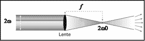
Reducing the laser beam diameter due to light beam convergence by means of a lens concentrates energy in a smaller area, as observed in Figure 1. Therefore, less power is required, resulting in more accurate surgical incisions and less thermal damage to adjacent tissues4,5,6.
Since the beam waist is determined by 2w0 = lf /pw2, where 2w is the beam diameter in the lens aperture, and l means CO2 wavelength7, if we reduce focal length, we obtain smaller beam waists.
Trying to reduce power used during laser application in the larynx, we developed a device using shorter f lenses, delivering the beam at short distance from the lesion and using angled videolaryngoscopy8 (Figure 2). Thus, we produced beam diameters smaller than those currently used. The preliminary results are presented in this study.
OBJECTIVE
To introduce an optical convergence system to deliver laser beams at short distance from the human larynx that is applicable to endolaryngeal surgeries.
METHODOLOGY
Instrument design
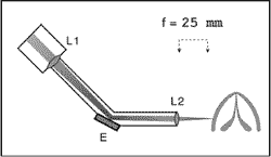
We used a device composed of two metallic cylindrical shafts with 6.0-mm internal diameter, articulated at a 135º angle from the horizontal (Figure 3) to reduce 2w0. In this joint, a selenium-sulfate-coated mirror with high reflex rate for CO2 light laser wavelength beams is positioned and deflects the light path. Both the design and angle used to build the prototype were based on previous studies9.
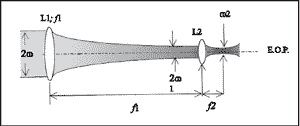
Considering that light beam reflex in a flat mirror does not change its properties, its modifications after crossing the lenses could be studied in a linear manner (Figure 4). Hence, a circular transversal symmetry and 2w-diameter beam strikes on the surface of L1 aperture f 1 lens. When crossing the lens, the beam is focused on L1 focal plan and produces a 2w1-beam waist. Beam diameter is minimal in this position. L2 lens is positioned there and produces a new f2 beam waist, referred as 2w2, and used as our cutting beam.
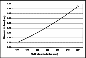
The beam waist obtained here in relation to L1 and L2 positioning is the 200-250-mm interval, as shown in Figure 5 (the relative L1 and L2 positioning inside the prototype was obtained by interactive interpolation of focal lengths f 1= 25 and f 2= 250 mm, through a processor Origin 6.00). Numerically calculated focus depth was 23mm (l CO2 = 10.6 mm). The focus depth (PF) of the prototype built was determined by the following equation: PF= pw2/ l.
Laboratory studies
The studies performed laser application at pre-established distances on wooden spatulas, vegetal paper and animal protein. We used 0.5-8.0 W power and 0.05-0.2 second exposure time.
Surgical technique
Three patients with vocal fold polyp and surgical indication for exeresis were treated with the above-mentioned device. The preoperative assessment included videolaryngoscopy and pictures of the lesion were taken at outpatient's setting. The patients gave their informed consent; the standards and criteria set by the Research Ethics Committee for this study were met.
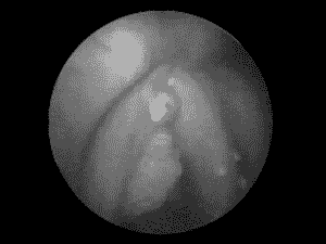
The prototype was coupled to the laser-articulated arm and glottis exposure was performed by angled videolaryngoscopy under general anesthesia. The prototype and mechanic instruments were introduced in the laryngeal cavity with light and image capture provided by an endoscope. A piece of cotton embedded with saline solution was placed in the infraglottis to protect the airways and the endotracheal tube balloon (Figure 6). The beam laser was directed at the target and CO2 laser was triggered for exeresis of the lesion. Excessive mucosa was removed using microscissors and triangular forceps for traction.
Suctioning secretions was performed by 1.0 and 2.0 mm-diameter metallic cannulae and continuous vapor suction by 1.5-mm canal inside the metallic endoscope body. The equipment was set to 0.5-2.0 W power, according to exeresis depth and 0.05 second exposure time. We observed possible difficulties in glottic exposure, quality of image provided by videolaryngoscopy system, focus depth of the cutting beam, vapor suction, bleeding of surgical wound, and carbonization of exeresis surrounding tissues.
In the postoperative period, the patients were assessed at weekly intervals for two months to observe tissue alterations resulting from surgery, that is, if scarring left no retractions in the sites laser was applied.
Figure 1. Beam waist obtained after a 2w-diameter-light beam crossed a focal length (f) lens.
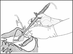
Figure 2. Application of CO2 laser using angled videolaryngoscopy.
Figure 3. Distal convergence of light beams through a f 25mm lens, delivered close to the glottis by means of the prototype developed.
Figure 4. Production of a 2w2-beam waist by convergence of two lenses inside the prototype developed.
Figure 5. Graph showing the waists produced by convergence of laser beam through two f 250 mm and f 25-mm lenses located inside the prototype.
Figure 6. Application of CO2 laser in vocal fold polyp; observe a piece of cotton embedded in saline solution to protect the endotracheal tube balloon.
RESULTS
The glottic exposure was performed with no difficulties and the procedure resulted in no lesions. The images of laryngeal structures obtained by endoscopic magnification were adequate for laser application. The beam waist was kept in focus for a more accurate dissection.
Introducing and positioning the laser end in the glottic region was easily performed and enabled appropriate polyp resection. We did not observe bleeding that could interfere in the procedure or formation of carbonized tissue in surgical wound margins.
In the follow-up of patients at outpatient's clinic, we noticed the surgical wounds had healed quickly, and there were no excessive tissue retractions. Subjective clinical vocal improvement occurred in the first two weeks.
DISCUSSION
The use of the shortest diameter beam waists available is important in vocal fold surgeries, since the shorter the diameter, the less thermal damage to the adjacent normal structures10. Using the prototype developed for CO2 laser application, with power set at 0.5-2.0 W, we achieved a beam waist comparable to the beam diameters produced by the second-generation micromanipulators currently available.
The distal end of the prototype built and the dissection instruments used in surgeries were laid on the glottic exposure system and in the patient, thus preventing or attenuating trembling or abnormal movements of the operator. These movements appear or are intensified during suspension microlaryngoscopy, because handling microinstruments causes 7-fold greater vibration in their bodies11.
The literature in Otorhinolaryngology has described difficulties, complications or even impossibility to obtain a stable and adequate view of the glottis with microlaryngoscopy12. Rischtsmeyer & Scher reported 8% (3/36) complications resulting from direct laryngoscope, and 5% of these (2/36) had fracture of upper incisive teeth, and 3% (1/36) presented transient vocal fold paralysis13. One year later, the same authors reported that for lesions located out of the observer frontal plan, such as in the hypopharynx wall, laryngeal ventricles and posterior laryngeal commissure, the linear approach using microlaryngoscopy would be extremely difficult, and the telescope would be paramount in laryngeal approach14. Unfortunately, frequent painful wounds in the palate and pharynx walls were described15.
Authors in favor of using laser to treat vocal fold carcinoma consider as one of the major criteria for its application the ability to expose the larynx with the widest possible lumen laryngoscopes. However, some patients present an unfavorable anatomy and treatment is unfeasible16,17. Since the system used exposes the larynx with no neck hyperextension and follows the natural path of the upper aero-digestive tract, it becomes an alternative to apply laser in patients whose laryngeal exposure is considered difficult.
Comparing microlaryngoscopy using Kantor-Berci laryngoscope18 with Dedo laryngoscope19, the authors did not succeed in performing the examination in 12.5% (3/24) of the patients with the first equipment, and laryngeal exposure was considered insufficient in other 16.6% (4/24). Using the second laryngoscope they did not succeed in 4.2% (1/24) of the patients, and the examination was considered difficult in another patient20. Dedo described his device and warned about difficulties in direct laryngoscopy, and reported impossible examination in 2.3% (2/85) of the patients. The technique here described enabled a dynamic examination, with no limitations associated with microlaryngoscopy, and it could make treatment of lesions located in the infraglottis, pyriform sinus, ventricles and other anatomical regions of difficult access easier.
Potential complications related to improper laser beam collision or its reflex in the path from the generating system to the target organ were also reported21,22,23,24,25. Such incidents are less likely to occur when using the technique described in the present study, since the cutting beam is immediately delivered on the lesion.
Our experience with laser delivery close to vocal folds is at the initial phase; histological studies and their application in a large number of patients are necessary. However, we could infer that the technique is feasible to treat other laryngeal conditions based on the accurate polyp removal here described.
CONCLUSION
We developed a device to apply CO2 laser in the larynx at short distance from the lesion. The beam diameter here produced is comparable with those currently used, and demonstrated it is feasible to treat vocal fold polyps.
ACKNOWLEDGMENTS
The authors acknowledge Dr. Hélio Ozawa and Mr. Eleutério Rocha Lima for building the prototypes.
REFERENCES
1. Strong MS, Jako GJ. Laser surgery in the larynx: early clinical experience with continuous laser. Ann Otol Rhinol Laryngol 1972,81:791-8.
2. Shapshay SM, Wallace RA, Kveton JF et al. New microspot micromanipulator for CO2 laser applications in otolaryngology-head and neck surgery. Otolaryngol Head Neck Surg 1988;98:179-181.
3. Ossoff RH, Werkhaven JA, Raif J, Abraham M. Advanced microspot microslad for the CO2. Otolaryngol Head Neck Surg 1991;105:411-14.
4. Reinisch L, Ossoff RH. Laser applications in otolaryngology. The Otolaryngol Clin North Am 1996;29(6):891-2.
5. Van Der Speck AFL, Spargo PM, Norton ML. The physics of lasers and implications for their use during airway surgery. Br J Anaesth 1988;60:709-29.
6. Benninger MS. Microdissection or microspot CO2 laser for limited vocal fold benign lesions: A prospective and randomized trial. Laryngoscope 2000;110:1-17.
7. Silvfast WT. Laser fundamentals. Cambridge University Press; 1966. p. 329-40.
8. Morgado PF, Abrahão M. Angled telescopic surgery, an approach for laryngeal diagnosis and surgery without suspension. São Paulo Med J 1999;117(5):224-6.
9. Morgado PF. Microcirurgia da laringe utilizando endoscópio e pinças anguladas. [Tese de Doutorado]. Universidade Federal de São Paulo. São Paulo-Brasil. 2000. p. 1-50.
10. Courey MS, Ossoff RH. Laser applications in adult laryngeal surgery. The Otolaryngol Clin North Am 1996;29(6):973-85.
11. Hurzeler D. Microsurgery of the larynx: an instrumental aid. Arch Otolaryng 1971;93:521-4.
12. Grundfast KM, Strong MS, Vaughn CW, De Vos P. Suspension microlaryngoscopy in the Boyce position with a new suspension Gallows. Ann Otol Rhinol Laryngol 1978;87:560-66.
13. Rischtsmeier WJ, Scher RL. Endoscopic Staple-Assisted Esophagodiverticulostomy for Zenker's Diverticulum. Laryngoscope 1996;106:951-6.
14. Rischtsmeier WJ, Scher RL.-Telescopic laryngeal and pharyngeal surgery. Ann Otol Rhinol Laryngol 1997;106:995-1001.
15. Kleinsasser O. Microlaryngoscopy and Endolaryngeal Microsurgery. Stuttgart: Schattauer Verlag 1990. p. 5-25.
16. Shapshay SM, Hybels RL, Bohigian KR. Laser Excision in early vocal cord carcinoma: indications, limitations and precautions. Ann Otol Rhinol Laryngol 1990;99:46-50.
17. Steiner W. Experience in endoscopic laser surgery of malignant tumors of the upper aero-digestive tract. Adv Oto-Rhino- Laryng 1988;39:135-144.
18. Kantor AND, Berci G, Partlow AND, Paz-Partlow M. A completely new approach to microlaryngeal surgery. Laryngoscope 1991;101:676-9.
19. Dedo HH. A fiberoptic anterior commissure laryngoscope for use with the operating microscope. Trans Am Acad Ophtalmol Otolaryngol 1976;82(1):91-2.
20. Yanagisawa AND, Horowitz JB, Yanagisawa K, Mambrino LJ. Comparison of new telescopic video microlaryngoscopic and standard microlaryngoscopic techniques. Ann Otol Rhinol Laryngol 1992;101:49-60.
21. Fried PM. A survey of the complications of laser laryngoscopy. Arch Otolaryngol 1984;110:31-4.
22. Alberti PW. The complications of CO2 laser surgery in otolaryngology. Acta Otolaryngol 1981;91:375-81.
23. Meyers A. Complications of CO2 laser surgery of the larynx. Laryngoscope 1981;90:132-4.
24. Ossoff RH, Hotaling AJ, Karlan MS, Sisson GA. The CO2 laser in otolaryngology-head and neck surgery: a retrospective analysis of complications. Laryngoscope 1983;93:1287-9.
25. Ossoff RH, Duncavage JA, Shapshay SM, Krespy YP, Sisson GA. Endoscopic laser arytenoidectomy revisited. Ann Otol Rhinol Laryngol 1992;99:764-71.
1 Physicist, Master in Otorhinolaryngology, Ph.D. in Medicine, Federal University of São Paulo - UNIFESP-EPM.
2 Bachelor and Master in Physics, State University of Campinas - UNICAMP.
3 Specialization under course, Department of Otorhinolaryngology and Human Communication Disorders, Federal University of São Paulo - UNIFESP-EPM.
4 Faculty Professor, Department of Otorhinolaryngology and Human Communication Disorders, Federal University of São Paulo - UNIFESP-EPM.
Address correspondence to: Rua Leandro Dupret, 459 - Vila Clementino -São Paulo - SP - Brazil. CEP 04025 - 011 - E-mail: pfmorgado@ig.com.br
Article submitted on November 19, 2001. Article accepted on January 10, 2002.
Print: ![]()