
Year: 2009 Vol. 75 Ed. 6 - (7º)
Artigo Original
Pages: 814 to 820
Reproducibility of the threedimensional endoscopic staging system for nasal polyposis
Author(s): Marcelo Castro Alves de Sousa1, Helena Maria Gonçalves Becker2, Celso Gonçalves Becker3, Mariana Moreira de Castro4, Nicodemos José Alves de Sousa5, Roberto Eustáquio dos Santos Guimarães6
Keywords: neoplasm staging, nasal polyps, endoscopy, steroids.
Abstract:
Nasal Polyposis is a chronic inflammatory process of the nasal mucosa, characterized by multiple and bilateral nasal polyps. Different drugs have been used in its treatment. In order to study the results of different treatment modalities it is necessary to have some kind of staging. Aim: to present a new endoscopic staging method, based on nasal endoscopy and on the three-dimensional nasal polyp assessment; and compare its reproducibility with that from two other systems already established in the literature. Study design: Cohort study. Material and methods: Three experts assessed the exams of 20 patients with nasal polyposis at different times, before, at 15 and at 30 days after the start of oral prednisone, 1 mg/kg/day, during 7 days. We assessed the agreement rate among the experts, using Kappa for statistic analysis. Results: The three methods were reproducible, and the method hereby proposed had the least agreement among the examiners. Conclusions: the three-dimensional staging system proposed proved reproducible, despite showing less agreement among the examiners than the other as the other two methods.
![]()
INTRODUCTION
Nasal polyposis (NP) is a chronic inflammatory process of the nasal mucosa, characterized by the presence of multiple and bilateral nasal polyps. Its pathophysiology is still unclear, with numerous theories described in the literature.
It is a clinical manifestation of diseases with different etiologies, such as: non-allergic eosinophilic rhinitis, asthma, aspirin intolerance, cystic fibrosis, Kartagener syndrome, Young and Churg-Strauss, among others. Its prevalence in the general population varies between 0.5 and 4 % according to some authors1. NP tends recurr2, which makes its treatment a challenge for ENTs.
NP treatment involves the use of different drugs, especially topical and systemic steroids and surgical procedures. Many literature papers already show the efficacy of steroids in its treatment3-6. Treatment goal is to reduce polyp size or, if possible, eliminate them, with consequent symptom relief, especially of nasal obstruction, hyposmia and anosmia, as well as to reduce the frequency of infections and improve associated lower airway symptoms, besides preventing complications such as mucoceles and orbit involvement. Steroids are also indicated in the preparation of these patients for surgery. Surgery is reserved for cases of clinical treatment failure.
Some type of NP staging is recommended in order to follow disease evolution in these patients, just as for the comparison between different types of treatment. For NP staging it is necessary to use the endoscope.
The literature describes numerous ways of staging NP using nasal endoscopy and there is still no method of universal consensus15-21. Most of them classify nasal polyps in a two dimensional way in the nasal cavities and in relation to the middle meatus and outside of it.
Studies have been carried out in an attempt to compare the agreement of different tomographic staging for Chronic Rhinosinusitis7-10 and only one study compared endoscopic staging among different observers6. In our setting, Stamm proposed a staging system based on CT Scan.
The staging to be tested in the present study, proposed by one of the authors, is based only on nasal endoscopy and it is a tridimensional evaluation of the polyps, in three spatial planes: horizontal, vertical and antero-posterior. It has been used in the ENT department of the Medical School Hospital of the Federal University of Minas Gerais for many years and it is believed to be the one which bests represents the real extension of nasal polyposis, besides precisely telling the location of nasal polyps in the nasal cavities.
OBJECTIVES
The goal of the present study was to show a new type of NP endoscopic staging system, assess its reproducibility among different examiners and compare it with the other two methods described in the literature (Lund- Mackay and Johansen)12,13.
MATERIALS AND METHODS
We selected 20 patients from the ENT Department of Federal University of Minas Gerais Medical School, bearers of eosinophilic nasosinusal polyposis. All the patients were volunteers, were educated about the procedure and signed the free and informed consent form. This study was approved by the Ethics Committee of the institution where it was done, under protocol number ETIC 171-6.
We excluded patients with some contraindication to use systemic steroids, as well as diabetics or hypertensive patients without proper control of their diseases. We also excluded those patients previously submitted to nasal surgeries.
The patients were previously submitted to polyp biopsy for the diagnosis of eosinophilic polyposis and should be at least 30 days without the use of oral or systemic steroids. The biopsies were carried out with a cup forceps with the help of an endoscope in cases of small polyps, or anterior rhinoscopy in cases of large polyps.
After the biopsy result, the patients were submitted to three endoscopic exams in their nasal cavities. After the first exam, the patients were medicated with 1mg/kg/day of oral prednisone up to the maximum dose of 60 mg, for a period of seven days. The second exam was held on the 15th and the 30th days after the first.
Thus, we assessed a total of 20 patients with bilateral polyposis, at three moments, corresponding to a total of 120 nasal cavities studied.
The patients were previously submitted to anterior rhinoscopy with vasoconstriction of the inferior turbinate through a cotton ball soaked in naphazoline.
The endoscopies were always carried out by one single examiner, with a 4mm and 30° rigid endoscope and in cases of anatomical alterations, such as septal deviation, we also used the 3.2mm flexible nasal fiberscope. The exams were recorded in a VHS video system for further assessment.
The exam started in the right nasal cavity, inspecting its floor, all the way to the choana. Whenever possible, we visualized the spheno-ethmoidal recess, then the middle meatus and the superior region of the nasal cavities, trying to see the polyps in the three planes.
After doing all the exams, they were copied to a single DVD disk, of which copies were made and handed over to the other two examiners simultaneously.
Staging
1) Tridimensional Staging:
This staging provides information on the location of the polyps in the nasal cavity in the three dimensions of space, that is, in the antero-posterior, horizontal and vertical planes.
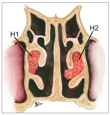
In the horizontal plane (H), polyps were classified as (Figs. 1 and 2):
Figure 1. NP staging in the horizontal plane - H1 - polyps restricted to the middle meatus. H2 - Polyps getting out of the middle meatus without touching the septum
Figure 2. NP staging in the horizontal plane - H0 - No polyps. HT - Polyps extending beyond the middle meatus and touching the septum
- H0 - No polyps
- H1- Polyps restricted to the middle meatus
- H2 - Polyps expand beyond the middle meatus, without touching the nasal septum.
- HT - Polyps expand beyond the middle meatus and touch the septum.
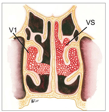
In the vertical plane (V), the polyps are classified as (Figs. 3 and 4):
Figure 3. NP staging in the vertical plane - V1 - Polyps in the middle meatus only. VS - Polyps extending superiorly to the middle meatus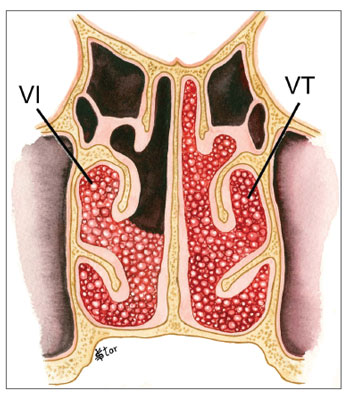
Figure 4. NP staging in the vertical plane - VI - Polyps extending inferiorly to the middle meatus. VT - Polyps occupying the entire vertical aspect of the nasal cavity
- V0 - No polyps - V1 - Polyps in the middle meatus only
- VI - Polyps extending inferiorly to the middle meatus, going beyond the upper border of the inferior turbinate
- VS - Polyps extending superiorly to the middle meatus, between the septum and the middle turbinate
- VT - Polyps occupying the entire vertical aspect of the nasal cavity
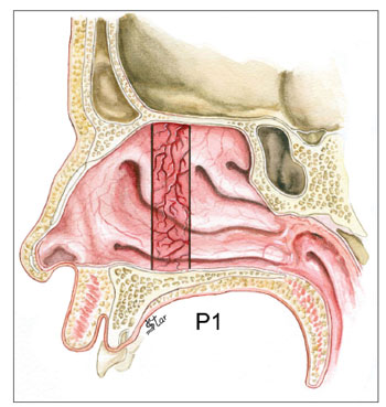
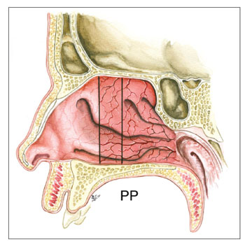
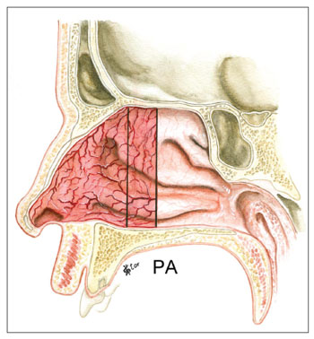
In the antero-posterior plane (P), the polyps are classified as (Figs. 5, 6, 7 and 8):
Figure 5. NP staging in the antero-posterior plane - P1 - Polyps in the middle meatus region only
Figure 6. NP staging in the antero-posterior plane - PP - Polyps extending posterior to the middle meatus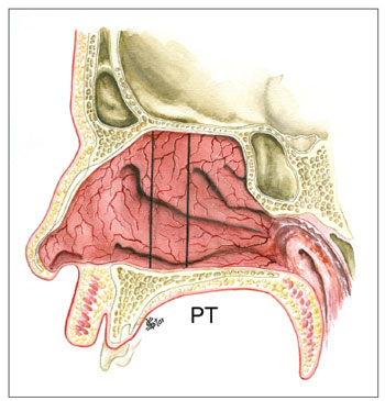
Figure 7. NP staging in the antero-posterior plane - PT - Polyps occupying the entire antero-posterior aspect of the nasal cavity
Figure 8. NP staging in the antero-posterior plane - PA - Polyps extending anteriorly to the middle meatus
- P0 - No polyps
- P1 - Polyps in the middle meatus only
- PA - Polyps extending anteriorly to the middle meatus, reaching the head of the inferior turbinate
- PP - Polyps extending posterior to the middle meatus, reaching the tail of the inferior and middle turbinate
- PT - Polyps occupying the entire antero-posterior aspect of the nasal cavity
2) Lund-Mackay12 Staging
In this endoscopic evaluation, polyps are classified as:
0- No polyps
1 - Polyps in the middle meatus only
2 - Polyps expand beyond the middle meatus
3) Johanssen Staging13
In this staging, the polyps are assessed in relation to the middle meatus and inferiorly on the following way:
0 - No polyps
1 - Polyps restricted to the middle meatus (Mild polyposis)
2 - Polyps expand beyond the middle meatus, without going beyond the lower border of the inferior turbinate (moderate polyposis)
3 - Polyps expand beyond the lower border of the inferior turbinate (Severe polyposis)
Statistical Analysis
The three classifications were assessed for the right and left sides, by three examiners for the 20 patients.
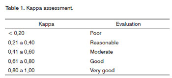
In order to assess the degree of agreement among the examiners, we used the Multiple Kappa coefficient. Such coefficient can be interpreted as being a mean value of the agreement coefficients among examiners two by two. Based on sample values, as estimated by the Multiple Kappa, as well as its respective 95% confidence interval and we also calculated the p-value. When there was agreement among examiners, its level varied according to the classification below.
The classifications of the calculated coefficients correspond to the ones presented on Table 1:
The statistical analyses were carried out using the software R - of public domain, and the conclusions were drawn from results obtained considering a 5% level of significance.
RESULTS
The estimates of the Kappa coefficients and the respective confidence intervals are presented on Table 2. We notice that for the evaluation of the right side, the Lund-Mackay method did not present significant agreement among the examiners at moments 1 and 3 and, regarding the horizontal tridimensional method, the agreement was not significant at moment 3.

