
Year: 2003 Vol. 69 Ed. 2 - (6º)
Artigo Original
Pages: 181 to 184
Microbiological study of core and surface of palatine tonsils in children with repetitive pharyngotonsillitis and adenotonsillar hypertrophy
Author(s):
Felipe Neiva Costa[1],
Odimara Santos[2],
Luc Louis Maurice Weckx[3],
Shirley S. N. Pignatari[4]
Keywords: bacteriology, pharyngotonsillitis, tonsil hypertrophy.
Abstract:
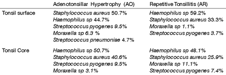
Objective: Bacterial pharyngotonsillitis and tonsil hypertrophy are extremely frequent in childhood. This study aims to identify and verify the frequency of the bacterial colonization in tonsils of children with history of recurrent tonsillitis and tonsil hypertrophy. Casuistic and Methods: Ninety children, both female and male, ages between 2 and 6 years (pre-school children) and 6 and 12 years (scholars) scheduled for adenotonsillectomy; 27 with history of recurrent pharyngotonsillitis (AR), and 63 with obstructive adenotonsillar hypertrophy (AO), assisted at Division of Pediatric Otolaryngology, Federal University of Sao Paulo, were evaluated from 1999 to 2002. Material from the surface of the tonsils was taken with swabs at the moment of the surgery. After tonsil removal, material was also taken from the tonsil core. The obtained material were submitted to culture and analyzed according to bacterial growth. Results: Overall, regardless the age and the diagnostic group, the most prevalent pathogenic isolated bacteria were Haemophilus sp, 50.5% in the AO group and 59.2% in the group AR; S. Aureus, 50.7% in the AO group, and 33.3% in group AR; S pyogenes, 9.5% in the AO group and 7.4% group AR; S. pneumoniae, 4.7% in the AO group, and 0% in group AR; and Moraxella sp, 6.3% in the AO group and 11.1% in group AR. No significant difference was noted between the bacteria isolated from surface and the tonsil core. S aureus was more frequent in the AO group compared to the group AR in both, school children and scholars. Scholars presented higher incidence of S pyogenes in the AR group, and although also present in the pre-school children tonsils, it was only isolated in the AO group. S pneumoniae was only isolated in children with obstructive adenotonsillitis (AO). Conclusion: The results of our study suggest that the surface and core bacterial colonization of the tonsils is similar. It also seems that the prevalence of S pyogenes colonization is high, around 10%, and that S aureus is more prevalent in children with hypertrophic adenotonsillitis compared to the group with recurrent infections.
![]()
INTRODUCTION
The knowledge of the bacterial flora that grows in palatine tonsils is becoming increasingly more important both due to its connection with recurrent infections and its likely association with tonsil hypertrophy. 5,11. It is believed that prevalence of S. pyogenes growth in the oropharynx is approximately 25% among the population in general13. Recent studies reported that this percentage increases significantly with infectious pharyngotonsilitis12. According to the study conducted by the Federal University of São Paulo in 1966 and 2000 approximately 30% of pharyngotonsillitis had positive test for Streptococcus pyogenes 12, which still remains the most important etiological agent. Its importance is based upon the fact that such infection results in complications with high morbidity rate, such as abscesses, rheumatic fever, glomerulonephritis etc.2,14, 15.
In connection with infectious processes it is believed that recurrent events and many times unsuccessful treatment with penicillin might be related to local growth of b-lactamase-producing microorganisms hindering both S pyogenes eradication and infection treatment 13. Although SBHGA (Streptococcus b-hemolytic of Lancefield Group A) is still universally considered as penicillin sensitive, one of the major concerns pursuant bacterial infections of the upper airways is sensitiveness change of classical anti-microbial agents presented by various other microorganisms. Penicillin-resistant Pneumococcus is becoming more frequent, as well as b-lactamase-producing Haemophilus influenzae4.
The authors researched and compared bacterial growth on the surface and core of the tonsils of pediatric patients in our population with recurrent pharyngotonsillitis and tonsil hypertrophy. They wanted to study bacteriological growth of the oropharynx in children, and to check the differences in the microflora both on the surface and in the core of tonsils, in addition to studying the relation between recurrent infectious events and hypertrophy.
MATERIAL AND METHOD
Ninety patients, boys and girls, aged from 02 to 12 years were assessed in the Department of Pediatric ENT of Hospital Sao Paulo, Federal University of Sao Paulo from April 1999 through January 2002. The group included 27 patients with recurrent tonsillitis (RT) and 63 with obstructive adenoid and tonsil hypertrophy (AO) with surgery indication.
Patients were divided into 2 age groups: pre-school and elementary school students (aged 2 to 6 & 6 to 12 years).
Material collection and processing:
The first material was collected, during the surgery with the patient under general anesthetic drug (orotracheal intubation), from the surface of the 2 tonsils with an alginate swab and was immediately seeded in blood and brown agar culture media supplemented with V and X factors (Columbia base, Oxoid, 5% of sheep blood). After surgical removal of the first tonsil, it was subjected to a saline solution spray and excessive blood was removed, then dived in Chlorexidine for 3' to be immediately seeded in a blood Agar plaque by the rolling method. The same cleaning sterilization procedure was performed in the second tonsil with the help of an electrical knife. Material was then collected from the core using a sliding alginate swab performed between the crypts inside the tonsil and was seeded in blood and brown Agar plaques.
Finally, seeded plaques were forwarded to the Microbiology laboratory of Sao Paulo Hospital and were incubated in microbiological oven with CO2 5% for 18 to 24 hours.
Identification was performed after bacterial isolation and was made according to the guidelines of the American Microbiological Association.
RESULTS
Sixty-four point four (64.4%) percent out of ninety (90) patients were boys and thirty-five point six percent were girls, sixty-six (66%) percent aged from 2-6 years and 44 percent aged from 6 to 12 years.
Generally, in all groups of pediatric patients both with recurrent tonsillitis (AR) and adenotonsillar hypertrophy (AO) bacterial percentages of pathogenic bacteria in upper airways (Haemophilus sp, S. aureus, S. pyogenes, S pneumoniae e Moraxella) were high. Prevalence on the surface and in the core according to groups is shown in Chart 1. Bacterial isolation was not significantly different both on tonsil surface and core.
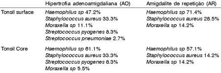
S aureus was more frequent both in pre-school and elementary students with hypertrophy (AO) against those with recurrent infections (AR). Higher prevalence of S. pyogenes was detected in elementary students of AR group; however, it was also found in pre-school students of AO group and it was not isolated in children with recurrent infection. S. pneumoniae was isolated only in children with adenotonsillar hypertrophy. It can be observed in Charts 2 and 3.Chart 1. Distribution of Bacteria on the Surface and in the Core of Tonsils in the 2 Studied Groups (AO e AR).
Chart 2. Frequency and distribution of bacteria on the surface and in the core of tonsils of pre-school children (2 to 6 years) in the AO and AR groups.
Chart 3. Frequency and distribution of bacteria on the surface and in the core of tonsils of school children (6 to 12 years) in the AO and AR groups.
DISCUSSION
High prevalence of pharygotonsillitis and adenoid and tonsil hypertrophy in the pediatric population is one of the major reasons for using anti-microbial agents and performing palate tonsillectomy 2,6. Some authors believe that the bacterial flora presented in palatine tonsils is closely related to recurrent infection events, treatment failure and even to excessive bacterial growth 1,5,8,11.
Along the years, swab studies from the oropharynx and rhinopharynx in pediatric population reported a decrease in S. pyogenes prevalence and an increase in S. aureus. Since S. aureus has been commonly isolated in oro and rhinopharynx in population in general in the absence of infections, its role in the etiology of infections remains uncertain.3, 7.
In our study with AO group and AR group, pathogenic bacteria prevalence was high both on the surface and in the core of palatine tonsils similarly to those reports by other authors 8,9,11. Many researchers have been trying to set up a connection between microorganisms on tonsil surface and micro flora in tonsil core. Although some reported discrepancy between bacterial growth in both sites, similarly as in our study, Kielmovitch et al, back in 1989, found some common aspects between bacterial micro floras in both sites.
According to studies by Brook & Foote, in 1992, the prevalence of b-lactamase-producing microorganisms tends to be higher in children with recurrent tonsillitis report than in children from the population in general, especially with S. aureus and H. influenzae8. In our study we also found a trend of higher bacterial growth by H influenzae in children with reports of recurrent infection against the AO group, especially in pre-school children. The same did not occur, however, with S. aureus, which had higher prevalence in AO group tonsils.
Overall, regardless of group and age range, it was interesting to find out that S. pyogenes growth was high (approximately 10%), but it was not as high as in international reports (approximately 25%)13.
In children with recurrent infections, particularly younger children, we found that the presence of S. pyogenes was considerably lower when compared against elementary school students. It shows that although children had recurrent infections, S. pyogenes was not an important bacterium in this age range. Would S. pyogenes be a less frequent etiological agent in pre-school children? Would infection treatment (common use of antibiotics) be effective in eliminating this pathogenic agent? It maybe that likelihood of recurrence is lower in younger children.
It was found out that pre-school children had a trend to present higher prevalence of Moraxella than elementary school children. Would gram-negative bacteria have a more important role in younger children?
In terms of tonsil growth, according to Barr and Crombie, the size of palatine tonsils seemed not to be significantly different among children with recurrent tonsillitis and children with isolated hypertrophy 1.
It is believed that this hypertrophy could be related to local presence of microorganisms that would stimulate the growth of lymphoid elements, which in turn would be the major cause of such tonsil hypertrophy and not the increase of stroma tissue itself. These studies reported that the amount of T-helper, T-suppressor and B cells is considerably higher in patients with tonsil hypertrophy than in control patients 5, 9.
Regardless of the fact that some studies suggested a connection between H. influenzae and tonsil hypertrophy5, in this study the presence of S. aureus on tonsil surface and in the core in all children with adenoid and tonsil hypertrophy within all age ranges studied was more significant than in children with recurrent infections suggesting a likely link between these bacteria and excessive tonsil growth.
The authors believe that further studies on oropharynx microbiota in children from the population in general, with no history of recurrent infection or hypertrophy, could be important to understand such difference.
CONCLUSION
The results in our study suggested that bacterial flora that grows on tonsil surface is similar to that in the tonsil core; S. pyogenes prevalence in oropharynx of children is approximately 10%; S. aureus is more frequent in children with adenoid and tonsil hypertrophy than in those with recurrent infection.
REFERENCES
1. Brodsky L, Moore L, Stanievich J. The role of Haemophilus influenzae in the pathogenesis of tonsils hipertrophy in children. Laryngoscope 1988;98: 1055-1060.
2. Kielmovitch IH, Keleti G, Bluestone CD, Wald ER, Gonzales C. Microbiology of obstructive tonsillar hypertrophy and recurrent tonsillitis. Arch Otolaryngol Head Neck Surg 1989;115:721-724.
3. Pichichero ME. Group A beta-hemolitic streptococcal infections. Pediatr Rev 1998; 19(9):291-302.
4. Paes O, Pignatari AC, Weckx LLM, Pignatari SN. Detection of BHSGA by using three different methods: culture, rapid test and molecular biology assay. Otolaryngol Head Neck Surg (special issue). Poster 147 2002;127(2):248.
5. Bluestone CB. Current Indications for tonsillectomy and adenoidectomy. Ann Otol Rhinol Laryngol 1992;101: 58-64;.
6. Weckx LLM, Teixeira MS. Amigdalites: Aspectos Imunológicos, Microbiológicos e Terapêuticos. JBM 1997;73: 94-101.
7. Weckx LLM & Weckx LY. Como diagnosticar e tratar amigdalites. Ver Bras Med 1988;45:27-32.
8. Brandileone MCC, DI Fabio JL, Vieira VSD, Zanella RC, Casagrande ST, Pignatari AC, Tomasz A. Geographic distribution of Penicillin resistance of Streptococcus pneumoniae in Brazil: Genetic relatedness. Microbial Drug Resistance 1998;4:209-217.
9. Brodsky L. Modern Assessment of Tonsils and Adenoids The Pediatric Clinics of North America 1989;36(6): 1551-69.
10. Barr GS, Crombie IK. Comparison of size of tonsils in children with recurrent tonsillitis and in controls. BMJ 1989;298:804.
11. Brook I, Foote PA. Comparison of the microbiology of recurrent tonsillitis. Arch Otolaryngol Head Neck Surg 1992;118: 507-508.
12. Brandão KG, Pereira RCC, Bordasch A, Pignatari SSN. Bacteriologia da hipertrófica e normal em crianças. Rev Paul Ped 1996;14: 37-42.
13. Brodsky L, Koch RJ. Bacteriology and Immunology of normal and diseased adenoids in Children. Arch Otolaryngol Head Neck Surg 1993;119:821-9.
14. François M, Bingen E, Soussi Th. Bacteriology of tonsils in children: Comparison between Recurrent Acute Tonsillitis and tonsils hypertrophy. Adv Otorhinolaryngol Baser 1992;47:146-150.
15. Kertesz DA, DI Fabio JL, Brandileone MCC et al. Invasive Streptococcus pneumoniae infection in Latin America children: results of the Pan American Health Organization Surveillance Study. Clin Infect Dis 1998;16;1355-61.
1 Undergraduate, Medical School, Federal University of São Paulo/Escola Paulista de Medicina (UNIFESP/EPM).
2 Microbiologist, Discipline of Pediatric Otorhinolaryngology, UNIFESP/EPM.
3 Full Professor, Head of the Discipline of Pediatric Otorhinolaryngology, UNIFESP/EPM.
4 Joint Professor, Discipline of Pediatric Otorhinolaryngology, UNIFESP/EPM.
Address correspondence to: Dra. Shirley S.N.Pignatari - Rua Vergueiro 3645 Apto 808 - Vila Mariana S Paulo 04101300
Tel: 55794502 - E-mail: pigna@terra.com.br
Article submitted on November 06, 2002. Article accepted on February 07, 2003.


