

Year: 2003 Vol. 69 Ed. 1 - (24º)
Relato de Caso
Pages: 142 to 144
 PDF PT
 Vascular hamartoma of the nasal: case report
Author(s):
Washington L. C. Almeida,
Paulo S.L. Perazzo,
César A. L. dos Anjos,
Márcio R. de Freitas Souza,
Márcio Silva Carvalho,
Marcelo Rosa Lima,
Washington L. C. Almeida,
Paulo S.L. Perazzo,
César A. L. dos Anjos,
Márcio R. de Freitas Souza,
Márcio Silva Carvalho,
Marcelo Rosa Lima
Keywords: vascular hamartoma, nose hamartoma.
Abstract:
Vascular hamartoma is a nonmalign malformation, very rarely found in the otorrhinolaringologic practice. We report a case of hamartoma of the nose cavity, specifically at the inferior turbinate in a patient who had no nasal complain, with further literature review.
![]()
Introduction
The term hamartoma (from the Greek, Hamartia - error, Oma - tumor growth)2,3,4 was introduced in 1904 by Eugem Albrecht to describe tumors characterized by abnormal growth in a set of native tissues from a specific area, originated from any of the germinative layers1, 3, 4, 5, 6, 7, 8 - ectoderm, mesoderm or endoderm, frequently with the predominance of one type1, 7, 9. It is a benign tumor, rarely found in ENT practice1, 7, 8, 10 with self-limited growth interrupted by puberty8, 10.
There are few cases related in the international literature and we found only seven cases to describe here. We report a case of hamartoma located in the right inferior nasal concha of a 59-year-old patient and we present the literature review about the pathology.
Case Report
Patient initials: APA. 59-year-old Caucasian female patient came to our service complaining of bilateral episodic tinnitus for about 2 years, without nasal complaints. In the clinical history, she reported diabetes mellitus type II (using Glibenclamide), hypertension (using captopril) and thyroidectomy performed two years before.
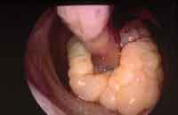
We conducted a routine ENT physical examination resulting in normal otoscopy and oroscopy. Endoscopic rhinoscopy showed a whitish, vegetating lesion, with the aspect of grapes, in the right inferior nasal concha (Figure 1).
Pure tone audiometry revealed bilateral mild sensorineural hearing loss and normal immitanciometry.
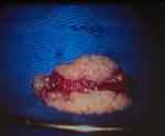
The surgical indication was made, the patient was submitted to endoscopic partial turbinectomy of right inferior nasal concha, with exeresis of the whole lesion. The tumor mass was very bleeding upon touch, but there were no other transoperative events.
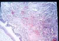
The resected lesion (Figure 2) was submitted to clinical pathology, which revealed the diagnosis of vascular hamartoma (Figure 3).
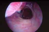
We conducted nasal endoscopic exam 3 months after the surgery and there were no signs of recurrence (Figure 4).
Figure 1. Photo of the preoperative lesion.
Figure 2. Photo of the surgical specimen.
Figure 3. Photo of the clinical pathology slide.
Figure 4. 3-month postoperative photo.
Discussion
Hamartomas are lesions characterized by excessive proliferation of one or more specific cellular components of a tissue type. They have been described as non-neoplastic tumors of non-inflammatory nature, resulting in exaggerated growth of normal tissue structure7.
As differential diagnosis, Eichel and Hallberg (1966) included teratoma and dermoid cyst and demonstrated that they are spontaneous, self-limited tumor growth derived from the pluripotential cells, with a cell structure foreign to the region in which they are located. Teratomas are formed by cells originated from the three germinative layers, whereas dermoid cysts present cells that originated only from mesoderm and ectoderm. Both are clinically difficult to differentiate from hamartoma, which behave as self-limited growth of a determined native tissue of the region in which it is located. In the case presented here, after clinical pathology analysis, we noticed absolute predominance of the vascular tissue1-5, 7, 11.
There are still other pathological entities commonly confused with hamartomas, such as: angiofibroma, lymphoma, hemangioma, and polypoid inflammatory lesions5, 7. As to vascular tumors (capillary, cavernous and venous hemangiomas) that may be found in the nasal cavity, paranasal sinuses and rhinopharynx, the differential diagnosis for the case presented should be especially made with venous hemangioma, whose clinical pathological presentation is very similar to that of hamartoma12.
By analyzing the rare cases of nasopharynx hamartoma published in the international literature, it seems that there is preferential location for these tumors. The symptoms are varied and include nasal obstruction, sneezing episodes, rhinorrhea, epistaxis, up to completely asymptomatic clinical pictures, such as those found in lesions accidentally diagnosed in ENT routine examinations, which was exactly what happened in the case reported here. In one of the published cases, there was recurrence of the lesion 4 months after the excision, but it was a lipomatous hamartoma 1.
References
1. Mahindra S, Daljit R, Sohail A, Malik G. Hamartoma of the nose. J Laryngol Otol 1978;92:57-60.
2. Graeme F, Pilch B. Hamartomas of the nose and nasopharynx. Head Neck 1992;14:321-327.
3. Kacker S, Dasgupta G. Hamartomas of ear and nose. J Laryngol Otol 1973;81:801-805.
4. Zarbo R, Mc Clatchey K. Nasopharyngeal hamartoma: report of a case and review of the literature. Layngoscope 1983;93:494-497.
5. Arrante J, Franche G, Barra M, Saffer M. Radiology forum: imaging quiz case 3. Hamartoma of the nasopharynx. Arch Otolaryngol Head Neck Surg 2000;126:1032-1036.
6. Day L, Arnold G. Rare tumors of the ear, nose and throat. Laryngoscope 1971;81:1138-1174.
7. Kaneto C, Inokuchi A, Kimitsuki T, Kumamoto Y, Shinokuma A, Natori Y, Komiyama S. Huge hamartoma with inverted papiloma in the nasal cavity: Eur Arch Otorhinolaryngol 1999;256:33-37.
8. Kim D, Low W, Billman G, Wickersham J, Kearns D. Chondroid hamartoma presenting as a neonatal nasal mass - case report. Int J Pediat Otorhinolar 1999;47:253-259.
9. McDermott M, Ponder B, Dehner, L. Nasal chondromesenchymal hamartoma - An upper respiratory tract analogue of the chest wall mesenchymal hamartoma. Am J Surg Pathology 1998;22(4):425-433.
10. Mahindra S, Malik G, Bais A, Logani K. Vascular hamartoma of the paranasal sinus. Acta Otolaryngol 1981;92:379-382.
11. Terris M, Billman G, Pransky S. Nasal hamartoma: case report and review of the literature. Int J Pediatr Otorhinolaryngology 1993;28:83-88.
12. Kato K, Ijiri R, Tanaka Y, Hara M, Sekido K. Nasal chondromesenchymal of infancy: the first Japanese case report: Pathology International 1999;49:731-736.
1 Otorhinolaryngologist, Clínica Otorrinos, Doctorate studies under course, Discipline of Otorhinolaryngology, Medical School, University of São Paulo.
2 Otorhinolaryngologist, Clínica Otorrinos. Master studies under course, Discipline of Otorhinolaryngology,
Santa Casa de Misericórdia de São Paulo, Assisting Professor of Speech and Language Pathology and Audiology, UNEB.
3 3rd Year Resident Physician in Otorhinolaryngology, Clínica Otorrinos.
4 1st Year Resident Physician in Otorhinolaryngology, Clínica Otorrinos.
5 2nd Year Resident Physician in Otorhinolaryngology, Clínica Otorrinos.
6 3rd Year Resident Physician in Otorhinolaryngology, Clínica Otorrinos.
Study conducted at Clínica Otorrinos: Rua Barão de Cotegipe, 1141 Centro, Feira de Santana BA 44025-030 - Brazil - Tel (55 75) 623.4455 Fax (55 75) 223.4117 - E-mail: otorrinos@gd.com.br
Address correspondence to: Washington Luís de Cerqueira Almeida - Rua Barão de Cotegipe, 1141 - Centro, Feira de Santana BA Brazil 44025-030 - Tel. (55 75) 623.4455 - Fax (55 75) 223.4117
Article submitted on August 26, 2002. Article accepted on September 28, 2002.
Print: ![]()