
Year: 2006 Vol. 72 Ed. 6 - (3º)
Artigo Original
Pages: 731 to 736
Immittance measures in individuals with Moebuis Sequence
Author(s): Mariana Cabral de Albuquerque Bezerra1, Silvana Maria Sobral Griz2, Graziela de Souza Azevedo3, Liana Ventura4, Angela Revoredo5
Keywords: facial palsy, acoustic reflexes, moebius sequence.
Abstract:
oebius Sequence has been described as a pathology which involves the VI and VII cranial nerves, causing facial palsy. Acoustic reflexes are elicited by a high intensity stimulation of the stapedius and the tensor tympani muscles. The VII cranial nerve is responsible for innervating the stapedius muscle. No acoustic reflexes are expected for individuals with this Sequence. Aim: To describe immittance findings in a series of individuals with Moebius Sequence. Materials and Methods: We had 17 individuals with Moebius Sequence of both gender, with age raging from 3 to 13 years, who were submitted to otoscopy and immittance measures. Results: The results of this study indicated a Type A tympanometry in the majority of the analyzed ears, demonstrating normal function of the stapedius muscle. For the contralateral acoustic reflexes we observed it present in 50% of the ears. The ipsilateral acoustic reflexes were absent in the majority of the ears. Conclusion: The results of the acoustic reflexes suggested that this measure could help in the prognosis of VII cranial nerve lesions, since half of the individual presented those reflexes.
![]()
INTRODUCTION
The Moebiüs Sequence was first described in 1880, by von Graaefe, who reported the case of a patient with facial nerve paralysis (VII cranial nerve)1-6. However, it was in 1888 that Paul Moebiüs described an individual with congenital bilateral facial weakening, malformation of the great pectoris muscle, syndactilya and lack of abduction. At this time, Paul Moebiüs broadened even further the signs and symptoms described previously by von Graaefe, including the paralysis of the abducent (VI cranial nerve)7.
The Moebiüs Sequence consists of a cranial nerve paralysis or palsy, associated to other anomalies, and in some studies5-8 is it considered the result of a fetal aggression, because of genetic and/or environmental factors, between the 4th and 5th gestational week.
The clinical manifestations of the Moebiüs Sequence have been researched in many areas, and, in current Speech and Hearing Therapy it is considered a challenge because of the other alterations present in their bearers9. Currently there are many studies5-6,8,10-12 which briefly report the incidence of such Sequence associated to the use of misoprostol (Cytotec), as a means to abortion. Notwithstanding, none of the studies mentioned above have reliable proof on etiology for this ailment. These studies only assume a relationship between the cause and the presence of the Moebiüs Sequence. It is believed that the current incidence is of 1:10,000 to 1:50,000 live births7. However, they reported equal prevalence of such disease in both genders3.
The population affected by such disease bears some clinical manifestations, the most frequent ones are: cranial nerve palsy, such as the VI (abducent) and the VII (facial), thus causing peripheral facial paralysis, usually bilateral and convergent strabismus. Other nerves may also be affected: III, V, VIII, X, XII, causing respectively: eyelid ptosis, sensitivity alterations, hearing impairment, dysphonia, dysphagia and tongue atrophy, which may happen in different combinations1-3,5-6,11,13.
As to the facial nerve paralysis, we observe a lack of eye and eyelid lateral movements, sialorrhea and sensitivity to loud noises. These facial alterations limit facial expression1-2,6,11,13-14. Because of this lack of facial movement, it is possible to notice a semi-open mouth and eyes that do not close - the Bell sign.
Another very relevant factor found in patients with Moebiüs Sequence is related to alterations in language and joints1-2,6. As to language, some authors1-2,6 affirm that their understanding is better than their expression, the articulation pattern may be impaired, specially the uttering of bilabial phonemes - dependent on lip sealing, and the articulation is imprecise, poor and restricted to the movement of half the tongue against the articulating points. Communication may also be impaired, specially because of hearing loss of different levels.
Conductive-type hearing loss has been the one most commonly described, because of predisposing factors such as perioral hypotonia, supplemental oral breathing pattern and possible paralysis of the soft palate muscles, which are important in the physiological mechanism of Eustachian tube contraction and pressure regulation in the middle ear. A study6 on the auditory status of patients with Moebiüs Sequence has revealed altered otoscopic results and varied degrees of tympanic membrane retraction and/or thickening, justifying that the otologic sequels seem to be unavoidable. Alterations have been seen already in the first years of life, with conductive hearing loss caused by constant Eustachian tube obstruction. Besides conductive hearing loss, we found mixed hearing alterations. The tympanometric alterations most found in this study were types B and C curves. There were no descriptions regarding the acoustic reflex investigation results.
The acoustic reflex represents an involuntary muscle contraction (stapedius and tensor tympani muscles) in the middle ear, in response to a high intensity sound stimulus15-16. In order to have the acoustic reflex, it is necessary to have an intact arc reflex, with intact auditory afferent and motor efferent pathways. This means intact VIII (auditory afferent via) and VII cranial (motor efferent via) nerves. The motor function of the stapedius muscle, innervated by the facial, has its innervation emerging from the cranial base, going through the inner auditory meatus in the temporal bone, together with the vestibulo-cochlear nerve (VIII cranial nerve), it follows along towards the face, going through the parotid gland, and ending in the facial movement muscles17.
Since the motor function of the stapedius muscle, innervated by the facial nerve, may be compromised in cases of Moebiüs Sequence, it is expected to find an alteration in this pattern of presence or absence, depending on the lesion site. Therefore, in facial paralysis cases, the acoustic reflex has been very successfully used in the topodiagnosis of the VII cranial lesion, located close or far from the facial nerve stapedius branching, and in its evolutional follows up18-19. Thus, the acoustic reflex presence indicates a distal lesion, in other words, one below the emergency of the stapedius branch; and a lack of reflex has a probable proximal location. This fact makes the stapedius reflex information very useful in the topodiagnosis, since it provides information on neural function20.
In the clinical assessment of facial paralysis, by observing the stapedius muscle function, we see that this function is one of the first to be recovered, starting from the very lack of acoustic reflex, and going through present responses in high sound intensities and, following that, we see the acoustic reflexes under normal sound stimulus 10.
Since the Moebiüs Sequence has been little studied by the speech and hearing sciences, and most specifically in the field of audiology, this paper aimed at describing the immittance results from Moebiüs Sequence patients, thus contributing for a better understanding of the audiologic profile of this population.
Thus, as with the other individuals, those with Moebiüs Sequence, immittance studies offer conditions to assess conductive hearing alterations6, through tympanometry, and it also offers information on the alterations that happen due to the paralysis of the VII cranial nerve. The acoustic reflex alterations in this population may occur due to predisposing factors, such as perioral hypotonia and the supplemental oral respiration pattern, as well as factors related to the paralysis of some cranial nerves that may directly or indirectly affect the arc reflex in these patients.
METHODS
This study was carried out as part of a Project from the Altino Ventura Foundation, approved by the Research Ethics committee under protocol # 012/05, from the Ethics Committee for Research of the Altino Ventura Foundation. It is study of cases, for which we have the description of the immittance characteristics of individuals with Moebiüs Sequence.
We had 17 Moebiüs Sequence patients (diagnosed by the genetic test - genotype) come to the Speech and Hearing Therapy School of the Catholic University of Pernambuco (UNICAP), 11 females and 6 males, with average age of 6 years and 8 months, age ranging between 3 and 13 years, without anatomical alterations of the external ear that would prevent the immittance exam. However, only 13 participants went through the immittance test, which consisted of the tympanometry and the acoustic reflex study by means of an AZ 7 Interacaoustics immittance meter. Before each exam, the patients' guardians signed an informed consent form with the study objectives and the other necessary information. After that, the participants underwent an otoscopic exam followed by an immittance exam. The procedure used for the tympanometry was based on descriptions15,16,19,18 and their results reported according to the Jerger15 classification. For the cases in which the tympanograms did not show maximum compliance and middle ear pressure (type B curves) we did not measure the acoustic reflex, since it has to be carried out at the balance pressure point between the external auditory meatus and the tympanic cavity, not seen in cases of type B tympanometric curves. In cases of tympanometry measurements equal to curves types A, As, Ad and C, the acoustic reflex may be studied contra and ipsilaterally for the frequencies of 500, 1000, 2000 and 4000Hz and of 1000 and 2000Hz, respectively16. The acoustic reflex threshold investigation was carried out based on the pressure point at which we observed maximum compliance, with intensities varying between 90 and 120dB HL, for contralateral stimuli, and between 80 and 110dB SPL, for ipsilateral stimuli, according to the specifications of the equipment used. Since the study took the design of a case series study, the data were analyzed based on descriptive statistical analysis.
RESULTS AND DISCUSSION
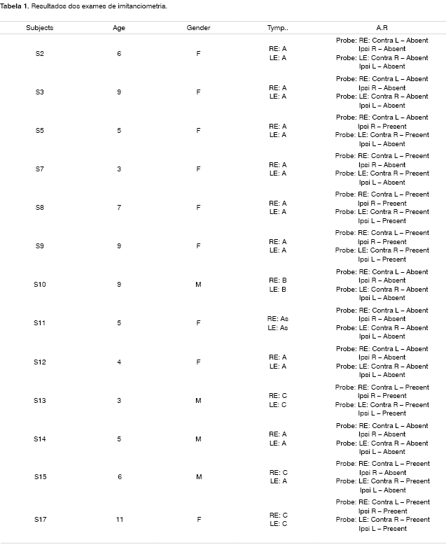
Results were obtained through the analysis of each ear individually (Table 1).
We observed results where there are Type A tympanograms and the lack of acoustic reflexes. This type of curve is commonly seen in normal ears18, with no clear middle ear pathology that would justify the lack of such reflex. In these cases, we believe such lack of reflex may have occurred due to a paralysis of the VII cranial nerve, which is compromised in individuals with Moebiüs1-6 Sequence. In cases of type A tympanograms, the acoustic reflex may be altered due to alterations seen in the efferent via of the arc reflex, such as ossicular rigidity18.
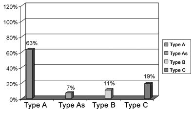
Chart 1 shows all the tympanometric curve types in the studied ears. We noticed that 63% (n=17) of ears presented Type A tympanograms, 19% (n=50) type C, 11% (n=3) with type B and 7% (n=2) with type As.
Chart 1. General result - tympanometry exam.
Type A tympanograms, in most of the patients, differs from results presented in the literature6, which the most common tympanometric curves with Moebiüs Sequence patients were types B and C. This type of alteration was expected since patients with this ailment have conductive and/or tube alterations, caused by the paralysis of the VI, VII, VIII cranial nerves or caused by soft palate muscle deficits, thus bringing about build up of secretion within the middle ear. Therefore, these characteristics 1-6, together with audiologic findings6, pointed to a typical pattern in the tympanogram results, in other words, As, B and C type curves. Our results suggest that there is no characteristic pattern able to demonstrate these conductive alterations. On the contrary, for the population studied, the tympanometric results revealed a characteristically normal functioning of the tympanic-ossicular system, analyzed based on the Type A tympanometry results.
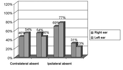
Graph 2 shows the results from the ipsilateral and contralateral acoustic reflexes. As we can see, 50% (n=13) of the ears studied showed no contralateral acoustic reflex and 50% (n=13) had the acoustic reflex. As to ipsilateral acoustic reflex, 73% (n=19) of the ears studied did not show such reflex and 27% (n=7) showed the acoustic reflex for ipsilateral stimulation.
Chart 2. General results of acoustic reflex
Despite the facial paralysis, we can see the acoustic reflex present in both situations of stimuli, ipsi and contralateral. It is known that in patients with the Moebiüs Sequence there may be total VII cranial nerve paralysis and nerve paresis; and this alone could justify both the presence and the absence of the acoustic reflex5-8.
From the anatomical standpoint, the facial nerve lesion may be above or bellow the branch that innervates the stapedius muscle18-19, impairing or not the motor function of this nerve21, seen both in the presence and absence of the acoustic reflexes. There are reports of VII nerve paralysis seen in patients with Moebiüs Sequence, associated to an aplasia of the facial nerve motor nucleus or even aplasia of the stapedius muscle22. Having said that, some studies1,6 suggest that there is no acoustic reflex in Moebiüs Sequence patients because of the facial nerve paralysis, however they do not present data to confirm such statements. Differently from what was seen in this study, the acoustic reflex was present in some cases, thus this test may be applied in order to better understand the level of impairment brought about by the disorder.
CONCLUSION
In most of the immittance results from these patients with Moebiüs Sequence, we found Type A curves. These results suggest that in this population studied there was no characteristic tympanometric pattern previously justified by the muscle alterations found in the patients with this Sequence.
As to the presence or absence of the contralateral acoustic reflex, we noticed that in both ears of these patients there was reflex presence and absence. As to the ipsilateral acoustic reflex, a majority of patients did not have it. These results are promising because they bring about a better understanding of the degree of facial nerve involvement, since the acoustic reflex presence in cases of facial paralysis may indicate both the site and degree of involvement, thus making the acoustic reflex investigation a powerful tool in the diagnostic and prognostic evaluation of patients with Moebiüs Sequence
Further studies should be carried out in order to better relate the presence or absence of the acoustic reflex with the degree of facial nerve involvement, so that the suggestions provided by the present study be corroborated.
REFERENCES
1. Boari C, Lima DRA, Brigagão GM, Moraes LMS, Toledo L, Gomes M, Pacheco VB, Limongi SCO. Intervenção fonoaudiológica precoce na seqüência de Moebiüs: relato de caso. Pró-fono 1996;8(2):55-60.
2. Gemignani E, Longone E, Guedes ZCF. Seqüência de Moebiüs - Relato de um caso clínico sob a luz da investigação fonoaudiológica e psicológica. Pró-fono 1996;25(1):51-4.
3. Roth MGM, Garcias GL, Ferreira FLS, Roth JM. Seqüência de Moebiüs. Arq. catarinenses de medicina 1996;25(1):61-4.
4. Da Cruz RL, Perim Júnior D, Radwanski HN. Síndrome de Moebiüs. Rev Bras Cir 1997;87(2):85-92.
5. Pupo Filho RA, Cardoso TAL, Martins JRO, Michelett JA, Carvalho Filho JF, Moraes MG, Duarte MSN. Síndrome de Moebiüs uma patologia emergente no Brasil. Rev Paul de Pediatria 1999;17(2):91-4.
6. Martins RHG, Nakanishi M, Dias NH Sousa JC, Tamashiro IA.
Seqüência de Moebiüs: manifestações clínicas e avaliação auditiva. Revista Brasileira de Otorrinolaringologia 2001;67(4):440-5.
7. Ventura LMVO. Seqüência de Moebiüs: estudo comparativo das anomalias e distúrbios funcionais em crianças com ou sem uso de misoprostol durante a gestação. Belo Horizonte: Universidade Federal de Minas Gerais; 2001.
8. Boudoux DD, Matos MAG, Gonçalves ED, Rocha M, Ventura LO, Hinrichsen SL. Síndrome de Moebiüs relacionada à ameaça de abortamento. Revista Brasileira de Oftalmologia 2000;59(3):173-7.
9. Carneiro MMS, Gomes ICD. O Perfil Morfo-Funcional Oral de Crianças Portadoras da Síndrome de Moebiüs. Revista CEFAC 22005;7(1):68-74.
10. Carvalho DR, Amorim GG, Arruda PSC, Coelho AF. Síndrome de Moebiüs e uso de misoprostol: relato de dois casos. Anais da faculdade medica 1999;44(2):126-8.
11. Fontenelle L, Araújo APQC, Fontana RS. Síndrome de Moebiüs: relato de caso. Arq Neuropsiquiatr 2001;59(3):1-6.
12. Vasconcelos GC, Silva FBD, Almeida HC, Boas MLMV, Álvares MG. Síndrome de Moebiüs:achados clínicos e cirúrgicos em 7 pacientes. Acesso em 07 Set. 2002. Online. Disponível na Internet http://www.abonet.com.br/abo/abo64302.htm
13. Araujo MP, Araujo MP, Araujo AJ. Síndrome de Moebiüs - Poland: relato de caso. Rev. Med. 1999;78(3):371-7.
14. Zurker RM. El sindrome de Moebiüs? Acesso em 05 Set. 2002. Online. Disponível na Internet http://www. Moebiüs.org/
15. Russo ICP, Santos TMM. Medidas da imitância acústica. In: Russo ICP, Santos TMM. A prática da audiologia clínica. São Paulo: Cortez; 1993. p. 123-58.
16. Frazza MM, Caovilla HH, Munhoz MSL, Silva MLG, Ganança MM. Imitanciometria. In: Munhoz MSL, Caovilla HH, Silva MLG, Ganança MM. Audiologia clínica. São Paulo:Atheneu;2000. p. 85-101.
17. Sanvito WL. Propedêutica Neurológica Básica. Rio de Janeiro: Atheneu; 2000. p. 121-7.
18. Frota S, Sampaio F. Logoaudiometria. In: Frota S. Fundamentos em Fonoaudiologia: audiologia. Rio de Janeiro: Guanabara Koogan; 1998. p. 63-68.
19. Wiley TL, Fowler CG. Acoustic immittance measures in clinical audiology. London; 1997.
20. Silman S, Silverman CA. Auditory Diagnosis: Principles and Applications. San Diego; 1991. Audi I.
21. Gray H, Goss CM. Gray anatomia. Rio de Janeiro: Guanabara Koogan; 1988. p.526.
22. Orobello P. Congenital and acquired facial nerve paralysis in children. Otolaryngologic Clinics of North America 1991;24(3):647-52.
1 Specialist in Clinical Audiology.
2 PhD in Cognitive Psychology, Professor at UFPE, FIR.
3 Specialist in Communication Disorders.
4 MD. PhD in Ophthalmology.
5 MS student in Language - UNICAP.
Universidade Católica de Pernambuco - Centro de Teologia e Ciências Humanas - Departamento de Psicologia - Curso de Fonoaudiologia.
Mailing Address: Mariana Cabral de Albuquerque Bezerra - Rua General Severiano 40 ap. 816 Rio de Janeiro RJ 22290-040.
Paper submitted to the RBORL SGP (Management Publications System) on September 21st, 2005 and approved on August 1st, 2006. cod. 1448.


