
Year: 2003 Vol. 69 Ed. 1 - (13º)
Artigo Original
Pages: 74 to 81
Transtympanic electrocochleography in patients with and without endolymphatic hydrops and hearing thresholds equal or major than 50 decibels
Author(s):
Letícia C. A. Soares,
Lara Sílvia O. Conegundes1,
Cláudia Fukuda,
Mário Sérgio L. Munhoz
Keywords: electrocochleography, hearing loss, Menière's disease.
Abstract:
Objectives: Through analysis of electrocochleographies realized in control individuals, in patients with endolymphatic hydrops with hearing thresholds equal or major than 50 decibels and in patients with labyrinth disorders without clinical signs of endolymphatic hydrops with hearing thresholds equal or major than 50 decibels, this study has the objectives of to determine statistical differences among the three groups in relation to the studied parameters; to assess what is the most sensitive parameter in the identification of the group with endolymphatic hydrops and to analyze if the association of parameters is useful to the laboratorial characterization of the group with endolymphatic hydrops. Methods: Retrospective study of 60 electrocochleographies. The relation of the percentage between the summating potential and the action potential (SP/AP), the amplitude between the first positive peak of the second component of the action potential and the base line (I/BSL), and the amplitude between the second positive peak of the second component of the action potential and the base line (II/BSL) were analyzed. Results: Analysis revealed different statistical behavior between the groups concerning the SP/AP ratio. The value of the SP/AP ratio equivalent to 46.0% presented indices of 75.0% of sensitivity and 60.0% of specificity to the identification of the group with endolymphatic hydrops. Conclusions: The groups presented different statistical behavior among themselves; the SP/AP ratio was the most sensitive and specific parameter to the identification of the patients with endolymphatic hydrops and the analysis of the association of the parameters was not possible.
![]()
INTRODUCTION
Endolymphatic hydrops is the pathophysiologic state in which there is distension of the endolymphatic compartment. The clinical picture of the condition comprises spontaneous episodes of recurrent vertigo, hearing loss and tinnitus, with or without aural fullness. The term Meniere's disease is applied to all cases of idiopathic endolymphatic hydrops 1. 2.
Electrocochleography consists of the recording of endocochlear potentials generated at the moment the sound stimuli are transduced to bioelectrical responses and the electrophysiology test employed for this end is the summating potential (SP) that is recorded as a small negative deflection that anticipates the potential of the acoustic nerve and is originated from the movement of outer hair cells and vibration asymmetry of the basilar membrane, and the action potential (AP), which is the algebraic sum of various individual action potentials, being recorded under the form of two negative peaks: the first one is denominated N1 and the second, of lower amplitude, is denominated N2. N2 is delimited by two positive deflections (I and II)3.
The SP/AP ratio can be modified by increase in SP amplitude or decrease in AP amplitude. In endolymphatic hydrops, in which the scala media is dilated, the recording of SP is greater than expected. AP is affected by the destruction of the hair cells and it may suffer adaptation in diseases that involve the auditory nerve and/or the brainstem auditory pathways, modifying the SP/AP ratio as well4. 5. To attenuate the great inter-subject response variability, we introduced a percentage relation between SP amplitude and AP amplitude, enabling safer assessment of electrocochleography 6.
In the advanced stages of endolymphatic hydrops it is common to find patients with auditory thresholds greater than 50 dB. These thresholds lead to a reduction in AP by destruction of hair cells and modify the SP/AP ratio, similarly to what happens in vestibular nerve tumors and degenerative diseases, when there is reduction of AP by adaptation7. 8.
Sensitivity and specificity of SP/AP ratio and the increment of this ratio by association with other parameters is still very controversial.
The analysis of deflections I and II would complement the assessment of electrocochleography. The criteria described for assessment of deflections I and II are subjective and very little studied9.
By analyzing the percentage correlation SP/AP, we can record the microvolt amplitude between the first positive peak and the second component of the action potential and the baseline (I/BSL), and the amplitude in microvolts between the second positive peak of the second action potential component and the baseline (II/BSL) by means of EcochG TT. To that end, we applied it in patients with clinical diagnosis of endolymphatic hydrops (CH) and subjective hearing thresholds equal or greater than 50 dB, in patients with labyrinth affections with no clinical evidence of endolymphatic hydrops (SH), and in subjective hearing thresholds equal or greater than 50 dB and one control group (CT). The purposes of the present study were:
1. to determine the statistical differences between the three groups concerning the studied parameters;
2. to assess which parameter is the most sensitive and specific one for the identification of the group with the clinical diagnosis defined as endolymphatic hydrops;
3. to analyze whether the association of parameters is possible and useful to characterize lab results of the group with clinical diagnosis of endolymphatic hydrops.
MATERIAL AND METHOD
We conducted a retrospective study developed by the analysis of electrocochleography exams performed at the discipline of Otoneurology at UNIFESP-EPM, after approval by the Medical Ethics Committee of the institution. We included 60 ears of 42 patients,. divided into 3 groups. Retrocochlear diseases were ruled out in all patients. During the investigation, one subject was excluded owing to diagnosis of intra-canicular vestibular Schwannoma.
Group CT: Control subjects (Table 1)
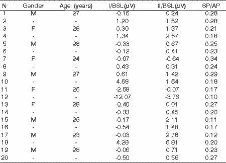
Twenty ears of 10 people, four women and six men, aged between 23 and 28 years. The inclusion criteria observed and suggested by the American ECG Society 199410 were: absence of otoneurological or neurological symptoms; no use of alcohol, illegal drugs or smoking three days before the test; otoscopy within the normal range; pure tone audiometry thresholds below 15dB hearing level in frequencies of 250. 500. 1.000. 2.000. 3.000. 4.000. 6.000 and 8.000 Hz, bilaterally; percentage speech recognition index equal or greater than 96%, bilaterally; type A tympanometric curve, bilaterally; presence of ipsilateral and contralateral stapedial reflexes in frequencies of 500. 1.000. 2.000 and 4.000 Hz, bilaterally; to sign the informed consent term.
Group CH: Subjects with clinical diagnosis of endolymphatic hydrops (Table 2)
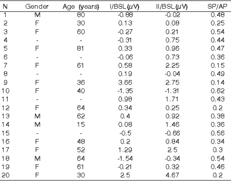
Twenty ears of 17 patients, 13 women and 4 men. The inclusion criteria, according to the suggestion made by the Committee on Hearing and Equilibrium 19952. concerning the defined diagnosis of Meniere's disease were:
to have a history of two or more spontaneous episodes of rotation or episodic vertigo, lasting 20 minutes or more; to refer tinnitus or ear fullness on the affected side and to have documented hearing loss. We also added the following inclusion criteria: otoscopy within the normal range; audiometric thresholds equal or greater than 50dB hearing level, calculated by the mean of frequencies of 500. 1.000. 2.000. 3.000 and 4000Hz; type A tympanometric curves, bilaterally; absence of concomitant neurological and/or otoneurological diseases; to sign the informed consent term.
Group SH: Subjects with labyrinth disease with no clinical evidence of endolymphatic hydrops (Table 3)
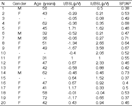
Twenty ears of 15 patients, 11 men women and 4 men. The inclusion criteria in the group were: history of dizziness episodes without the typical characteristics of episodic vertigo in Meniere's disease; not to report sensation of ear fullness, head pressure and fluctuating tinnitus; otoscopy within the normal range; to have a documented hearing loss and hearing thresholds equal or greater than 50dB hearing level, calculated by the means of frequencies 500. 1.000. 2.000. 3.000 and 4000 Hz; type A tympanometric curve, bilaterally; absence of concomitant neurological and/or otoneurological diseases; to sign the informed consent term.
All test were conducted in the device Navigator SE® by Bio-logical Systems Corp following the same protocol.
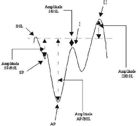
The examiner marked the points BSL, SP, AP, I and II and the device was programmed to provide amplitudes in mV between SP and BSL; AP and BSL; I and BSL and II and BSL (figure 1) and to calculate SP/AP ratio.
In the statistical analysis, we calculated the mean age in years, in the three groups and percentage distribution of the groups according to patients' gender.
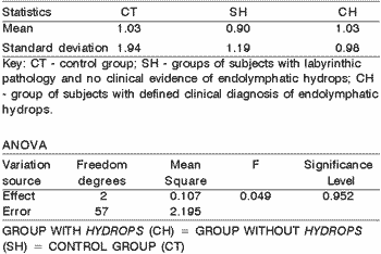
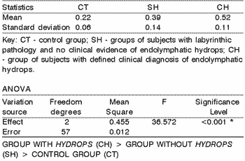
The parameters SP/AP, I/BSL, II/BSL were statistically analyzed and the variance analysis (ANOVA) was used to check the differences in behavior among the groups. The significance level defined was 5.0% and the values above this limit were identified by an asterisk (tables 4. 5 and 6).
The significant differences were assessed by means of multiple comparisons of Bonferroni in order to identify the groups of uneven behaviors11. Graphs 1. 2 and 3 present the confidence intervals. The confidence intervals presented here were calculated by the formula: confidence interval = mean ± 1.96 * standard deviation/Ö (n-1).
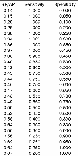
In order to measure the capacity of the parameters, we calculated the sensitivity and specificity indexes between the groups with and without endolymphatic hydrops for data that were considered statistically significant.
Sensitivity was defined as the capacity to identify among the patients with clinical diagnosis defined as hydrops, and specificity as the capacity of finding the subjects of the groups without clinical evidence of hydrops.
The efficiency of parameters was calculated by following the instructions advocated by Silva12. The purpose of efficiency was to check the possibility of sensitivity (S) and specificity (E) having similar and close values to one (100%). The value with the greatest balance between sensitivity and specificity to identify the groups with hydrops was marked in bold (Table 7).
RESULTS
The mean age of the control group was below those of the other groups (CT=26.50 years; SH=52.27 years and CH=48 years). There was prevalence of male gender in the control group (12:8), in the other two groups 75% of the patients were female.
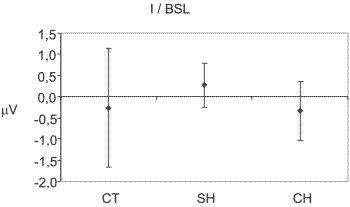
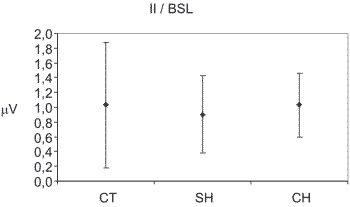
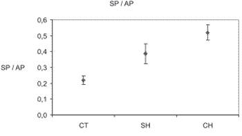
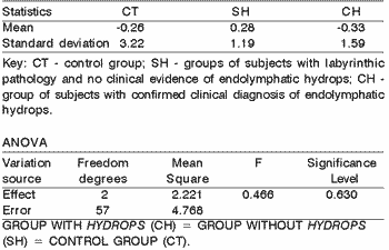
The ANOVA variance analysis and the multiple comparison test of Bonferroni demonstrated that the three groups presented statistical different behaviors when analyzing the SP/AP ratio, but there was no statistically significant difference between the three groups when the assessed parameters were I/BSL and II/BSL (Tables 4. 5 and 6 and graphs 1. 2 and 3).
Figure 1. Records of EcochG TT. The drawing shows the demarcation of the baseline (BSL), summation potential (SP), action potential (AP), peaks I and II, conducted by the examiner and of amplitudes of SP/BSL, AP/BSL, I/BSL and II/BSL provided by the software.
Graph 1. Demonstrative graph of the confidence interval containing the mean expressed in microvolts, amplitude between the baseline and the first peak that delimits the second component of the action potential (I/BSL). Key: CT - control group; SH - groups of subjects with labyrinthic pathology and no clinical evidence of endolymphatic hydrops; CH - group of subjects with defined clinical diagnosis of endolymphatic hydrops.
Graph 2. Demonstrative graph of the confidence interval containing the mean expressed in microvolts, amplitude between the baseline and the second peak that delimits the second component of the action potential (II/BSL). Key: CT - control group; SH - groups of subjects with labyrinthic pathology and no clinical evidence of endolymphatic hydrops; CH - group of subjects with defined clinical diagnosis of endolymphatic hydrops.
Graph 3. Demonstrative graph of the confidence interval of the mean SP/AP ratio expressed in microvolts. Key: CT - control group; SH - groups of subjects with labyrinthic pathology and no clinical evidence of endolymphatic hydrops; CH - group of subjects with defined clinical diagnosis of endolymphatic hydrops.
When we analyzed the values of SP/AP between the groups with clinical diagnosis defined as hydrops (CH) and no clinical evidence of hydrops (SH) to calculate the sensitivity and specificity rates, the value of 0.46 was the one that demonstrated the greatest balance between sensitivity and specificity (table 7).
Table 1 - Subjects in the control group (CT), divided by gender, age in years, amplitude in microvolts between the baseline and the first peak. and between the baseline and the second peak, according to the component of action potential (I/BSL and II/BSL) and relation between action and summation potential amplitudes (SP/AP). Key: N - number of examined ears; M - male; F- female; I/BSL - amplitude between the baseline and the first peak of the second component of the action potential; II/BSL - amplitude between the baseline and the second peak of the second component of the action potential; SP/AP - correlation between the summation and action potential amplitudes; µV - microvolts. Note: Negative values indicate that the first or second peak was below the baseline. Cells with an hyphen (-) mean that both ears of the same subject were assessed.
Table 2. Group of patients with labyrinth disorder with no clinical evidence of endolymphatic hydrops (SH), divided by gender, age in years, amplitude in microvolts between the baseline and the first peak and between the baseline and the second peak, according to the component of action potential (I/BSL and II/BSL) and relation between action and summation potential amplitudes (SP/AP). Key: N - number of examined ears; M - male; F- female; I/BSL - amplitude between the baseline and the first peak of the second component of the action potential; II/BSL - amplitude between the baseline and the second peak of the second component of the action potential; SP/AP - correlation between the summation and action potential amplitudes; µV - microvolts. Note: Negative values indicate that the first or second peak was below the baseline. Cells with an hyphen (-) mean that both ears of the same subject were assessed.
Table 3. Group of patients with clinical diagnosis of endolymphatic hydrops (CH), divided by gender, age in years, amplitude in microvolts between the baseline and the first peak. and between the baseline and the second peak, according to the component of action potential (I/BSL and II/BSL) and relation between action and summation potential amplitudes (SP/AP). Key: N - number of examined ears; M - male; F- female; I/BSL - amplitude between the baseline and the first peak of the second component of the action potential; II/BSL - amplitude between the baseline and the second peak of the second component of the action potential; SP/AP - correlation between the summation and action potential amplitudes; µV - microvolts. Note: Negative values indicate that the first or second peak was below the baseline. Cells with an hyphen (-) mean that both ears of the same subject were assessed.
Table 4. Mean and Standard deviation, in microvolts, of the amplitude between the baseline and the first peak that delimits the second component of the action potential (I/BSL), in groups CT (control), CH (defined clinical diagnosis of endolymphatic hydrops) and SH (no clinical evidence of endolymphatic hydrops). The joint assessment of the 3 pieces of information was not possible, since the information of I/BSL and II/BSL was not statistically different in the group comparisons.
Table 5. Mean and Standard deviation, in microvolts, of the amplitude between the baseline and the second peak that delimits the second component of the action potential (II/BSL), in groups CT (control), CH (defined clinical diagnosis of endolymphatic hydrops) and SH (no clinical evidence of endolymphatic hydrops)
Table 6. Mean and Standard deviation of the percentage ratio between the amplitude of action and summation potentials (SP/AP) in the in groups CT (control), CH (defined clinical diagnosis of endolymphatic hydrops) and SH (no clinical evidence of endolymphatic hydrops).
Table 7. Sensitivity and specificity indexes of SP/AP ratio defined for the groups SH (ears with no clinical evidence of endolymphatic hydrops) and CH (clinical defined diagnosis of endolymphatic hydrops). Key: SP/AP - percentage ratio between amplitude of action and summation potentials. In bold, the value of greatest balance between sensitivity and specificity (S=E=1).
Discussion
Portmann reported the great challenge that the study of Meniere's disease was and everything that was associated with it when he first defined it as a disease of great variability of clinical presentation, imprecise diagnosis and ineffective treatment13. All these characteristics associated with high prevalence14 justify the high number of research studies.
The difficulties in the defined diagnosis, which can only be made by histology studies after death, have led to the development of various clinical criteria for the definition of the diagnosis. All these criteria emphasize the association of clinical signs and symptoms with audiometric findings2.
Electrocochleography is an exam employed for the diagnosis of endolymphatic hydrops and various studies confirmed its efficacy in detecting the electrophysiological abnormalities of the inner ear15. 16. However, the reports found in the literature about the ideal parameters are very different, which also applies to the value for the diagnosis of endolymphatic hydrops and specificity and sensitivity of the diagnostic method in Meniere's disease17. 18.
The study of patients with auditory thresholds equal or greater than 50dB is justified by the high prevalence of this severe hearing loss in patients with endolymphatic hydrops, especially in advanced stages of the disease.
The hearing level equal or greater than 50dB was chosen to study the influence of AP in SP/AP ratio. AP would be the potential influenced by the destruction of hair cells, found in auditory diseases greater than 50dB and by adaptation, present in diseases that involve the auditory nerve and/or the brainstem auditory pathways5. 19.
The destruction of hair cells and adaptation modify the amplitude of AP determined by the alteration of the SP/AP ratio, not by increase in SP, but rather by decrease in AP. This reduction is justified by lack of synchrony in AP-forming neuron firing7 and was observed by other authors8.
Transtympanic electrocochleography was the test method used in our study since it is reported to be efficient in the literature as the method that enables the best response, with little intra-test and inter-test variability 20. 21.
The choice of SP/AP ratio as the main assessment parameter was based on the statement made by Coast that proposed the assessment of this relation to reduce the great inter-subjects variability of SP and AP amplitudes22.
The assessment of amplitude of both peaks that delimit N2 (I/BSL and II/BSL) was based on the hypothesis that the association of these parameters and the SP/AP ratio could help the differentiation between the two groups, following the concept of parameter association that is widely used in the definition of clinical diagnosis of the disease23.
The inclusion of the group with labyrinth disease with no clinical evidence of hydrops arose from the need to compare the groups with similar hearing thresholds, since the tested hypothesis would be of abnormal SP/AP ratio by reduction of AP, in patients with destruction of hair cells; to that end, we were extra careful to exclude patients with neurological and central otoneurological diseases.
The mean age of the group with hydrops was 48 years. This age is within the age range with the highest prevalence of the disease, as reported in the literature24.
The prevalence in female subjects in the group without hydrops and with hydrops was expected since labyrinth diseases are more frequent among women14. 24.
There were no statistically significant differences between the groups in I/BSL and II/BSL that delimited N2 of AP, differently from the data obtained by Munhoz23 that studied this association in patients with hearing thresholds below 50dB, obtaining an increment in sensitivity and specificity of the method with the combined analysis of values SP/AP and I/BSL. The amplitude of N2 has already been studied by other authors, with no detection of significance for the differential diagnosis with labyrinth disorders7.
The three groups presented different behaviors when we assessed the ratio SP/AP. The control group presented very different behaviors from the other two groups and we did not carry on the statistical analysis since it was only the control group. The main interest was to obtain values capable of differentiating, by lab results, the patients with diagnosis of endolymphatic hydrops from the patients in the group with no clinical evidence of hydrops, based on clinical relevance of these two groups.
By analyzing the group with defined diagnosis of endolymphatic hydrops and the group with no clinical evidence of hydrops, the value of 0.46 in SP/AP ratio showed the greatest balance between sensitivity and specificity (sensitivity of 75% and specificity of 60%). We did not find in the literature specific studies with patients whose thresholds were equal or greater than 50 dB to compare the results, but many studies have already recorded the influence of hearing loss in electrocochleographic findings5. 25. Some authors did not report difficulties in making the differential diagnosis between groups similar to ours upon the analysis of SP/AP ratio26. but Coats22 did not manage to differentiate the groups upon the analysis of SP/AP ratio only.
Munhoz23 recorded sensitivity and specificity rates of 100% in determining the presence of endolymphatic hydrops when using the value of 0.35 for SP/AP ratio, following the same technique proposed here, but he studied patients whose thresholds were below 50 dB. Our study demonstrated that it is extremely important to differentiate between patients with thresholds above and below 50dB; it has also served to justify why there is great variability of general rates of sensitivity and specificity in relation to SP/AP for the diagnosis of endolymphatic hydrops found in the literature, since these two groups have never been studied separately.
The issue of electrocochleographic diagnosis in patients with hearing thresholds greater or equal to 50dB was not solved by the association of parameters, as proposed in the present study (SP/AP,. I/BSL and II/BSL), but the solution can lie on the use of different techniques, with stimuli that enable better selection of frequency18. 27. 28. or even a change in the way the assessment parameters are obtained in this test22. Sass emphasized the diagnostic efficiency of electrocochleography with 1kHz tone burst. The association of this technique with the assessment of SP/AP evoked by clicks elevated the sensitivity in diagnosis of endolymphatic hydrops from 62% to 82.0%, with no change in specificity18.
Conclusion
1. The three groups presented different statistical results.
2. The SP/AP ratio was the most sensitive and specific parameter to identify the patients with confirmed clinical diagnosis of endolymphatic hydrops.
3. The association of parameters was not possible and therefore, it is not useful to be used for lab characterization of the patients in the group with confirmed clinical diagnosis of endolymphatic hydrops.
REFERENCES
1. Alford BR. Report of Subcommitte on Equilibrium and its Measurements. Menière's disease: criteria for diagnosis and evaluation of therapy for reporting results. Trans Am Acad Ophthalmol Otolaryngol 1972;76:1462-4.
2. Committee on Hearing and Equilibrium. Committee on Hearing and Equilibrium guidelines for the diagnosis and evaluation of therapy in Meniere´s disease. Otolaryngol Head Neck Surg 1995;113:181-85.
3. Munhoz MSL, Silva MLG, Ganança MM, Caovilla HH, Frazza MM. Eletrococleografia. In: Munhoz MSL, Caovilla HH, Silva MLG, Ganança MM (editores). Audiologia clínica. São Paulo: Atheneu; 2000b. p. 173-190.
4. Eggermont JJ. Basic principles for Electrocochleography Acta Otolaryngol Suppl 1974;316:7-16.
5. Eggermont JJ. Summating potentials in Meniere's disease. Arch Otorhinolaryngol 1979;222:63-75.
6. Staller SJ, Goin DW, Asher DL, Mischke RE. Summating potential in Meniere's disease. Laryngoscope 1982 Dec;92(12):1383-9.
7. Marangos N, Laszig R. Differential diagnosis of enhanced summating to compound action potential ratio. In: Arenberg IK (editor). Surgery of the Inner Ear. Amsterdam: Kugler; 1991. p. 263-7.
8. Levine SC, Margolis RH, Fournier EM, Winzenburg SM. Tympanic electrocochleography for evaluation of endolymphatic hydrops. Laryngoscope 1992 Jun;102:614-22.
9. Arenberg IK, Gibson WPR, Höhmann D, Mihalco LL. International standards for transtympanic electrocochleography. In: Höhmann (editor). ECoG, OAE and intraoperative monitoring. Amsterdam: Klugler; 1993. p. 115-8.
10. American ECG Society. Clinical evoked potentials guide lines. Recommended standards for normative studies of evoked potentials, statistical analysis of results and criteria for clinically significant abnormality. J Clin Neurophysiol 1994;11:47-7.
11. Downing D, Clark J. Estatística aplicada. São Paulo: Saraiva; 1992.
12. Silva MCM. Testes de diagnóstico ou screening tests. In: Silva MCM (autora). Estatística aplicada à psicologia e ciências sociais. Lisboa: McGraw-Hill; 1994. p. 161-180.
13. Potmann G. The old and new in Menière's disease - over 60 years in retrospect and a look to the future. Otolaryngologic Clinics of North America 1980;13(4):567-75.
14. Anadão CA. Doença de Menière: revisão. Acta Awho 1993;12(2):44-50.
15. Gatland DJ, Billings RJ, Youngs RP, Johnson NP. Investigation of the physiological basis of summating potential changes in endolymphatic hydrops. Acta Otolaryngol 1988 Mar-Apr;105 (3-4):218-22.
16. Orchik DJ, Shea JJ, Ge NN. Summating potential and action potential ratio in Meniere's disease before and after treatment. Am J Otol 1998 Jul;19(4):478-82.
17. Dornhoffer JL. Diagnosis of cochlear Menière's disease with electrocochleography. ORL 1998;60:301-05.
18. Sass K. Sensitivity and specificity of transtympanic electrocochleography in Meniere's disease. Acta Otolaryngol 1998;118:150-6.
19. Ozdamar O, Dallos P. Synchronous responses of the primary auditory fibers to the onset of tone burst and their relation to compound action potentials. Brain Res 1978 Oct 20;155(1):169-75.
20. Densert B, Arlinger S, Sass K, Hergils L. Reproducibility of the electric response components in clinical electrocochleography. Audiology 1994;33:254-63.
21. Roland PS, Rosenbbloom J, Yellin W, Meyerhoff W. Intrasubject tese-retest variability in clinical eletrococheography. Laryngoscope 1993;103:963-66.
22. Coats AC. The summating potential and Meniere´s disease. I. summating potential amplitude in Meniere and non-Meniere ears. Arch Otolaryngol 1981 Apr;107(4):199-208.
23. Munhoz MSL. Da sensibilidade e especificidade da eletrococleografia transtimpânica em pacientes com e sem hydrops endolinfático (tese). São Paulo: Universidade Federal de São Paulo - Escola Paulista de Medicina; 2001.
24. Campos CAH. Principais quadros clínicos no adulto e no idoso. In: Ganança MM (editor). Vertigem tem cura? São Paulo: Lemos editorial 1998. p.49-57.
25. Ferraro J, Lovar GB, Arenberg IK. The use of eletrocochleography in the diagnosis, assessment and monitoring of endolymphatic hydrops. Otolaryngol Clin N Am 1983;16(1):69-82.
26. Filipo R, Barbara M. Natural history of Menière's disease: staging the patients or their symptoms? Acta Otolaryngol (Stockh) 1997;526 Suppl:10-3.
27. Conlon BJ, Gibson WPR. Electrocochleography in the diagnosis of Meniere's disease. Acta Otolaryngol 2000 Jun;120(4):480-3.
28. Sass K, Densert B, Magnusson M, Whitaker S. Electrocochleographic signal analysis: condensation and rarefaction click stimulation contributes to diagnosis in Menière's disorder. Audiology 1998;37:198-206.
1 Master degree under course, Discipline of Otoneurology, Federal University of São Paulo - Escola Paulista de Medicina.
2 Doctorate studies under course, Discipline of Otoneurology, Federal University of São Paulo - Escola Paulista de Medicina.
3 Full Professor, Discipline of Otoneurology, Federal University of São Paulo - Escola Paulista de Medicina.
Address correspondence to: Letícia Clemente A. Soares Av. 11 de junho 1006/63 - Vila Clementino - 04041-003 - São Paulo - SP - E-mail: leticiaclemente@ig.com.br
Article submitted on October 23, 2002. Article accepted on February 07, 2003.
