

Year: 2003 Vol. 69 Ed. 1 - (10º)
Artigo Original
Pages: 53 to 62
 PDF PT
 Revision surgery in 74 cases of stapedectomy/stapedotomy
Author(s):
Luiz Rogerio Pires de Mello,
Andrea Pires de Mello Azevedo
Keywords: otosclerosis, conductive deafness, surgery, stapedectomy, stapedotomy.
Abstract:
Aim: The aim of this study is to identify the various reasons for a new surgical procedure to be decided on. In spite of several technical alterations, such as total platinectomy, partial platinectomy, bone interposition, fenestration in the footplate and the use of different types of prosthesis, there are still many problems concerning the improvement of impaired hearing, partly due to the insufficient training resident doctors undergo at our University Centres, where the number of stapedial surgeries has been decreasing from year to year. This creates a situation in which our resident doctors have little opportunity to gain experience in this area. Form of Study: Retrospective Evaluation. Material and Method: The study was based on 74 cases of revision stapedectomy, performed on 68 patients, 47 ears having been submitted to a revision, all having been previously operated on by the first author above mentioned, out of a population of 725 stapedectomies performed at the Antonio Pedro University Hospital (HUAP) - "Fluminense" Federal University (UFF) / Niterói-RJ, from July 1980 to June 1999. We added 21 more cases of revision surgery originating elsewhere. As far as obliteration of the oval window was concerned, for which we had to use cutting drills, there were 04 cases of variable sensorineural disacusis and 02 cases of anacusis. The cases of vertigo and / or loss of balance with a possible perilymphatic fistula were solved by means of the replacement of the long prosthesis by a smaller one, the vertigo and/or loss of balance being thus eliminated. We conducted the audiologic accompaniment of all operated-on ears 06 months after the operation and of 66 operated-on ears 01 year after the operation, 08 patients not having undergone a second examination. Auditory postoperative results depend on the surgical pathology found, not only because of the presence or absence of the incus but also because of the osseous reobliteration of the oval window. Conclusion: Considering all revision surgery cases, the auditory gains obtained were the following: for 78,3% of the patients, the auditory threshold was as low as 20db, for 10,8% of the patients, it ranged between 20db and 25db, and for 8,2% of the patients, it was only above 25%; there occurred anacusis in 2,7% of the patients. The standard audiologic evaluation included puretone threshold for air and bone condition at 250, 500, 1000, 2000, 4000 and 8000Hz, speech recognition threshold and speech discrimination score. The PTA at 500, 1000 and 2000 was calculated for air and bone curves from both preoperative and postoperative audiograms.
![]()
INTRODUCTION
Based on our experience, stapedectomy revisions are not uncommon and depend a lot on the experience of the surgeon in the first surgery.
It is known that the number of otosclerosis surgeries has decreased gradually in teaching centers, and in the USA, resident physicians currently perform five stapedectomies during their surgical internship, which is not enough for their daily practice in the future.
We are constantly in dilemma whether we should use cutting drills or not in the presence of obliteration of the oval window niche by thickened bone tissue, leading to advanced sensorineural hearing loss and even to anacusis in some cases. Another important issue is to know whether we should open or not the whole oval window with scaring tissue and multiple adherence when facing a wire prosthesis, tilted laterally to the margin in primary stapedectomy or whether we should open it totally or partially to place a new prosthesis.
More experienced surgeons 1-7 can present better postoperative hearing results by closing the air-bone gap that is below or equal to 10 to 20dB in 18% to 80% of the cases, being that our figures were about 78.3% up to 20dB. The statistics presented showed that sensorineural hearing loss can be absent or affect up to 20% of the re-operated cases.
Audiometry 8, 9 considers pre and post-operatively mean differences of air and bone pathways in frequencies of 500, 1000 and 2000Hz, which are the speech frequencies, plus the quantification of the bone structures, which represents the cochlear reserve 10, 11.
In such revisions we did not use laser 12-16 and maintained the traditional surgical technique 1, 2, 17-20 using specialized hooks, but rarely sharp cutting drills to open the stapes footplate. The prosthesis used in most revision surgeries was usually made of 0.4 to 0.6mm thick teflon and in some cases we used steel and teflon prostheses. In few cases, we used only metallic prostheses, when there were cases of obliterative otosclerosis.
Follow-up audiometry was made 06 months and 12 months after surgical revision, and 8 patients did not come to the second hearing test.
MATERIAL AND METHOD
Seventy-four (74) stapedectomy/stapedotomy revisions were conducted in 68 patients, being that 6 of them were submitted to a second revision in the period between July/1980 and June/1999 at the private practice and at the Teaching Hospital Antonio Pedro/Niteroi-RJ. Out of the total, there were 43 women (47 ears) and 23 men (27 years) whose ages ranged from 20 to 76 years.
Revisions were conducted under local anesthesia and supported by intravenous sedation with sublingual Dormonid® (midazolam), one hour before the surgery, and we used transcanal access with 6/7mm incision at the tympanic border.
After opening of the tympanic cavity, we noticed the mobility of malleus and incus upon palpation, and in case of fibrous adherence, we removed them carefully up to the niche of the oval window, removing the poor positioned prosthesis, if present, mostly commonly made of wire. In order to assess the harmful mobilizations to auditory recovery, in many situations we cut 2/3 of the prosthesis and left a small portion adhered to the oval window border and decided to place a new prosthesis to restore hearing levels. In many cases we did not completely open the oval window niche and only made it whenever there was multiple adherence that penetrated into the inner ear. We tried to create a small bed to place the prosthesis. When there was erosion of long process of incus or if we removed it entirely, we sometimes used it to sculpture the piece for inter-positioning between the oval window and malleus anterior process, or still we used 61/2mm wire prosthesis or the 6mm teflon prosthesis.
Audiometry 8, 9, 11, 18 performed preoperatively included pure tone audiometry and speech discrimination, immittanciometry and SRT, all of them repeated postoperatively to check auditory gain10, 11. During some stapedectomy revisions we used 512Hz and 1024Hz tuning forks after the surgery to check if the prosthesis was in place, with the confirmation by the good hearing skills presented by the patients.
Revisions were performed whenever the patients complained about hearing loss of at least 20dB or if they had dizziness with instability, cases in which we considered the presence of perilymphatic fistula.
We re-operated on five cases that had been previously submitted to fenestration of lateral semicircular canal with wire prosthesis from the malleus anterior process to the oval window, cases that we did not include in the present statistics.
Table 1. Revision of stapedectomy according to age, gender and side.
Female patients - 43 (47 years)
Male patients - 25 (27 years)
Age 19 to - 76 years
Right side - 41
Left side - 33
Table 2. Age of patients in the revision surgery of stapedectomy/stapedotomy.
Age / N cases
Up to 20 years - 02
21 - 40 years - 36
41 - 60 years - 21
> 61 years - 15
Total - 74
Table 3. Time to revision surgery of stapedectomy/stapedotomy.
Time to revision / N cases
AUp to 01 year - 12
01 - 05 years - 29
06 - 10 years - 11
11 - 15 years - 17
> 16 years - 05
Total - 74
Table 4. Prostheses found during revision surgery of stapedectomy/stapedotomy.
Types of Prosthesis / N cases
Wire - 29
Teflon - 21
Polyethylene - 11
Steel + Teflon - 11
Sculptured incus - 02
Total - 74
Table 5. Prostheses employed during revision surgery of stapedectomy/stapedotomy.
Types of prostheses / N cases
Teflon - 41
Steel + Teflon - 14
Wire - 08
Sculptured incus - 06
Steel - 05
Total - 74
Table 6. Time of revision after stapedectomy/stapedotomy.
Time / N cases
Up to 01 year - 12
01 - 05 years - 29
06 - 10 years - 11
11 - 15 years - 17
> 16 years - 05
Total - 74
Table 7. Time between first and second revision of stapedectomy/stapedotomy.
Table 8. Revision surgery of stapedectomy/stapedotomy caused by prosthesis displacement - 23 cases.
Material of the prosthesis found / N cases
Wire - 11
Polyethylene - 05
Teflon - 05
Teflon + Steel - 02
Total - 23
Table 9. Management in re-operations of stapedectomy/stapedotomy caused by prosthesis displacement - 23 cases.
Prosthesis used / N cases
Teflon - 18
Steel + Teflon - 05
Total - 23
Table 10. Revision surgery of stapedectomy/stapedotomy caused by erosion of long process of incus - 1st revision - 9 Cases.
Material of the prosthesis found / N cases
Wire - 03
Polyethylene - 02
Teflon - 02
Teflon + Steel - 02
Total - 9
Table 11. Management in re-operations of stapedectomy/stapedotomy caused by erosion of the long process of incus - 1st revision - 9 cases.
Management used / N cases
Incus sculpture - 04
Wire (6.5mm) - 03
Steel and Teflon (6.5mm) - 01
Steel + Teflon - 01
Total - 09
Table 12. Management in re-operations of stapedectomy/stapedotomy caused by erosion of long process of incus - 2nd revision - 02 Cases.
Type of Case / N Cases / Management
Sculptured incus - 01 - 6.5mm wire prosthesis
Damaged incus - 01 - Damaged incus structure
Table 13. Revision surgery of stapedectomy/stapedotomy caused by fibrous adherence- 09 Cases.
Material of the prosthesis found / N Cases
Wire and fat - 03
Wire and connective tissue - 02
Teflon - 02
Teflon + Steel - 02
Total - 09
Table 14. Management in re-operations of stapedectomy/stapedotomy caused by fibrous adherence - 09 Cases.
Management used / N Cases
TTeflon - 07
Steel + Teflon - 02
Total - 09
Table 15. Revision surgery of stapedectomy/stapedotomy caused by subluxation of incus- 1st revision - 05 Cases.
Material of the prosthesis found / N Cases
Wire - 01
Teflon - 02
Teflon + Steel - 02
Total - 05
Table 16. Management in re-operations of stapedectomy/stapedotomy caused by subluxation of incus - 1st revision - 05 Cases.
Management used / N Cases
Incus sculpture - 02
Wire (6.5mm) - 02
Steel + Teflon - 01
Total - 05
Table 17. Management of re-operations of stapedectomy/stapedotomy caused by incus subluxation - 2nd revision - 02 Cases.
Type of Case / N Cases / Management
Teflon in the non-eroded portion- 01 - 6.5mm wire prosthesis
Sculptured incus - 01 - 6.5mm wire prosthesis
Table 18. Revision surgery of stapedectomy/stapedotomy caused by obliteration of oval window - 1st revision - 04 Cases.
Material of the prosthesis found / N Cases
Teflon - 03
Steel + Teflon - 01
Total - 04
Table 19. Management in re-operations of stapedectomy/stapedotomy caused by obliteration of oval window - 1st revision - 04 Cases.
Management Used / N Cases
Steel + Teflon - 02
Teflon - 02
Total - 04
Table 20. Management in re-operations of stapedectomy/stapedotomy caused by new obliteration of oval window - 2nd revision - 02 Cases.
Type of Case / N Cases / Management
Steel + Teflon - 02 - Steel
Table 21. Revision surgery of stapedectomy/stapedotomy caused by perilymphatic fistula - 05 Cases.
Material of the prosthesis found / N Cases
Polyethylene - 02
Wire - 03
Total - 05
Table 22. Management in re-operations of stapedectomy/stapedotomy caused by perilymphatic fistula - 05 Cases.
Management Used / N Cases
Teflon - 05
Total - 05
Table 23. Revision surgery of stapedectomy/stapedotomy caused by short prosthesis - 04 Cases.
Material of the prosthesis found / N Cases
Wire - 02
Teflon - 02
Total - 04
Table 24. Management in re-operations of stapedectomy/stapedotomy caused by short prosthesis - 04 Cases.
Management Used / N Cases
Teflon - 04
Total - 04
Table 25. Revision surgery of stapedectomy/stapedotomy caused by long prosthesis - 03 Cases.
Material of the prosthesis found N Cases
Teflon - 02
Steel + Teflon - 01
Total - 03
Table 26. Management in re-operations of stapedectomy/stapedotomy caused by long prosthesis - 03 Cases.
Management Used / N Cases
Teflon - 03
Total - 03
Table 27. Revision surgery of stapedectomy/stapedotomy caused by fluctuating footplate - 02 Cases.
Material of the prosthesis found / N Cases
Wire - 01
Teflon - 01
Total - 02
Table 28. Management in re-operations of stapedectomy/stapedotomy caused by fluctuating footplate - 02 Cases.
Management Used / N Cases
Teflon - 02
Total - 02
Table 29. Revision surgery of stapedectomy/stapedotomy caused by narrowed oval window niche - 01 Case.
Material of the prosthesis found / N Cases
Wire - 01
Total - 01
Table 30. Management in re-operations of stapedectomy/stapedotomy caused by narrowed oval window niche - 01 Case.
Management Used / N Cases
Teflon - 01
Total - 01
Table 31. Revision surgery of stapedectomy/stapedotomy caused by facial nerve damage - 01 Case.
Material of the prosthesis found / N Cases
Teflon - 01
Total - 01
Table 32. Management in re-operations of stapedectomy/stapedotomy caused by facial nerve damage - 01 Case.
Management Used / N Cases
Facial nerve flap and Teflon - 01
Total 01
Table 33. Revision surgery of stapedectomy/stapedotomy caused by fixed stapes footplate - 01 Case.
Material of the prosthesis found / N Cases
Polyethylene and vein (stapes footplate fragmentation technique) - 01
Total - 01
Table 34. Management in re-operations of stapedectomy/stapedotomy caused by fixed stapes footplate - 01 Case.
Management Used / N Cases
Teflon - 01
Total - 01
Table 35. Revision surgery of stapedectomy/stapedotomy caused by elimination of teflon prosthesis - 01 Case.
Material of the prosthesis found / N Cases
Teflon - 01
Total - 01
Table 36. Management in re-operations of stapedectomy/stapedotomy caused by teflon prosthesis elimination - 01 Case.
Management Used / N Cases
Steel - 01
Total - 01
Table 37. Main causes of failure in the first surgery of stapedectomy/stapedotomy.
Table 38. Statistics of auditory results of revision surgery of stapedectomy/stapedotomy.
Table 39. Auditory gain six months after revision surgery of stapedectomy/stapedotomy - 74 Cases.
RESULTS
Our results were based on 74 operated ears, being that 68 were first revisions and six were second revision surgeries (02 cases of oval window obliteration, 02 cases of long process incus erosion and 02 cases of subluxation of incus).
In the case of perilymphatic fistula suspicion (05 cases) we were based on the imbalance of patients, which suggested perilymphatic fistula. The revisions showed patients operated on with placement of polyethylene tubes fixed to the lenticular apophysis.
The age of patients at revision is described in Table 2.
The mean time between the first surgery and the revision surgery was eleven years, being that 12 cases were re-operated within 12 months.
The type of prosthesis found was polyethylene tube, wire, steel and teflon, pure steel and teflon.
The types of prostheses used in our revisions were mostly teflon, steel and teflon, steel and wire.
Sealing of oval window was made using tympanic connective tissue, fat and gelfoam and/or perichondrium.
We did not confirm the presence of problems with sensorineural hearing loss and use of gelfoam, as advocated by Sheehy23, who interrupted its use in 1968.
As to re-operation and period of revision, we detected what is shown in Table 6.
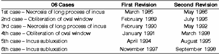
As to the six cases submitted to a second revision the time to surgery is described in Table 7.
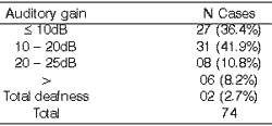
Hearing improvement was detected in 78.3% of the patients in up to 20dB in the first audiometry made 6 months after revision surgery and in 19 cases the gap was closed - equal or below to 10dB.
There were also six cases of variable sensorineural hearing loss in low frequencies, especially in cases with thickened stapes footplate; in primary surgery, we faced difficulties to open these footplates and it was necessary to use sharp cutting drills. In the revision surgery, we had to use them again, and the auditory result was poor, resulting in two cases of anacusis.
Prosthesis displacement - 23 Cases (31.08%)
Prosthesis displacement, with or without associated fibrous adherence, was the most common cause for the revision. In the revision surgeries, we found that in the displacement cases the lower portion of the central part of the oval window was displaced to the lateral portion or there was poor contracture of the high portion of the long incus apophysis. Sometimes it was found dropped on the bottom of the cavity.
Erosion of the long process of incus - 9 Cases
As to necrosis of long process of incus there were nine cases of first revision, two of them operated with polyethylene, three with wire, two with steel and teflon, and two with teflon prostheses. We reused incus and sculptured it in 4 cases. In one case, since the bone damage was small, we reused the incus as a whole, placed the steel and teflon prosthesis in the longer portion of the apophysis with no erosion. In the 4 cases we used 61/2mm wire prosthesis from the malleus anterior process to the oval window (in 3) and in another case we used the steel and teflon prosthesis from the malleus anterior process to the oval window, since the malleus was a little vertical. We sealed the window with connective tissue, perichondrium and gelfoam.
We had two cases of second revision in which hearing worsened after surgery and in the first case, we removed the incus and placed a steel and teflon prosthesis from the malleus anterior process to the oval window.
There were 2 cases of second revision in which hearing worsened after the proposed surgery, and in the 1st case, we removed the reused incus and placed a steel and teflon prosthesis from the malleus anterior process to the oval window.
Fibrous Adherence - 9 Cases (12.16%)
There were 9 cases of fibrous adherence, being four cases after use of wire, two cases of steel and teflon, and two cases of teflon prostheses in the first surgery. In all cases, were released the adherence, removing the prostheses partially or totally. In the case of wire prosthesis, we replaced it for teflon prostheses. In other four cases, we placed two teflon and two steel and teflon prostheses.
Incus Subluxation - 5 Cases
The author operated on two cases of incus subluxation, and it was very difficult to place the prosthesis in the initial stapedectomy, resulting in poor auditory results. In two cases initially operated with wire prostheses, we removed the prostheses and the subluxation incus and placed a 6.5mm wire prosthesis from the malleus anterior process to the oval window, recovered by connective tissue. In the other three cases of external patients, we removed the two prostheses of teflon and one of wire that were lateralized, since the incus was luxated, reusing two incus that were sculptured and replaced between the malleus anterior process and the oval window recovered with connective tissue. In the third case we used steel and teflon prosthesis from the malleus anterior process to the oval window, with connective tissue sealing.
There were two cases in which hearing was poor postoperatively and we decided to have a second revision surgery. In the first case, we placed the teflon prosthesis in the non-eroded portion of incus, and it was found on the bottom of the cavity during the revision. We removed the eroded incus and placed a wire prosthesis from the malleus anterior process to the oval window. In the second case, we had made an interposition of sculpted incus between the malleus anterior process and the oval window niche and during revision we saw that it was on the bottom of the cavity. We decided to use 6.5mm wire prosthesis from the malleus anterior process to the oval window for hearing recovery.
Obliteration of oval window by neoformed bone - 06 Cases (8.10%)
There were four cases of bone obliteration in the first revision, being three cases with use of teflon and one with use of steel and teflon prosthesis, and two cases of second revision - cases that owing to the initial difficulty of opening the very thick footplate, forced us to excessively manipulate it using sharp microcutting drills, which enabled the opening of 6 to 08mm. We conducted the revision in these cases by placing in two steel and teflon prostheses and in the other two, teflon prostheses. The re-operated cases with steel and teflon were submitted to a second revision for new re-obliteration of the oval window, using two steel prostheses. As to hearing, four cases presented variable sensorineural hearing loss and two cases had complete deafness. All patients presented vertigo since the initial stapedectomy surgery.
Perilymphatic fistula - 05 Cases (6.88%)
After some time, two patients that underwent stapedectomy presented dizziness, imbalance and progressive hearing loss, leading us to suspicion of labyrinthic fistula, confirmed by audiologic assessment. The patients had been operated and polyethylene tubes were used as prostheses. They were re-operated, the prostheses were removed as well as the adherence tissue in the oval window niche, sealing the window with connective tissue and placing teflon prosthesis. The other 3 cases of external patients had been operated with wire prostheses; two cases had implant of steel and teflon prostheses; one had received the same previous technique of revision surgery with teflon prosthesis. We have also placed teflon prostheses in these three cases after having sealed the oval window with connective tissue from the tympanum.
Short prosthesis - 04 Cases (5.40%)
Of the four cases, two were external patients. We found adherence throughout the whole base, in the oval window niche, pulling the vertical portion of the prosthesis and leading it to lateral border of the oval window (wire prosthesis). In the other 2 cases in which we operated the first stapedectomy, we cleaned scarred tissue, removed the teflon prostheses, sealed the oval window with connective tissue in one case and perichondrium in the other. We replaced the prostheses found for four teflon prostheses.
Long prosthesis - 03 Cases (4.05%)
Of the three re-operated cases, two were external patients and one was the author's private patient. The main complaints were dizziness, imbalance and difficulty to walk. We conducted revision surgery as fast as possible (90 to 150 days) in order to correct the labyrinthic fistula. We found two cases of teflon prosthesis, with previous sealing of the oval window. After the surgery, dizziness and imbalance disappeared and hearing improved.
Fluctuating Footplate - 02 Cases (2.70%)
We found in these patients the whole footplate on the oval window, and it forced us to make a very difficult maneuver, opening the promontorium laterally and using a straight hook to remove the stapes footplate. We found one wire prosthesis and another teflon prosthesis. We removed them both, sealed the oval window with connective tissue and placed two teflon prostheses, resulting in good hearing recovery. See Table 28.
Narrowed oval window niche - 01 Case (1.35%)
We found the wire prosthesis outside the oval window with a small opening. We proceeded to opening the posterior portion of the stapes footplate and placed a 0.4mm thick teflon prosthesis over connective tissue, replacing the previous prosthesis.
Facial nerve damage - 01 Case (1.35%)
Facial nerve dehiscence hindered visualization of stapes footplate, causing damage to it and placement of teflon prosthesis over the abnormal facial nerve fibers.
We re-operated the patient with a facial nerve graft and placement of a 0.4mm thick teflon prosthesis.
Fixed stapes footplate - 01 Case (1.35%)
The patient was operated when stapedectomy first started, using footplate fragmentation technique and polyethylene tube and vein. We removed the polyethylene tube, reopened the stapes footplate, sealed the oval window with connective tissue and placed a teflon prosthesis for hearing recovery.
Elimination of teflon prosthesis - 01 Case (1.35%)
We had a unique case of a patient operated on with teflon prosthesis that had been displaced and was eliminated five years later through epytympanic opening. We performed a revision surgery and placed a steel prosthesis because we thought it could be a case of prosthesis rejection.
DISCUSSION
Revisions of stapedectomy in the period between July 1980 and June 1999 were not rare, owing probably to total opening of oval window or by the material initially used, such as polyethylene tubes, wire and gelfoam.
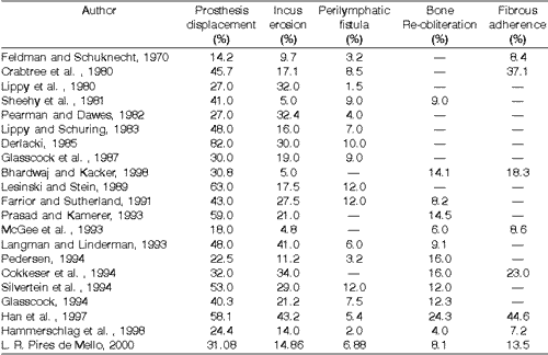
As we can see in Table 37, the various statistics presented by different authors showed a great variation in the most important causes of failure in the first surgery.
Of the various studied authors, we noticed great statistic variation concerning displacement of prosthesis from the oval window niche, ranging from 14.2% reported by Feldman and Schuknecht (1970)21, to 82% in the statistics by Derlacki (1985)22. In our sample of 74 re-operated patients, we found 31.8% of prosthesis displacement cases. As to incus erosion, the statistics ranged from 4.8% in patients operated by Mcgee et al. in 199320, 5% by Sheehy et al. in 198123, 5% by Bhardwaj and Kacker in 199817, to 41.0% in the statistics of Langman and Linderman in 19933 and 43.2% in the data by Han et al.9 in 1997. In our cases, the figure was 14.86%.
As to perilymphatic fistula, the variation was from 1.5%, reported by Lippy et al. in 198324, to 12% in the findings reported by Lesinski and Stein in 198912, Farrior and Sutherland in 199120 and Silverstein et al. in 199415. We found in our cases 6.88% of perilymphatic fistulae. As to oval window bone obliteration, the figures ranged from 4.0% in the findings reported by Hammerschlag et al. in 19888, to 24.3% in the statistics by Han et al. in 19979. Our data resulted in 8.1% of re-operated cases and two patients were submitted to a second revision.
Finally, as to fibrous adherence, the ranges varied from 7.2% operated cases by Hammerschlag et al. in 19988, to 44.6% of cases reported by Han et al. in 19979. We found 13.5% of re-operated cases because of fibrous adherence.
Based on the statistics presented in Table 37, we once again emphasize the great variation in figures of re-operations reported by various authors.
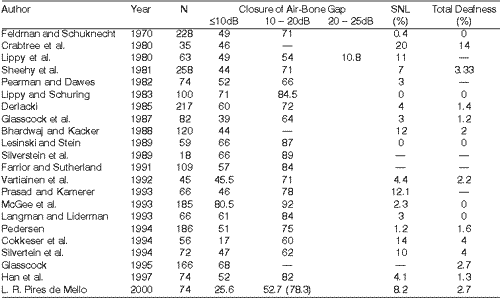
Concerning the closure of air-bone gap, we noticed a variation among the statistics reported by authors such as Hammerschlag et al. in 19988, in 306 cases, Feldman and Schuknecht in 197021, in 228 cases, Sheehy et al. in 198123, in 258 cases, Derlacki in 198522, in 217 cases, up to Crabtree in 198019 in 35 cases.
Air-bone gap closure equal or below 10dB varied from 80.5% in McGee13 cases to 17% in Cokkeser18 et al.15,16. In our revised cases, we had 52.7% of patients within this range.
In cases of sensorineural hearing loss (SNL) statistics ranged from 0% in Lesinski and Stein12, and Lippy and Schuring24,25, to 0.4%, in the cases reported by Feldman and Schuknecht21, to 20% in the cases by Crabtree19 et al., being that our statistics for this aspect was that 8.17% of the cases presented sensorineural hearing loss.
As to anacusis, statistics ranged from 0% in Feldman and Schuknecht21 cases, Lippy and Schuring24,25, Lesinski and Stein12, McGee13 et al., Langman and Linderman3, to 14% in Cabtree et al.19 cases. In our re-operations, the figure for total deafness was 2.6% of the sample.
CONCLUSION
In our total of 74 cases of revision surgery of stapedectomy/stapedotomy, operated on between July 1980 and June 1999, there were 27 cases (36.4%) of gap closure (£ 10dB) and 31 cases of 10dB to 20dB gain (41.9%), 08 cases of 20dB to 25dB (10.8%), 06 cases over 25dB (8.2%), and 02 cases of total deafness (2.7%).
The most common findings in our revision were displacement of prosthesis - 23 cases (31.08%), incus long process erosion - 11 cases (14.86%), fibrous adherence - 09 cases (12.6%), incus subluxation - 07 cases (9.45%), oval window re-obliteration - 06 cases (8.10%), perilymphatic fistula - 05 cases (6.88%).
We concluded that the revision surgeries conducted by experienced surgeons are prone to produce excellent results, which may reach 80.6% of gap closure, as shown by McGee in 1993.
REFERENCES
1. Glasscock ME, Storper IS, Haaynes DS, et al. Twenty-five years of experience with stapedectomy. Laryngoscope 1995;105:8999-904.
2. Glasscock ME, Mckennan KX, Levine SC. Revision stapedectomy surgery. Otolaryngol Head Neck Surg 1987;96:141-8.
3. Langman AW, Linderman RC. Revision stapedectomy. Laryngoscope 1993;103:954-8.
4. Pearman K, Dawes DK. Post-stapedectomy conductive deafness and results of revison surgery. J Laryngol Otol 1982;96:405-10.
5. Pederson CB. Revision sugery in otosclerosis-operative findings in 186 patients. Clin Otolaryngol 1994;19:446-50.
6. Prasad S, Kamerer DB. Result of revision stapedectomy for conductive hearing loss. Otolaryngol Head Neck Surg 1993;109:742-u7.
7. Vartiainen E, Nutinen J, Virtaniemi J. Long-term results of revisión stapes surgery. J Laryngol Otol 1992,106:971-3.
8. Hammerschlag PE, Fishman A, Scheer AA. A review of 308 cases revision stapedectomy. Laryngoscope 1998;108:1794-1800.
9. Han WW, Incesulu A, Mckenna MT, Rauch SS, Nadol JB, Glynn RJ. Revision stapedectomy: intraoperative findings, results and review of literature. Laryngoscope 1997;107:1185-92.
10. Berliner KI, Doyle KJ, Goldenbeerg RA. Reporting operative hearing results in stapes surgery: does choice of outcome measure make a difference? Am J Otol 1996;17:521-8.
11. Committee on Hearing and Equilibrium. Committee on hearing and equilibrium guidelines for the evaluation of result of treatment of conductive hearing loss. Otolaryngol Head Neck Surg 1995;113:186-7.
12. Lesinski SG, Stein JÁ. Stapedectomy revision with CO2 laser. Laryngoscope 1989;99:13-19.
13. McGee TM, Diaz-Ordaz EA, Kartush JM. The role of KTP laser in revision stapedectomy. Otolaryngol Head Neck Surg 1993;109:893-43.
14. Rauch SD, Barley ML. Argon laser stapedectomy: comparison to traditional fenestration techniques. Am J Otol 1990;13:556-60.
15. Silverstein H, Bendet E, Rosenberg S, et al Revisions stapes surgery with and without laser: a comparison. Laryngoscope 1994;104:1431-8.
16. Silverstein H. Rosenberg S, Jones R. Small fenestra stapedectomies with and without KTP laser: a comparison. Laryngoscope 1989;99:485-8.
17. Bhardwaj BK, Kacker SK. Revision stapes surgery. J Laryngol Otol 1988;102:20-4.
18. Cokkeser Y, Naguib M, Aristegui M, et al. Revisión stapes surgery: a critical evaluation. Otoryngol Head Neck Surg 1994;111:473-7.
19. Crabtree JA, Britton BH, Powers WH. An evaluation of revision stapes surgery. Laryngoscope 1980;90:224-7.
20. Farrior J, Sutherland A. Revison stapes susrgery. Laryngoscope 1991;101:1155-61.
21. Feldman BA, Schuknecht HF. Experience with revision stapedectomy procedures. Laryngoscope 1970;80:1281-91.
22. Derlacki EL. Revison stapes surgery: problems with some solution. Laryngoscope 1985;5:1047-53.
23. Sheehy JL, Nelson RA, House HP. Revision stapedectomy: a review of 258 cases. Laryngoscope 1981;91;43-51.
24. Lippy WL, Schuring AG. Stapedectomy revision of the wire-gelfoam prosthesis. Otolaryngol Head Neck Surg 1983;91:9-13.
25. Lippy WH, Schuring AG. Stapedectomy revision. Am J Otol 1980;2:15-21.
26. Scheer AA. A new method of incus bypass in stapedectomy. Arch Otol 1974:100(4)322-3.
1 Professor Titular de Otorrinolaringologia da Faculdade de Medicina da Universidade Federal Fluminense - Niterói/RJ
2 Mestranda da Fundação Oswaldo Cruz - FIOCRUZ/ENSP
Médica Otorrinolaringologista responsável pelo Setor de Otoneurologia da Clínica Luiz Pires de Mello
Endereço para correspondência: Praia de Icaraí, 341 apt. 1301 bloco A Icaraí - Niterói RJ 24230-005
Telefax. (0xx21) 2612-2288 - E-mail: rpmello@urbi.com.br
Trabalho apresentado no 35º Congresso Brasileiro de ORL realizado em Natal, no ano de 2000.
Article submitted on June 01, 2001. Article accepted on September 19, 2002.
Print: ![]()