

Year: 2003 Vol. 69 Ed. 1 - (7º)
Artigo Original
Pages: 34 to 38
 PDF PT
 Technical associations combined for snoring and peripheral OSA surgical repair
Author(s):
Jeferson Sampaio D'Avila,
Carlos R. T. Gois,
Ronaldo C. Santos Jr.,
João C. Todt Neto
Keywords: snoring, obstructive sleep apnea, uvulopalatoplasty.
Abstract:
Introduction: Associating those classical surgical techniques, it's aimed to offer, in a safer way, what each technique offers better, introducing the Optical Microscopy in all procedures. Material and Method: The practical peripheral topodiagnosis was made with well-defined criteria, isolating only the ones labeled as Level 1 of Fujita. Three specific types of techniques were associated. Group A: LAUP (Kamami) + U.P.F.P.. Partial (modified Fujita). Group B: LAUP + cryptolisis (Krespi) + U.P.F.P. Partial (modified Fujita). Group C: LAUP + Tonsils Microsurgery (Andréa/Dias) + U.P.F.P. Partial (Modified Fujita). The surgical identification for each one are the following: Group A: Uvula Hypertrophy + palate low placement + lack of Tonsils (range I). Group B: anatomical parameters equal to the Group A + Tonsils trophy or hypertrophy (range I). Group C: Uvula and tonsils hypertrophy (range II and III) + pharynx redundancy. In 38 months period, there has been 60 operated cases in a proportion of: Group A: 20; Group B: 12 e Group C: 28. Result: Clinical improvement occurred in 46 patients (76,67%). The best results were detected in groups A and C. Conclusion: Selection in groups of patients with snoring and SAOS (peripheral) labeled as Level 1 of Fujita, considering anatomical and clinical parameters, offer more safety in the choice of surgical methods. Thus, with this proposal of associating those techniques, we've been accomplishing better results and so we suggest the effective use of this basic protocol in other services.
![]()
Introduction
Clinical and/or surgical repair of snoring and obstructive sleep apnea (OSA) has been a constant reason for carrying out new studies. The understanding of the physiological and pathogenic aspects of these disorders is in full evolution and increasingly better defined criteria tend to be presented in a more straightforward and methodic way 1-7.
A statically increase of better outcomes has been clearly observed, which are primarily surgical for the treatment of snoring and OSA of peripheral nature. This is due to particularly more stringent studies of their etiology. Obstructive levels such as classic Fujita levels, serve to identify obstructive areas. 8-11.
The Fujita levels are classified in three levels: Level I is represented by obstructive processes that involve the oropharynx, more specifically related to the uvula-palate region. Level II is represented by the obstructive processes that involve the tongue base, be it due to hypertrophy or muscle hyperplasia and lingual tonsil, or associated with obstructive processes of the oropharynx region, in other words, those from level I of Fujita. Level III is the last one and is represented by obstructive processes exclusive of the region of the tongue base (hypopharynx)2,10,12,13.
This peripheral topodiagnosis is essential for the good outcome of therapy. Clinical evaluations such as flexible rhinolaryngoscopy, and cephalometry with simple radiological study of the aerial space of the hypopharynx and the recording in cassette tapes or the sleep video are basic aspects for the indication of surgery. Methods such as polysomnography and cephalometry are essential to confirm the topodiagnosis and to study the complications of this syndrome12-15.
There are some therapeutic surgical methods to correct snoring and OSA (peripheral) and among these the most important are the following: classical uvulopalatopharyngoplasty (UPPP) (Fujita, 1981) with the purpose to enlarge the air space of the pharynx through soft tissue exeresis using sutures with several threads. LAUP (Laser Assisted Uvulo-Palatoplasty) is also an effective surgery, which uses CO2 laser as the most important device (Kamami, 1994). There is also tonsil microsurgery (TM.) (Andrea; Dias, 1993), in addition to other methods such Cryptolysis (Krespi, 1994), to reduce palate tonsils 4,6,16-22.
All surgical methods, if proper indicated, have well-defined and acknowledged values.
Objective
The objective of this study is to analyze screening criteria and post-operative outcomes of patients that have undergone combined surgical techniques for the treatment of snoring and OSA (peripheral) and the choice of surgical methods depended on anatomic-clinical factors.
MATERIAL AND METHOD
After establishing a positive peripheral topodiagnosis for snoring and peripheral OSA, without setting to a secondary plane the study of nasal cavities and rhinopharynx as likely zones for associated obstructive sites, we performed 60 surgeries in 36 months with patients exclusively ranked as Fujita's Level I.
Optical pharyngeal microscopy was included in all procedures (Andrea and Dias, 1993). It was performed in association with conventional techniques that were grouped together and classified in Groups A, B, or C.
In Group A, LAUP (Kamami, 1994) was associated with. Partial UPPP (Modified Fujita - FM). For the latter this variation of the terminology was designated to justify the basic principle of the technique (Fujita, UPPP) would be kept, and only variations in sutures were performed with fewer amounts of stitches. The patients that have undergone surgeries in Group A were those that fit in clinical cases of uvula hypertrophy, lowering of the palate and atrophy or absence of palate tonsils (post former tonsillectomy) (Figures 1 and 2).
In Group B, LAUP (Kamami, 1994), Cryptolysis (Krespi, 1994) and UPPP. (FM) were associated. The anatomical and clinical picture of the patients that have undergone surgeries in this group was similar to those clinical pictures of Group A, associated palate tonsil hypertrophy levels II and I (Figures 3 and 4).
In Group C, the associated surgical techniques were: LAUP, Tonsil Microsurgery (Andrea and Dias) and UPPP (FM). The group of patients that fit in this group was those with uvula hypertrophy, tonsil hypertrophy (level III) and redundant tissue in the posterior wall of the pharynx (Figures 5 and 6).
This analysis is easier to be understood with comparative observation of the anatomical-clinical-surgical picture of the patients (Table 1).
Table 1. Anatomic-clinical-surgical.
All procedures were performed under general and regional anesthesia under oral intubation and patients stayed in the hospital for only 1 day. Regional anesthesia used was marcaine 0.5% with adrenaline. The microscope used was a D.F. Vasconcelos with 300 mm magnification.
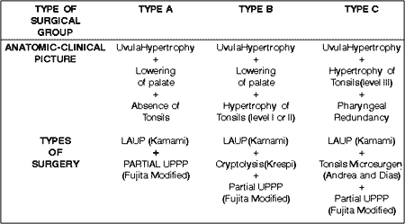
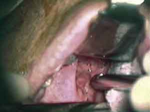
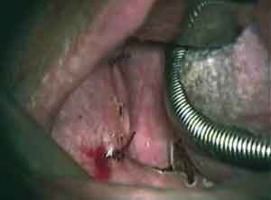
Figure 1. Anatomical Type A: dropped palate with absence of tonsils.
Figure 2. Surgical Type A: uvulopalatoplasty with CO2 Laser.
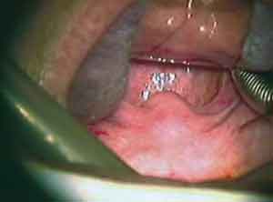
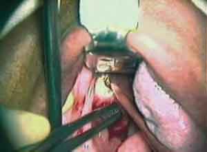
Figure 3. Anatomical Type B: Hypertrophy of palate tonsils (Level I).
Figure 4. Surgical Type B: Modified Fujita technique.
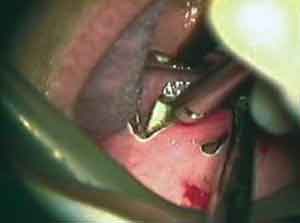
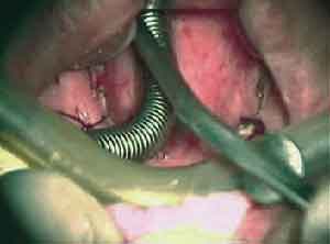
Figure 5. Anatomical Type C: Hypertrophy of palate tonsils (Level III).
Figure 6. Surgical type C: The final aspect of technique association.
Results
Sixty surgeries were performed in a period of 38 months following the approach of technique association, and they were distributed as follows: Group A: 20 surgeries performed, Group B: 12 and Group C: 28.
The objective of this surgical system was to promote in a less traumatic and safer way the increase of the air space in the pharyngeal site in Fujita region I. Therefore, we were able to control patients' breathing, and this was confirmed by patients' satisfaction with the improvement of their sleep time, well being and energy during day time, elimination or relative control of snoring in over 76.6% of the cases, or 46 cases. Among all groups, the one that had better results was Group A; with significant improvement of 16 out of 20 patients that were operated on (80%). In relation to Group C, 22 out of 28 patients (78.5%) reported they were satisfied with the clinical improvement. More modest results were obtained with Group B, with only 8 patients (66.67%) reporting improvement.
Discussion
Stringent screening for surgery indication and the performance of well-defined peripheral topodiagnosis resulted in an exclusive case selection of obstructive processes classified as Fujita's level I. It had a dramatic favorable impact in our results, since the techniques used are known as conventional for this kind of mechanical respiratory obstruction2,3,10.
Magnification provided by optical microscopy in all procedures ensured safety of the surgery, since the preservation of noble structures and the understanding of the anatomical, physiological and pathogenic aspects of the patient at the moment of tissue exeresis, together with the ability to perform the sutures in specific sites, were fundamental for the success of the procedure16,20,22.
These associations of surgical techniques made us use the better of each one all together in an innovative way. This diagnostic and therapy standardization resulted in satisfactory outcomes of all our cases. 17,18.
As to result differences among the groups, it is believed that the fact that the most successful cases were in Groups A and C can be explained by anatomical and functional issues. In group A we had all those patients with uvula hypertrophy and lowering of soft palate as the only obstructive factors. Therefore, partial resection of uvula associated with restructuring of the palate velum provided a major increase of air space.
In case of Group C, the elimination of the excessive uvula, palate hypertrophy tonsils and pharyngeal redundancy not only made the air space larger but also decreased laxity of the tissues of the posterior wall of the pharynx, which played a major role in causing snoring. Meanwhile, in Group B, the sparing of intermediate-sized tonsils could be the major reason for therapeutic failures among those patients. These facts have been encouraging us to perform tonsillectomy in patients with snoring symptoms and moderate tonsil hypertrophy, when they need to undergo uvulopalatopharyngoplasty.
Conclusion
The selection of the groups of patients with snoring and peripheral OSA (Fujita's level I) taking into account anatomical and clinical parameters provided increased safety in the proper choice of the surgical approach method, with a global success rate of 76.6%. Therefore, with this proposal of technique association we have been able to improve our results and we suggest that this protocol becomes effective in other services.
References
1. Berg S, Cole P, Hoffstein V, Haight JS. Upper airway pressures in snorers and nonsnorers during wakefulness and sleep. J Otolaryngol 2001 Apr;30(2):69-74.
2. Henk Boot, Robert van Wegen, René ML. Long-Term Results of Uvulopalatopharyngoplasty for Obstructive Sleep Apnea Syndrome. Laryngoscope 2000;110:469-475.
3. Friedman M, Tanyeri H. Clinical Predictors of Obstructive Sleep Apnea. Laryngoscope 1999;109:1901-1907.
4. Pinto JA, Paupério A. O uso do Laser de CO2 em Otorrinolaringologia. Ars Cvrandi Set/1981.
5. Pontes PA, Gregório LC. O laser de CO2 em Otorrinolaringologia. Aplicações clínicas. Parte II. Acta Awho mai/ago 1990;9(2).
6. Schechter MS. Technical report: diagnosis and management of childhood obstructive sleep apnea syndrome. Pediatrics 2002 Apr;109(4):e69.
7. Viner S, Szalai JP, Hoffstein V. Are history and physical examination a good screening test for sleep apnea? Ann Intern Med 1991;115:356-359.
8. Barnes M, Houston D, Worsnop CJ, Neill AM, Mykytyn IJ, Kay A, Trinder J, Saunders NA, Douglas McEvory R, Pierce RJ. A randomized controlled trial of continuos positive airway pressure in mild obstructive sleep apnea. Am J Respir Crit Care Med 2002 Mar 15;165(6): 773-80.
9. Brown DJ, Kerr P, Kryger M. Radiofrequency tissue reduction of the palate in patients with moderate sleep-disordered breathing. J Otolaryngol 2001 Aug;30(4): 193-8.
10. Fujita S, Conway W, Zorick F, Roth T. Surgical correction of anatomic abnormalities in obstructive sleep apnea syndrome: uvulopalatopharyngoplasty. Otolaryngol Head Neck Surg 1981;89:923-934.
11. Seemann RP, DiToppa JC, Holm MA, Hanson J. Does laser-assisted uvulopalatoplasty work? Na objective analysis using pre-and postoperative polysomnographic studies. J Otolaryngol 2001 Aug;30(4):212-5.
12. Goldberg AN, Schwab RJ. Identifying tha patient with sleep apnea: upper airway assessment and physical examination. Otolaryngol Clin North Am 1998;31:919-930.
13. Richardson MA, Seid AB, Cotton RT, Benton C, Kramer M. Evalution of tonsils and adenoids in sleep apnea sysdrome. Laryngoscope 1980;90:1106-1110.
14. Finkelstein Y, Stein G, Ophir D,Berger R, Berger G. Laser-assisted uvulopalatoplasty for the management of obstructive sleep apnea: myths and facts. Arch Otolaryngol Head Neck Surg 2002 Apr;128(4):429-34.
15. Nieminen P, Lüppünen T, Tolonen U, Lanning P, Knip M, Lüppünen H. Growth and biochemical markes of growth in children with snoring and obstructive sleep apnea. Pediatrics 2002 Apr;109(4): e55.
16. Andréa M. Microsurgical Bipolar Cautery Tonsilectomy. Laryngoscope 1993;103:1177-8.
17. Conway W, Fujita S, Zorick F et al. Uvulopalatopharyngoplasty: one year follow-up. Chest 1985:88:385-387.
18. Kimmelman PC, Levine B, Shore ET, Millman RP. Uvulopalatopharyngoplasty: a comparison of two techniques. Laryngoscope 1985;95:1488-1490.
19. Krespi, Yosef P, Keidar. Anat. Laser-assisted Uvulopalatoplasty for The Treatment of Snoring. Operative Techniques in Otolaryngology - Head and Neck Surgery 1994;5(4) (DEC); p. 228-234.
20. Kujawski O, Dulguerov P, Gysin C, Lehmann W. Microscopic tonsilectomy: a double blind randomized trial. Otolaryngology Head and Neck Surgery 1997 (in press).
21. Rodenstein DO. Assessment of uvulopatatopharyngoplasty for the treatment of obstructive sleep apnea syndrome. Sleep 1992;15:s56-s62.
22. Yanagiswa E, Isaacson G, Andréa M. Techniques of Tonsilectomy (video). American Academy of Otolaryngoscopy Head and Neck Surgery, Continuing Education with Television.
1 Ph.D. in Otorhinolaryngology, Medical School, University of São Paulo.
2 Scientific Director, Centro de Estudos Avançados em ORL e Fonoaudiologia, in Sergipe.
3 Ph.D. in Otorhinolaryngology, Medical School, University of São Paulo.
4 Master studies in Otorhinolaryngology under course, Medical School of Ribeirão Preto, University of São Paulo.
Affiliation: Centro de Estudos Avançados em ORL e Fonoaudiologia de Sergipe (CEAOF/SE).
Address correspondence to: Av. Gonçalo Prado Rollemberg, 211. Cj. 409 - São José Aracaju SE 49010-410
Tel (55 79) 246-3315 (R)/ 211-0609/ 211-1047 (C) - Fax (55 79)211-0978 - Mobile 9988-1231. - E-mail: jefersondavila@bol.com.br
Article submitted on September 17, 2002. Article accepted on January 17, 2003.
Print: ![]()