

Year: 2002 Vol. 68 Ed. 1 - (15º)
Artigo Original
Pages: 86 to 90
 PDF PT
 Reflux esophagitys and reflux laryngitis: Different stages of the same disease?
Author(s):
Fabiano B. Gavazzoni 1,
André L. de Ataíde 1,
Francisco Herrero Júnior 2,
Evaldo D. de Macedo Filho 3
Keywords: gastroesophageal reflux disease, oropharynx globus, retro-esternal, dysfagia, oropharyngeal pirosis, cough.
Abstract:
Introduction: Gastroesophageal reflux disease (GRD) is an entity due to anatomical or functional failure of contention mechanisms of gastric content. Otolaryngology manifestations of GRD are oropharynx globus, dysphonia, oropharyngeal pirosis, dysfagia, chronic cough. The research intends to evaluate the otolaryngology symptoms of reflux in patients with surgical indication of hiatal hernia and compare the patients' complaints. Study design: prospective clinical randomized. Material and method: The patients were divided in two groups. Group A, with 18 patients admitted in the General Surgery Service with surgical treatment indication for esophagitys, degrees III and IV and group B, with 40 patients of the Otolaryngology Service with reflux laryngitis complaints. Patients were submitted to a protocol, videolaryngoscopy and upper endoscopy (UE). Obtained data were tabulated and compared to the literature. Results: Symptoms referred by the patients of group B were the same of the group A, but dysphonia, cough, oropharynx pirosis were more prevalent in group B. The laryngoscopic evaluation of group B has shown more altered findings than group A. Altered UE were more prevalent in patients of the group A. Considering the results it is possible to verify that the degree of esophageal disease has no correlation with laryngeal disease. Conclusion: The comparison among the two groups allowed concluding that, despite the similarity of the clinical picture and fisiopatology, the gastroesophageal reflux and the laryngeal reflux must not be considered different stages of the same disease.
![]()
1 Otorhinolaryngologist, Service of Otorhinolaryngology, HC-UFPR.
2 Resident physician, Service of Otorhinolaryngology, HC-UFPR
3 Professor, Service of Otorhinolaryngology, HC-UFPR
Affiliation: Federal University of Paraná - Hospital de Clínicas - Sector of Health Sciences - Service of Perioral Endoscopy - Discipline of Otorhinolaryngology
Address correspondence to: Francisco Herrero Júnior - Rua 21 de Abril, 557 ap. 601A - Curitiba - PR - 80060-260 - Tel: (55 41) 360-1800 ext. 6588 - e-mail: fherrerojr@hotmail.com
Article submitted on July 17, 2001. Article accepted on August 15, 2001.
INTRODUCTION
Gastroesophageal reflux disease (GRD) is caused by anatomical and/or functional failure in the contention mechanisms of the gastric content. It is related to inferior esophageal sphincter insufficiency and ineffective esophageal clearance due to decreased peristaltic movements. Combined studies of pHmetry and manometry concluded that reflux occurs due to four factors: permanent low pressure of the inferior esophageal sphincter, increase in the intra-abdominal pressure, transient relaxation of the inferior esophageal sphincter after swallowing, and improper transient relaxation of the inferior esophageal sphincter, mainly during the night11.
According to their prevalence, the symptoms of gastroesophageal reflux are divided in typical and atypical. The most common typical symptoms are retroesternal pyrosis and regurgitation, which are the two specific GRD symptoms, but of low sensitiveness9. It is important to highlight that non-existence of such symptoms do not rule out the presence of pathological reflux, and their intensity do not help to diagnose esophagitis16. This study will address only laryngeal atypical symptoms. Some of the symptoms mentioned were dysphonia and other voice anomalies associated with posterior inflammation of the larynx and vocal folds16, globus in the oropharynx, oropharynx pyrosis, chronic cough and throat clearing13. Current literature does not present a clear relation between the level of esophageal disease and laryngeal manifestations. It encouraged the authors to study a group of patients with advanced stages of the disease that did not respond to clinical therapy, specially laryngeal symptoms, and a group that had only laryngeal manifestations without clinical esophageal disease to establish some relation between typical and atypical symptoms.
The GRD investigation depends on innumerable factors. It begins with the proper anamnesis that is likely to strongly indicate the diagnosis. EGD (esophagogastroduodenoscopy) is one of the most commonly used supplementary examinations.14 It can detect sphincter diseases such as hiatal hernia, which can cause or be an aggravating factor of the reflux. It also detects the changes of the esophageal mucosa. Some other examinations mentioned were esophageal manometry, to detect abnormal peristaltic movements and inadequate tonus of the inferior esophageal sphincter; and esophageal pHmetry to monitor the esophagus exposure to gastric acid during normal life and patient's habits. Videolaryngoscopy seemed to be an extremely useful supplementary examination to observe laryngeal changes resulting from reflux4,6.
The purpose of gastroesophageal reflux treatment should be to eliminate symptoms and correct triggering factors. Changes in life style and hygiene-dietary habits should always be guided. If those measures do not have adequate response they will require drug therapy. Current literature suggests that the initial drug therapy should be proton pump blockers. The effect appears with one single dose a day for 8 weeks2. The use of drugs that increase esophageal motility can be associated with proton pump inhibitors, however, their isolated effects hardly ever solve the complaints.
Surgical treatment is also part of the therapeutic tools and has well-established indications. Patients that did not have significant improvement with clinical therapy or presented reflux-related complications 8 or 12 weeks after the treatment are likely to be referred for surgery. The laparoscopy is the best approach to treat these patients because it has a significant advantage over open surgery8. The most commonly used method is Nissen's operation (fundoplication).
OBJECTIVES
Analyze the existence and prevalence of otorhinolaryngological symptoms in patients with gastroesophageal reflux and advanced esophageal disease and the indication for surgical treatment. Compare the findings in examinations of patients with only laryngeal symptoms of GRD with the results of patients with advanced esophageal disease. Establish, if possible, some relation between the groups. Present the existing relation between gastroesophageal reflux and laryngitis caused by reflux.
MATERIAL and Method
This prospective study selected two groups of patients: Group A, with 18 patients that came to General Surgery and Digestive System Surgery at the Clinical Hospital of Federal University of Parana, seeking healthcare from May to October 1998. Those patients had gastroesophageal-related complaints and, after investigation, they were diagnosed with esophagitis or hiatal hernia Level III or above, therefore, had an indication for surgery. Group B, with 44 patients referred to the outpatient otorhinolaryngology care in the same hospital with typical laryngeal complaints of GRD.
The investigation of both groups created the protocol that included major laryngeal clinical manifestation resulting from gastroesophageal reflux, EGD and videolaryngoscopy. The definitive diagnostic and surgical treatment for group A was based on EGD and on patient's symptoms. In group B, the definitive diagnostic and surgical treatment indication was based mainly on clinical history. Thus, according to the literature, lack of changes in videolaryngoscopy does not rule out GRD. The study excluded those patients with anatomical or functional laryngeal changes unrelated to gastroesophageal reflux. The study did not have any other additional discriminating factor.
The results were listed on a table, compared and subjected to statistical analysis. They were then presented and discussed.
RESULTS
Group A
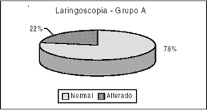
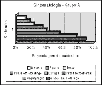
During the clinical assessment and laryngoscope examination of the patients the findings were: Only 4 out of 18 patients assessed or 22.22% presented some change in laryngoscope examination (thickening and/or hyperemia of the posterior commissure) - Graph 1. The most common symptoms reported by the patients were: pharyngeal globus, 16 patients or 88.88%; retroesternal pyrosis, 10 patients or 55.55%; dysphagia, 7 patients or 38.88%; oropharyngeal pyrosis, 6 patients or 33.33%; 4 patients or 22.22% complained about cough; throat clearing and dysphonia were reported respectively by 3 patients or 16.6% and by 2 patients or 11.11% - Graph 2.
The EGD results of Group A were not presented because they all had at least Level III esophagitis.
Group B
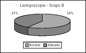
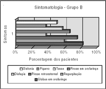
The results of clinical evaluation and laryngoscopy of Group B were: 25 out of the 44 patients assessed or 56.81% had some change in laryngoscopy (thickening or hyperemia/ulceration of the posterior commissure and hyperemia of vocal folds) - Graph 3. The symptoms reported were: pharyngeal globus, 34 patients or 77.27%; regurgitation, 25 patients or 56.81%; retroesternal pyrosis, 33 patients or 75%; dysphagia, 17 patients or 38.64%; oropharyngeal pyrosis, 28 or 63.64%; cough complaints, 24 patients or 54.54%; throat clearing, 17 patients or 38.64%; and dysphonia, 22 patients or 50% of the patients - Graph 4.
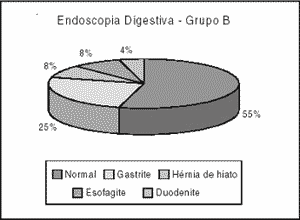
Patients of Group B also underwent EGD with the purpose to assess gastric and esophageal picture. The results were: 24 patients or 54.54% within the normal range; 33 patients or 25% with gastritis; 4 patients or 9.09% with hiatal hernia; 4 patients or 9.09% with esophagitis and 2 patients or 4.54% with duodenitis - Graph 5.
The statistical analysis using chi square test and p-values showed statistical difference between the two groups in laryngosgopy examination (c2: 6.1412 and p-value: 0.0132), pyrosis in oropharynx (c2:4.7364 and p-value: 0.0295) cough (c2:5.3890 and p-value: 0.3203); dysphonia (c2: 8.1428 and p-value: 0.0043); regurgitation (c2: 17.1376 and p-value: 0); globus in oropharynx (c2: 1.1043 and p-value: 0.2933), retroesternal pyrosis (c2: 2.2724 and p-value: 0.1317), dysphagia (c2: 0.003 and p-value: 0.9852) and throat clearing (c2: 2.8215 a p-value: 0.0930) were considered statistically equivalent between the two groups (Table 1).Graph 1. Laryngoscopy Results of Group A.Graph 2. Group A symptoms.Graph 3. Laryngoscopy Results of Group B.Graph 4. Group B symptoms.Graph 5.
DISCUSSION
The result analysis and comparison of data collected in groups A and B allowed us to differentiate several aspects.
Laryngeal gastroesophageal reflux (GER) symptoms have already been well-established in the literature and the most common symptoms are: dysphonia, cough, pharyngeal globus and throat clearing 5,7,10,12. The pharyngeal globus was also mentioned by Woo and is advocated as an unspecific symptom of laryngeal irritation and plays a major role in GRD etiology17. The symptoms of this disease gained higher significance in many cases and are more likely to be taken into account for the diagnosis than supplementary examinations 15,17. It is based on the fact that many times the confirmation diagnosis of GER is not obtained only with one examination. Most authors associate several techniques with clinical picture to make the diagnosis.5,7,13,15
The symptoms found in Group B confirmed literature data. Nevertheless, the findings of group A that were likely to be as or more noticeable than those of group B, such as esophageal symptoms, since they are more intense and have longer evolution, were not confirmed. Dysphonia was reported in 50% of the patients in Group B against only 11% in Group A. Cough was also prevalent in group B. The only laryngeal symptom that was proportionally prevalent in both groups was pharyngeal globus, 89% in Group A, 77% in Group B. Typical symptoms had similar prevalence in both groups, except for regurgitation.
Laryngoscopy is advocated as an essential examination for patients with GER complaints7,15. Other extremely important examinations are: pHmetry (currently performed with dual probe 17), manometry and EDG. This study used only laryngoscopy, with the purpose to find laryngeal changes (interarytenoid pachydermia, laryngeal edema, arytenoids hyperemia, mucous thickening17) and EDG with the purpose to correlate the gastric and esophageal findings of the two groups.
Again the differences of the two groups are highly evident. With respect to EDG, the study reported 54% examinations within the normal range in Group B against 100% of altered examinations in Group A. The findings were inverted in laryngoscopy. The authors found a higher number of changes in Group B in relation to Group A. Note that the group with a higher level of changes in the digestive tract presented considerably less laryngoscopic changes, thus, supporting the idea that there is no correlation between the level of esophageal disease and laryngological complaints.
Based on the above data, the authors suggested that, although these diseases have quite similar pathophysiology and sometimes overlaid symptoms, the laryngeal changes caused by GRD and esophageal disease (esophagitis) due to reflux should not be considered different stages of the same disease. Laryngeal changes, which are many times more severe in patients with less esophageal disorder, are proof of it.
CONCLUSION
The results of this study showed significant correlation between otorhinolaryngological symptoms and gastroesophageal reflux. GRD is a digestive system pathology, which is frequently under diagnosed in our field. The comparison between a group with established and advanced esophageal disease with indication for surgery, and another group with mainly laryngeal complaints allowed us to conclude that, in spite of the pictures presenting a similar pathophysiology, they are not different stages of the same disease.
REFERENCES
1. BECKER, D.J. et al. - A comparison of high and low fat meals on postprandial esophageal acid exposure. Am J Gastroenterol, 84:782, 1989.
2. CASTELL, D.O. - The esophagus. second edition, New York, 1995.
3. CHERNOW, B.; CASTELL, D.O. - Diet and heartburn. JAMA, 241:2307, 1979.
4. CLARK, C.L. et al. - Complications of gastroesophageal reflux disease. Esophatis, acid laryngitis, and beyond. Postrad Med, 1996.
5. GAYNOR, E.B. - Otolaryngologic manifestations of gastroesophageal reflux. Am J Gastroenterol, 86(7):801-8 1991.
6. HANSON, D.G. et al. - Outcomes of antireflux therapy for the treatment of chronic laryngitis. Ann Otol Rhinol Laryngol, 1995.
7. HAWKINS, B.L. - Laryngopharyngeal reflux: a modern day "great masquerader". J Ky Med Assoc, 95(9):379-85 1997.
8. HINER, R.A. et al. - Laparoscopic Nissen fundoplication is an effective treatment for gastroesophageal reflux disease. Ann Surg 200:472, 1994.
9. KLAUSER, A.G.; SCHINDLBECK, N.E.; MÜLLER-LISSNER, A.S. - Symptoms in gastroesophageal reflux disease. Lancet, 325:205, 1990.
10. KOUFMAN, J.A. - The otolaryngologic manifestations of gastroesophageal reflux disease (GERD): a clinical investigation of 225 patients using ambulatory 24-hour pH monitoring and an experimental investigation of the role of acid pepsin in the development of laryngeal injury. Laryngoscope, 101 (4 Pt 2 Suppl 53):1-78 1991.
11. MINCIS, M. - Gastroenterologia e hepatologia. Primeira edição, São Paulo, 1997.
12. OLSON, N.R. - Laryngopharyngeal manifestations of gastroesophageal reflux disease. Otolaryngol Clin North Am, 24(5):1201-13 1991 Oct.
13. PAPARELLA, M.M.; SHUMRICK, D.A.; GLUCKMAN, J.L.; MEYERHOFF, W.L. - Otolaryngology. Third edition, Philadelphia, 1991.
14. RICHTER, J.E.; CASTELL, D.O. - Gastroesophageal reflux. Pathogenesis, diagnosis and therapy. - Ann Inter Med 97:93, 1982.
15. SHAW, G.Y. et al. - Subjective, laryngoscopic and acoustic measurements of laryngeal reflux before and after treatment with omeprazole. J Voice, 10(4):410-8, 1996.
16. WINTERS, C.; SPURLING, T.J.; CHOBANIAN, S.J. - Barrett's esophagus. A prevalent, occult complication of gastroesophageal reflux disease. Gastroenterology, 92:118, 1987.
17. WOO, P. et al. - Association of esophageal reflux and globus symptom: comparison of laryngoscopy and 24-hour pH manometry. Otolaryngol Head Neck Surg, 115(6):502-7 1996.
Print: ![]()