

Year: 2005 Vol. 71 Ed. 5 - (19º)
Artigo Original
Pages: 676 to 679
 PDF PT
  PDF EN
Anatomic and radiograph study of the persistence of Foramen of Huschke
Author(s): Rodrigo Costa Moreno1, Israel Chilvarquer2, Jorge E. Hayek3, Paulo Isaias Seraidarian4
Keywords: Foramen of Huschke, panoramic, submental vertex, corrected saggital linear temporal mandibular joint, tomography.
Abstract:
The aim of this study is to assess and locate the Foramen of Huschke using contrast material like gutta-percha and barium sulfate, through extraoral radiographs, such as panoramic, submental vertex and corrected saggital linear Temporal Mandibular Joint tomograms in four skulls where we clinically checked the existence of foramen of Huschke. The results proved that the foramen of Huschke can be observed in skulls submitted to contrast using radiographic techniques.
![]()
INTRODUCTION
Known as foramen of Huschke, the opening of the developing tympanic ring (Gardner et al., 1967) was first described by Emil Huschke, in 1889, as being a structure present during embryological development in the tympanic portion of the temporal bone that is normally closed by the age of 5 years (Faig-Leite, 1998).
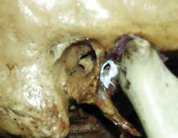
The persistence in adult subjects is known as an anatomical anomaly and it may be attributed to affections such as herniations of temporomandibular joint (TMJ), as well as otological problems in the external acoustic canal. Foramen of Huschke is located on the anterior wall of the external acoustic canal on the tympanic portion of the temporal bone, presenting a communication between the external acoustic canal and the mandibular fossa (Figure 1).
OBJECTIVE
The purpose of the present study was to identify and locate the Foramen of Huschke through three different extraoral radiographic techniques in four dry skulls and to assess which radiological techniques may be used to identify the persistence of Foramen of Huschke.
MATERIAL AND METHOD
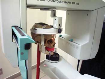
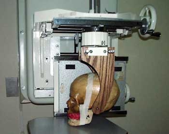
To assess the presence of Foramen of Huschke in radiological exams, we used 4 dry human skulls, 3 of adult subjects who had persistence of Foramen of Huschke bilaterally and one subject aged about 4 years, in which the presence of the foramen was within the normal range. The skulls were submitted to extraoral techniques of panoramic x-ray (Figure 2A) with Orthopantomograph OP-100 (Instrumentarium Imaging - Tuusula, Finland), submental vertex technique (inverted Hirtz), and lateral linear tomography corrected for TMJ (Figure 2B) with dental tomographer Quint Sectograph (Denar Corporation, Anaheim, Calif. USA) and then new exams were performed using contrast material such as gutta-percha (sealing the foramen) and barium sulfate (on the foramen edges), to evidence the Foramen of Huschke on the x-rays.
After the execution of the exams, we compared the images with and without contrast material.
RESULTS
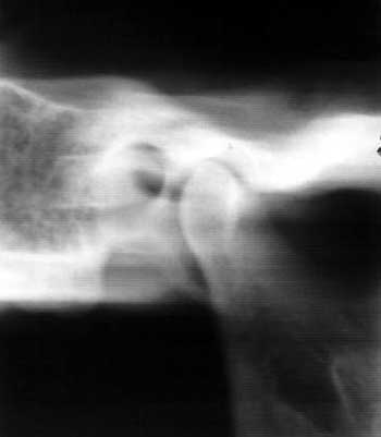
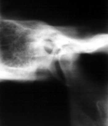
The results found after the corrected lateral linear tomography without contrast material showed the presence of radiolucent area of elliptical format (Figure 3A).
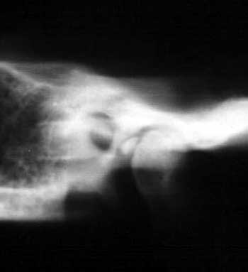
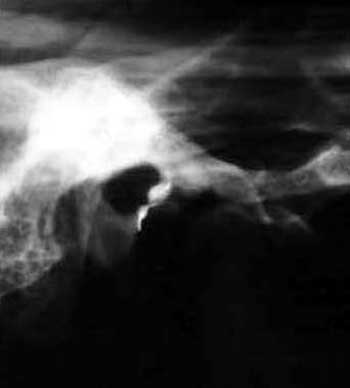
Even using barium sulfate, we observed radiolucent image of elliptical format with radiopaque contour (Figure 3B), and with gutta-percha, we detected the presence of radiopaque image of elliptical form (Figure 3C).
These images were located on the anterior wall of the external acoustic meatus on the tympanic portion of the temporal bone, presenting a communication between the external acoustic canal of the temporal bone tympanic portion, with the mandibular fossa of the temporal bone squamous portion.
We observed that in the panoramic technique performed with barium sulfate, two radiopaque points presented communication between the external acoustic canal of the temporal bone tympanic portion, with the mandibular fossa of the temporal bone squamous portion (Figure 4).
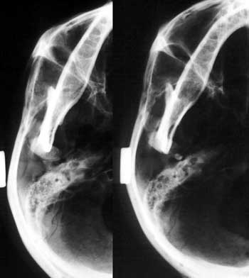
In the submental vertex technique, performed without contrast material (Figure 5A), we observed radiolucent image of elliptical form.
As to execution of images with gutta-percha, we observed elliptical radiopaque image (Figure 5B) and the images were located medially and posteriorly to mandible condyle, on the anterior wall of the external acoustic canal of the temporal bone tympanic portion.
The characteristics of the obtained radiographic images were the same for the four studied skulls.
Figure 1. Foramen of Huschke evidenced with barium sulfate.
Figure 2A. Panoramic technique.
Figure 2B. Corrected lateral CTs can.
Figure 3A. Corrected lateral CTs can - TMJ CT scan sections - section A without contrast material.
Figure 3B. Corrected lateral CTs can - TMJ CT scan sections - section B with barium sulfate.
Figure 3C. Corrected lateral CTs can - TMJ CT scan sections - section C with gutta-percha.
Figure 4. Panoramic technique, detail of the right TMJ region.
Figure 5. Submental vertex technique- detail of the right TMJ region; A. without contrast material, and B. with gutta-percha.
DISCUSSION
After the study of radiological images of four dry skulls we found the same results in the four skulls, observing the presence of Foramen of Huschke in the linear tomography technique. In submental vertex and panoramic techniques it was less evident as a result of the overlapping of bone structures.
Rosemberg and Graczik (1986) stated that the best radiological technique capable of identifying the structures that comprise the TMJ is corrected axial lateral tomogram, even though this radiological technique does not completely eliminate the overlapping of the TMJ region, because the structure that is located in the fulcrum of the rotation of the system will appear in more details and those that are below or above this point will seem to be blurred (Chilvarquer, 1995).
Owing to the overlapping of bone structures in the TMJ region, Holmlund et al. (1986) reported difficulty in examining TMJ through conventional radiograms. Therefore, they suggested that CT scan was the preferred method to avoid overlapping in this region.
Ali and Rubinstein (2000) detected in temporal bone CT scan a bone defect in the anterior aspect of the auditory canal, the defect known as Foramen of Huschke.
As to comparison of images initially obtained, with and without contrast (gutta-percha and barium sulfate), we observed that images described in the three radiological techniques are compatible with the anatomical location of Foramen of Huschke.
CONCLUSION
Foramen of Huschke could be observed after its evidence with contrast material in radiological images of 4 dry skulls using extraoral, panoramic, submental vertex (inverted Hirtz) and TMJ corrected lateral linear tomogram techniques.
We concluded that among the used techniques, linear tomography proved to provide the best results and the literature recommends that CT scan is the best technique to visualize the Foramen of Huschke.
REFERENCES
1. Melgaço CA, Penna LM, Seraidarian PI. O Forame de Huschke e suas implicações clínicas. In: Revista Brasileira de Otorrinolaringologia 2003; 3ª ed. 69.
2. Ali TS, Rubinstein JT. Rheumatoid arthritis of the temporomandibular joint with herniation into the external auditory canal. Ann Otol Rhinol Laryngol 2000; 109 (2): 177-9.
3. Anand VT, Latif MA, Smith WP. Defects of the external auditory canal: a new reconstruction technique. J Laryngol Otol 2000; 144 (4): 279-82.
4. Ars B. Le Foramem De Huschke. Acta Otol Rhino Laryngol 1988; 42 (5): 654-8.
5. Bento RF et al. Anatomia do osso temporal. In: Tratado De Otologia. São Paulo: Universidade de São Paulo; 1998. Cap. 1, p.17-21.
6. Cecire AA, Austin BW, Ng PK. Polyp of the external ear canal arising from the temporomandibular joint. A case report. J Otolaryngol 1991; 20 (3): 168-70.
7. Chilvaquer I. Imagenologia da ATM. In: Barros JJ, Rode SM. Tratamento das desordens crânio mandibulares. São Paulo: Santos; 1995. p.129-36.
8. Denar - Corporation Quint-Sectograph Tomograph System Instruction Manual Anaheim, CA, United States.
9. Faig-Leite H, Horta Jr JAC. Persistência do Forame de Huschke em crânios adultos. In. Reunião Anual Da Sociedade Brasileira De Pesquisa Odontológica, 1998, Águas De Lindóia. Anais Da Sociedade Brasileira De Pesquisa Odontológica, São Paulo; 1998. p. 145.
10. Freitas A, Panella J. Técnicas radiográficas extrabucais In: Radiologia Odontológica. 4a. Ed. São Paulo: Artes Médicas; 1998. p. 211-33.
11. Freitas A, Torres FA. Radiografias panorâmicas. In: Radiologia Odontológica. São Paulo: Artes Médicas; 1998. p. 211-33.
12. Gardner E, Gray DJ, O´Rahilly R. Anatomia - estudo regional do corpo humano. 2ª Ed. Rio De Janeiro: Guanabara Koogan; 1967. p. 858.
13. Genovese WJ, Freitas L, Samartini R. Exames complementares. In: Genovese WJ. Exame Clínico Em Odontologia. São Paulo: R.S.; 1985. p. 161-2.
14. Hawke M et al. Bilateral spontaneous temporomadibular joint herniation into the external auditory canal. J. Otolaryngol 1987 16 (2): 387-9.
15. Heffez L, Anderson D, Mafee M. Developmental defects of the tympanic plate: case reports and review of the literature. J Oral Maxilofac Surg 1989; 47 (12): 1336-40.
16. Herzog S, Fiese R. Persistent Foramen of Huschke: possible risk factor for otologic complications after arthroscopy of the temporomandibular joint. Oral Surg Oral Med Oral Pathol 1989; 68 (3): 267-70.
17. Holmlund A, Hellsing G, Wredmak T. Arthroscopy of the temporomandibular joint: a clinical study. Int J Oral Maxilofac Surg 1986; 15: 715-21.
18. Rabinov CR et al. Recurrent pleomorphic adenoma of the parotid gland involving the osseous external auditory canal. Ann Otol Rhinol Laryngol 1997; 106 (2): 589-93.
19. Reis HP. Estudo tomográfico da persistência do Forame Huschke: importância clínica - Tese De Mestrado Faculdade De Odontologia de São José dos Campos; 2000.
20. Sharma PD, Dawkins RS. Patent Foramen of Huschke and spontaneous salivary fistula. J Laryngol Otol 1984; 98: 83-5.
21. Sobotta J. Atlas de Anatomia Humana. V.1 20ª ed. Rio de Janeiro: Guanabara Koogan; 1995. p.60.
22. Swartz JD, Harnsberger HR. The external auditory canal. In: imaging of the temporal bone. 3rd ed. New York: Thieme; 1998. cap. 2, p.16-20.
23. Wang R et al. Persistence of the Foramen of Huschke in the adult. An osteological study. J Otolaryngol 1991; 20 (4): 251-34.
Dentist, major at Dental School, Universidade Ibirapuera. Intern, Discipline of Dental Radiology, Department of Stomathology, University of Sao Paulo.
Master, Ph.D. and Full Professor, FOUSP; Post-graduation at University of Texas at San Antonio - USA, Faculty Professor, Discipline of Imaging, Universidade Ibirapuera.
Master and Ph.D. in Mouth Diagnosis, FOUSP, Joint Professor of Imaging, Universidade Ibirapuera.
Master and Ph.D. in Restorative Dental Practice, major in Buccomaxillofacial Prostheses, coordinator of the Master Program in Dental Prosthesis, PUC-MG, Professor responsible for the Discipline of Occlusion and TMJ, Universidade Ibirapuera-Unib, Assistant Professor, Department of Dental Prosthesis, Dental School, São José dos Campos/UNESP.
Dental School, Universidade Ibirapuera.
Address correspondence to: Rua João de Souza Dias 43 apt 21 Campo Belo São Paulo SP 04618-000.
Tel (55 11) 5535-4789 - E-mail: moreno20@terra.com.br
The present article was submitted through SGP on 13/Apr/2005 and approved on 31/May/2005.
Print: ![]()