

Year: 2005 Vol. 71 Ed. 5 - (14º)
Artigo Original
Pages: 639 to 643
 PDF PT
  PDF EN
Intracranial complications of chronic suppurative otitis media in children
Author(s): Maurício S. Miura1, Rita C. Krumennauer1, José F. Lubianca Neto1,2
Keywords: chronic otitis media, intracranial complications, meningitis.
Abstract:
In spite of significant decrease after antibiotic advent, intracranial complications of otitis media still represent a jeopardizing situation, since its high mortality rate (36%). Most common presentations are meningitis, cerebral abscess, extradural abscess and lateral sinus thrombophlebitis. For early management, it is necessary a high index of suspiction. It is important identification of non-typical cases because they might be masqueraded by antibiotic use. Aim: Herein, we present six cases of intracranial complications due to otitis media in children and adolescents in past two years at Complexo Hospitalar Santa Casa de Porto Alegre. Study design: series review.
![]()
INTRODUCTION
In the pre-antibiotic era, there was significant incidence of mastoiditis and intracranial complications (ICC) caused by otitis media, which presented high rate of mortality. After the introduction of antimicrobial agents, there has been a reduction in the incidence from 2.3% to 0.04% 1,2 . However, nowadays, intracranial complications still represent a situation of risk given that mortality rate is high, reaching 36%3. The most common ICC are meningitis, cerebral abscess, extradural abscess and thrombophlebitis of lateral sinus (TLS) 4. In this series of cases, we present six patients who had ICC caused by chronic suppurative otitis media in the Service of Otorhinolaryngology, Complexo Hospitalar Santa Casa (CHSC), Porto Alegre for a period of 2 years.
CASE REPORT
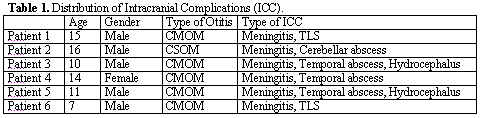
Within a period of 2 years (April 2000 to May 2002), we received 6 cases of chronic otitis media with intracranial complications (ICC) in the Service of Pediatric Otorhinolaryngology, Hospital da Criança Santo Antônio, Complexo Hospitalar Santa Casa, Porto Alegre. In all patients, intracranial complications were multiple, and some were followed by extracranial complications (neck abscess of Bezold - patient 6) (Table 1). The mean duration of the disease in these patients was of 5 years, since the onset of complaints of otorrhea. Even though it is not an accurate estimate of the disease duration, it represents the minimum time the disease was present, given that many came to medical services after the occurrence of otorrhea. Five patients (83.3%) presented ICC resulting from cholesteatomatous chronic otitis media and 1 case (16.7%) from simple chronic otitis media. As to age, ICC affected patients aged 7 to 16 years.
As to gender, we had 5 cases of boys (83.3%). As to race, we had 5 cases of Caucasian patients (83.3%).
The most frequent ICC was meningitis, which was detected in all patients. Five of them had abscesses, three in the temporal region and two in the cerebellar region. Three cases manifested as hydrocephalus. Lateral sinus thrombosis occurred in 2 patients.
In 100% of the cases, there was clinical presentation and cerebrospinal fluid (CSF) pattern compatible with meningitis. There was bacterial growth and positive CSF culture in 4 cases (57.1%), with presence of Proteus mirabolis, Pseudomonas sp. and Staphylococcus aureus. Our date were incomplete concerning culture for anaerobes.
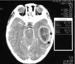
Abscesses were the second most frequent type of complication. There were 4 cases (66.7%), three with temporal location (Figure 1) and 1 with cerebellar location, which progressed to death.
Hydrocephalus was, together with lateral sinus thrombophlebitis, the third most common complication, both affecting 2 patients (33.4%). All patients with hydrocephalus presented association with abscess, even though the exclusively otogenic etiology was considered. In all 6 cases reported here, management comprised intravenous antibiotic therapy and open tympanomastoidectomy.
DISCUSSION
In the past, acute otitis media (AOM) and chronic otitis media (COM) represented severe risks to children owing to the potential of intra and extracranial complications. As a result of the advent of antibiotics, there has been significant decrease in incidence, with significant decrease in intracranial complications (ICC) from 2.3% to 0.04% and in mastoiditis from 20% to less than 0.5%. However, ICC still represent a risk situation because of high mortality rate (36%) 1-3. Even though it is less common than simple chronic otitis media, cholesteatomatous otitis media (CMOM) is normally associated with complications because of its invasive potential.4
Our cases do not differ a lot from the large series reported in the literature. Studies performed in Thailand showed a rate of 0.36% of ICC in 32 patients within a period of 13 years 5. We do not have the rate calculated based on number of cases of COM seen in our service, but in our sample of 6 ICC within 2 years there may be a higher incidence. We know that duration of the disease, time of diagnosis and early intervention reduce the number of complications 1-4. We should also consider the presence of cholesteatoma in about 60 to 70% of the studies in Thailand, South Africa and Brazil 4-7, which exceeds the American statistics of 27% cholesteatomas, a country in which antibiotic therapy and surgery are performed early, leading us to the conclusion that it may be the best option to prevent ICC 1-3. In fact, our reality is somewhat different. All the reported cases were from the public healthcare system, where there are long waiting lines for tympanomastoidectomy, and this delay is probably one of the main factors for the development of ICC in our country.
Complications of otitis media may be divided into intratemporal and extratemporal. The former include tympanic membrane perforation, conductive or sensorineural hearing loss, ossicle lesions, facial palsy, mastoiditis, labyrinthitis and petrositis. Extratemporal complications are subdivided into intracranial (abscess of the central nervous system, meningitis, lateral sinus thrombophlebitis and otic hydrocephalus) and extracranial complications (retroauricular, zygomatic and Bezold abscess).8
We should suspect of ICC when the patient has clinical manifestations such as persistent headache, malaise, fever, otalgia, lethargy, nausea/vomiting, neck rigidity, diplopia, hemyanopsia, papilla edema, blurred vision, ataxia, seizures, aphasia, intention tremor, dysmetry and/or dysdiadocokinesia. Currently, the use of antibiotics may mask some ICC symptoms, hindering the diagnosis. These patients present mild symptoms but for a prolonged period of time, in addition to high fever and variable degree of otological and neurological signs.8
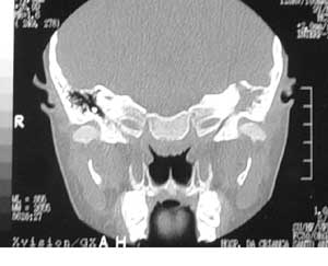
ICC secondary to chronic suppurative otitis media normally occur by the extension of the mucoperiosteum inflammatory process to the head cavity, developing in the brain, lateral sinuses and epidural, subdural and subarachnoid spaces. In most cases, ICC extend through bone dehiscence on the tegmen tympani (Figure 2) or in the antrum; through vascular canals directly to the lateral sinus, through the superior petrous sinus; vascular anastomosis; caroticotympanic canaliculi, pericarotid venous plexus; cavernous sinus; bone dehiscence on the cavum tympani; through the endolymphatic sac; optic capsule fistula, or they may result in sinudural angle or petrous apex osteitis; or empyema of cochlear aqueduct or perineural spaces of the inner acoustic meatus 9.
As to most common forms of presentation of ICC, there are some differences in the literature. Pennybacker et al., in 1961, reported 200 cases of ICC: they found 85 cases of temporal lobe and cerebellum abscess, 28 cases of otic hydrocephalus, 13 cases of meningitis, and 8 cases of lateral sinus thrombosis.10 Kuczkowski and Mikaszewski reported 503 cases of ICC (372 by COM and 131 by AOM); 80.7% of ICC were isolated and 19.3% were multiple. The most common ICC was meningitis with 177 cases (35.2%), extradural abscess with 122 cases (24.2%), cerebral abscess with 64 cases (12.7%), lateral sinus thrombosis with 90 cases (17.9%), cerebellum abscess with 35 cases (7.0%), hydrocephalus with 14 cases (2.8%), cavernous sinus thrombosis with 1 case (0.2%).11 Data from the second report were close to ours in relation to frequency of findings despite the relatively different distribution, which occurred mainly because our sample was small. It is important to highlight that there were also adults in the sample.
Meningitis was the most common ICC in our series, but there may be direct invasion of the disease; inflammation in areas close to the meninges (abscess, thrombophlebitis), or hematogenic dissemination from the infected ear (more frequent in AOM cases). Signs and symptoms include headache, fever, nausea/vomiting, malaise, and neck rigidity, as well as Kerning and Brudzinski signs. CSF shows pleocytosis, proteinorrhachy, and reduction of glucose. The most common bacteria are S. pneumoniae, S. pyogens and H. influenzae. It is important to perform a tympanic paracentesis for collection of material and drainage. Treatment consists of intravenous antibiotic therapy. In cases of recurrent meningitis, we may indicate middle ear and mastoid surgical exploration, trying to communicate with the central nervous system.8 All our patients presented meningitis, in agreement with the literature, as the most frequent ICC. Despite the fact that our data about the microbiology are incomplete, there was growth of Proteus mirabolis, Pseudomonas sp. and Staphylococcus aureus, which are not germs normally found in other samples of cases associated with chronic otological disease.
Abscesses may be divided into extradural (epidural), subdural, intracerebral or intracerebellar. The most common type is extradural. There may be bone destruction by cholesteatoma when reaching the dura, or by direct invasion. Symptoms include temporal headache, severe otalgia, low fever and malaise. The examination shows profuse otorrhea, pulsatile, thick and sometimes increasing jugular vein compression. Computed Tomography (CT scan) confirms the diagnosis and location of the abscess. Treatment consists of surgical drainage and intravenous antibiotic therapy. In subdural abscess, symptoms are similar, but as the abscess increases, there may be focal neurological signs indicating the location of the abscess, including hemiplegia. Drainage should be performed by a neurosurgeon, followed by exploration of otological focus. In general, it occurs by direct extension of the infection or rarely, by thrombophlebitis. It has high rate of mortality and neurological sequels in most survivals. Cerebral and cerebellar abscesses also occur by direct extension or thrombophlebitis and may be multiple. The most common form is cerebral (temporal) and the most lethal, cerebellar. It is the progression of an extradural abscess, which evolves to subdural, up to reaching the cerebral or cerebellar parenchyma. The increase in volume of the abscess may cause neurological signs such as aphasia (cerebral), ataxia and intention tremor (cerebellar). CSF analysis shows increase in pressure and concentration of proteins. CT scan confirms the diagnosis. Treatment consists of intravenous antibiotic. Surgical drainage is made by the neurosurgeon in those cases that do not progress well. If possible, we should perform a tympanomastoidectomy .8 In our series, there were 3 cases of temporal lobe abscess and 1 case of cerebellar abscess. In one case, CT scan showed a bulky abscess of the left temporal lobe and dehiscence of ipsilateral tegmen tympani (Figures 1 and 2). There was one case that progressed to death, presenting multiple cerebral abscesses (temporal and temporal-parietal right side), who arrived at the emergency service with evident neurological condition of mental confusion, behavior disorder and convulsive crises. We performed cranioectomy with drainage of 15ml of pus and radical mastoidectomy, in which we observed destruction of tegmen tympani by CMOM.
Otic hydrocephalus is the increase of intracranial pressure without affection of the CSF. The patient may present headache, 6th cranial nerve palsy, reduction of attention, lethargy, diplopia, nausea/vomiting and papilla edema. Treatment is based on intravenous antibiotics and mastoidectomy. Intracranial hypertension is handled by systemic corticoids. It may be submitted to repeated lumbar punctures, even though there is the risk of herniation. Recovery is slow, and it may persist for weeks to months 8. We detected 2 cases of otic hydrocephalus.
Lateral sinus thrombosis occurs by bone erosion of the mastoid over the sinus owing to the presence of cholesteatoma, granulomatous processes or coalescence, which form a perisinusal abscess. The formation of TLS may be considered a form of protection to limit the infection. The abscess puts pressure on the bone, causing necrosis on the anterior portion of the sinus and the intima, with adherence of fibrin, red blood cells and platelets, forming a mural thrombus. The thrombus may propagate towards the jugular vein bulb, and to other sites, for subcutaneous tissue 9,12 . Another less common cause of osteothrombophlebitis in early stage of acute otitis media. Concomitant complications are common such as meningitis and intracranial abscess. The thrombus may get infected and release infected emboli. Mortality ranges from 10 and 36%. Microbiology in general presents mixed flora with bacterioids, Streptococcus, Proteus and Pseudomonas. There may be high fever, retroauricular pain, thickness of perisinusal region, sternocleidomastoid muscle pain and papilla edema. CT scan shows the classification of delta sign by dura in cases of lateral sinus thrombosis, confirming the diagnosis13. Sensitivity of CT scan reaches levels of 87.2 to 100%. Magnetic resonance imaging (MRI) may show increase in intensity in the presence of the thrombus 9,14.
After the confirmation, we performed the opening of the sinus. Ligation of the internal jugular vein and the use of anticoagulants are controversial approaches that have generated literature discussion.12,15 There were two cases of TLS in our series, and they were both managed with open tympanomastoidectomy, without opening of the affected sinus and with good clinical progression.
Figure 1. Extensive cerebral abscess on the left temporal lobe.
Figure 2. Cholesteatomatous COM on the left with bone erosion and tegmen tympani dehiscence.
CONCLUSION
Even though less incident than in the past, ICC by otitis media continues to be a risk situation to patients with high mortality rate. High index of suspicion is essential for diagnosis and skilful management. It is important to identify the typical cases that may be masked by the use of antibiotics. Cholesteatomatous COM may be the main cause of ICC in our country, and it should be early managed with tympanomastoidectomy as a preventive measure of complications.
REFERENCES
1. Dowes JD. Complications of infections of the middle ear. In: Scott-Brown WG, Ballantyle, Groves.
2. Palva T, Virtamen H, Marine J. Acute and latent mastoiditis in children. J Laryngol Otol 1985 Feb; 99(2): 127-36.
3. Teichgraeber JF, Per-Lee JH, Turner JS Jr. Lateral sinus thrombosis: a modern perspective. Laryngoscope 1982 Jul; 92(7 Pt 1): 744-51.
4. Samuel J, Fernandes CM, Steinberg JL. Intracranial otogenic complications: a persisting problem. Laryngoscope 1986 Mar; 96(3): 272-8.
5. Kangsanarak J, Navacharoen N, Fooanant S, Ruckphaopunt K. Intracranial complications of suppurative otitis media: 13 years\' experience. Am J Otol 1995 Jan; 16(1): 104-9.
6. Barrionuevo, CE, Maniglia, JJ, Mocellin, M. Complicações intracranianas dos processos infecciosos crônicos do ouvido médio. Rev Bras Otorrinol; 1986.
7. Balbani ADS, Santos Júnior RC, Miziara ID. Abscesso cerebral como complicação de otite média crônica. Rev Bras Otorrinol; 1996.
8. Piza MRT. Complicações e seqüelas das otites médias. In: Otorrinolaringologia - princípios e práticas. Costa SS, Cruz LM, Oliveira JÁ.
9. Kuczkowski J, Mikaszewski B. Intracranial complications of acute and chronic mastoiditis: report of two cases in children. Int J Pediatr Otorhinolaryngol 2001 Sep 28; 60(3): 227-37.
10. Pennybacker J. Discussion on intracranial complications of otogenic origin. Proc R Soc Otol 1961; 54: 309-20.
11. Kuczkowski, K, Mikaszewski, B, Starzyska, A. Epidemiology of otogenic intracranial complications treated at the Otolaryngology Department of Medical University of Gdansk in the years 1948-1996. In: M Tos, Editor. Proceedings Third Extraordinary International Symposium On Recent Advances In Otitis Media Copenhagen 14 June, 1997, Kugler, Amsterdam (1997).
12. Lubianca Neto JF, Sant\'Anna GD, Saffer M. Complicaçðes Intracranianas da Otite Média Crônica Supurativa. Jornal Brasileiro de Neurocirurgia 1995; 4: 15-19.
13. Lubianca Neto JF, Saffer M, Rotta FT, Arrarte JLF, Brinckmann CA, Ferreira P. Lateral sinus thrombosis and cervical abscess complicating cholesteatoma in children: case report and review. International Journal of Pediatric Otorhinolaryngology 1998; 42: 263-9.
14. Kaplan DM, Kraus M, Puterman M, Niv A, Leiberman A, Fliss DM. Otogenic lateral sinus thrombosis in children. Int J Pediatr Otorhinolaryngol 1999 Aug; 20; 49(3): 177-83.
15. Lubianca Neto JF, Vieira LF, Dall\'Igna C, Linden A. Otite Média Supurativa e Tromboflebite do Seio Lateral: Relato de Um Caso e Revisão Bibliográfica. Rev Bras Otorrinolaringologia 1994; 60: 101-3.
Physician, Service of Otorhinolaryngology, Complexo Hospitalar Santa Casa, Porto Alegre (CHSC).
Joint Professor, Discipline of Otorhinolaryngology, Department of Ophthalmology and Otorhinolaryngology, Fundação Faculdade Federal de Ciências Médicas de Porto Alegre (FFFCMPA). Coordinator, Ambulatory of Pediatric Otorhinolaryngology, Hospital da Criança Santo Antônio de Porto Alegre - Complexo Hospitalar Santa Casa, Porto Alegre.
Affiliation: Fundação Faculdade Federal de Ciências Médicas de Porto Alegre, Service of Pediatric Otorhinolaryngology, Hospital da Criança Santo Antônio, Complexo Hospitalar Santa Casa de Porto Alegre.
Address correspondence to: José Faibes Lubianca Neto - Rua Dona Laura 320 9oandar Porto Alegre RS 90430-090.
E-mail: jlubianca@terra.com.br
Print: ![]()