
Year: 2005 Vol. 71 Ed. 5 - (8º)
Artigo Original
Pages: 598 to 602
Evaluation of inspiratory pressure in children with enlarged tonsils and adenoids
Author(s): Melissa Guerato Pires1, Renata Cantisani Di Francesco2, Anete Sevciovic Grumach3, João Ferreira de Mello Jr.4
Keywords: tonsil, adenoids, child, adenoidectomy, tonsillectomy.
Abstract:
Children with enlarged tonsil and adenoid usually present breathing abnormalities such as snoring, mouth breathing and sleep apnea. It is known that upper airway obstruction and consequent mouth breathing may result in pulmonary diseases. Aim: The goal of this preliminary study was to evatuate the inspiratory pressure in children with upper airway obstruction due to enlarged tonsils. Study design: clinical with transversal cohort. Material and Method: We evaluated 37 children (4 -13 years old, female/male) with enlarged tonsils who would be submitted a T&A surgery in the Department of Otolaryngology University of São Paulo Medical School at the same period. Control group was composed by 28 children without tonsillar disease submitted to the same tests. Inspiratory pressure was obtained using a manovacuometer. Results: We could observe lower inspiratory pressures in children with upper airway obstruction. The mean of inspiratory pressure in the upper airway obstruction group was 14,607cm/H2O and in the control group was 27,580cm/H2O. Conclusions: The enlarged tonsils and adenoids were associated with a poor inspiratory pressure and it results in an increased breathing effort and work of the involved muscles.
![]()
Introduction
Upper airway obstruction due to enlarged tonsils results in limited airflow. Such limitation is caused by a mechanical blockage that obstructs airflow, leading to mouth breathing, as it has lower resistance to air passage.1
Under normal conditions, nasal resistance is greater in childhood - nearly six times higher than that observed in adults. According to Barreto2, nasal airway accounts for 2/3 of total respiratory resistance.
Pulmonary repercussion due to tonsil enlargement is better understood if considered in the context of "united airways". In fact, disorders of upper and lower airways frequently coexist, as they present similar histology 3,4.
Several clinical complaints have been associated with upper airways obstruction, such as excessive day sleepiness, desynchronized sleep, day headaches, aggressiveness, restless sleep, deep sudoresis, night enuresis, weight-height deficit15. Some authors relate obstruction of upper airways to bruxism.6
Cazerta and Pacheco7 described two children with cor pulmonale, pulmonary edema and respiratory difficulty due to obstruction of airways by enlarged tonsils. After surgery, disorders regressed in one child, while the other child evolved to death due to important and irreversible hypertrophy of the right ventricle. Andrade& Britto reported five children with cardiac disorders due to enlarged tonsils, who presented regression of disorders in all cases8.
Studies designed to objectively assess pulmonary repercussion of enlarged tonsils have not been developed. Several evaluation methods have been proposed to quantify the strength of respiratory muscles9.
Inspiratory Pressure (IP), measured by a manometer and vacuum meter, is the most common and easy parameter. The manometer plus vacuum meter is used to quantify positive pressures (manometer) and negative pressures (vacuum meter).
Measurement of respiratory muscles strength may be widely employed, allowing diagnosis of respiratory insufficiency due to muscular failure, early diagnosis of respiratory muscle weakness, helping the evaluation of respiratory mechanics and indication of intubation, removal of artificial breather and patients' extubation.
Due to the importance of efficient breathing in children with enlarged tonsils and the lack of objective data on this disorder's repercussions, the present study aims at assessing IP in children with enlarged tonsils.
Material and Method
Thirty-seven children (age range of 4-13 years, female/male) with obstruction of upper airways by enlarged tonsils were evaluated and followed up in the Department of Otolaryngology, Medical School, University of Sao Paulo, from October 2002 to March 2003.
Two children were excluded - one female and one male - for not performing in the procedure, and 7 children for presenting grade 2 enlarged tonsils. Ten children were excluded, as they were under 6 years of age, since kids in this age were not evaluated in the control group.
Children evaluated in the control group came from Casa de Apoio Madre Clélia, were within the same age range of the studied group and did not present tonsil enlargement or other respiratory disorders. IP tests were performed similarly and following the same criteria as those used in the enlarged tonsils group.
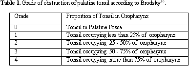
In the control group, 28 children were evaluated (11 females and 17 males) in the 4-13 age range. Three (3) children were excluded - 2 did not understand what was being asked and 1 presented tonsillitis at evaluation. Diagnosis of enlarged tonsil was obtained through paranasal sinuses radiography. Severity of palatine tonsillar obstruction was classified according to criterion described by Brodsky10 (Table 1), including patients with grades III and IV obstruction.
All the assessed children were in the group with surgery indication (adenotonsillectomy). Patients excluded from the group were those with asthma, neurological involvement, no surgery indication or those who did not understand what was being asked or did not help during complementary exams.
Inspiratory pressure (IP) is defined as a maximum negative pressure produced orally against occluded airway11, as well as the maximum negative pressure through the mouth after complete expiration of residual volume, followed by a single inspiration of maximum effort12 or diaphragm force index13. This measure was obtained by a MV-120 manometer plus vacuum meter (Ger-Ar-SP Com. Equip. Ltda.) by means of a tracheal and mouth connector. For final results, all children had 3 attempts and the highest value was considered (cm/H2O). IP value was obtained with the children comfortably seated, with no restrictions to pulmonary expansion, such as tight clothes and orthodontic braces, among others.
The protocol was submitted and approved by the Ethics and Research Committee of the Otorhinolaryngology Department, Hospital das Clinicas. Parents' permission term was signed before evaluation.
Results
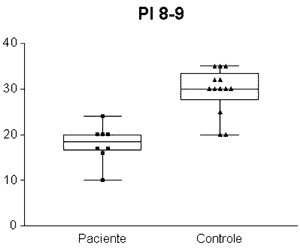
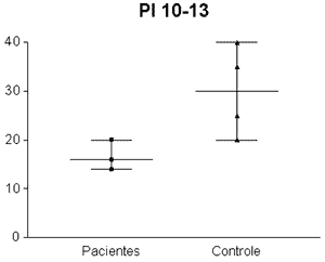
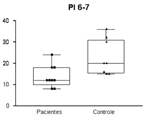
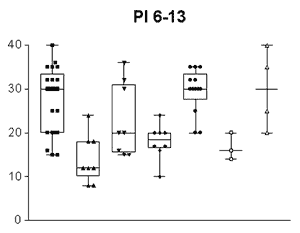
The IP value obtained was significantly different between the groups: 14.607cm /H2O ± 7.3321 (8-24 cm/H2O) in the enlarged tonsils group and 27.580/H2O ± 4.7791(15- 40.00 cm/H2O) in the control group (n= 0.001) (Graph 1). Standard error was of 1.4664 in the enlarged tonsils group and of 0.8880 in the control group. Groups' assessment according to age ranges presented the following: PI = 13.777cm/H2O ± 5.2387 (8-24cm/H2O) in the enlarged tonsils group (n=8) and 23.0 cm/H2O ± 8.4007 (15-36cm H2O) in the control group (n =7) among children between 6 and 7 years of age, with statistically significant difference (p= 0.003) (Graph 2). Inspiratory pressure analyzed in the 8-9 age range presented an IP of 18.00cm/H2O ± 4.106 (10 - 24.00 cm/H2O) in the enlarged tonsils group (n=8) and IP of 29.54cm/H2O ± 5.043 in the control group (n=13) (p= 0.001) (Graph 3). The enlarged tonsils group in the age range of 10-13 years presented IP of 16.67cm/H2O ± 3.055 (14 - 20.00cm /h2O) (n=3). Statistical difference was not significant between both groups (p= 0.293) (Graph 4).
Discussion
Tonsils enlargement is one of the main causes of respiratory disorders during sleep. Oral breathing and snoring are very common symptoms in children14.
There are no reports describing the use of IP for evaluation of respiratory muscle strength in children with enlarged tonsils. Therefore, we searched for groups of study on different pathologies in which IP measurement was applied in other diseases as a parameter of secondary pulmonary involvement.
Iandelli assessed patients with neuromuscular diseases and verified several factors that may influence the IP value, such as: different techniques, personal motivation, cooperation and facial muscular weakness. However, despite these variations, this parameter may be applied in the evolution process of disease, as well as in the response to therapy.15
In myasthenia gravis, the evaluation of 23 patients demonstrated that respiratory muscle strength remained normal, with no restrictive standard; however, the strength of respiratory muscles was reduced.16 In patients with multiple sclerosis, inspiration pressure is jeopardized by fatigue, muscular spasticity, and lack of coordination of respiratory and facial musculature12. In normal subjects, where the efficacy of postoperative incentive inspirometer was assessed, IP value was a good way to measure gain of the respiratory activity and, consequently, respiratory muscle strength17.
In the present study, children with enlarged tonsils presented lower IP than those of the control group, which suggests reduced respiratory activity and, consequently, lower oxygenation, resulting in chronic hypoxia, as reported by Cazerta.7
Children with enlarged tonsils presented difficulty to mechanically breathe through the upper airways and, to survive, they chose a less resistant air passage, that is, the mouth. However, with time, they make less effort to breathe, demanding less force from respiratory muscles, which leads to muscle weakness. This mechanism explains the different pressures among the groups.
The IP difference is more evident in younger age ranges. It is known that 90% of the craniofacial growth occurs up to 12 years old5. Therefore, at this age, when tonsil enlargement occurs, adjustment of bone structures regarding palatine and/or pharyngeal tonsils have already occurred. Children with enlarged tonsils present lower inspiratory pressure, which in our study was more evident among those at ages of 6 and 7 years.
The children included in our study, as well as the patients evaluated by Diez in a study conducted at Hospital La Paz - Madrid, in which 23 patients with clinically stable Myasthenia Gravis were assessed16 , did not present respiratory fatigue, despite the weak musculature. In both groups, IP and EP values were analyzed.
We concluded that children with enlarged pharyngeal and palatine tonsils presented lower inspiratory pressure. These children must be treated, once tonsillar enlargement, in the long term, leads to facial tonus alterations, morphological craniofacial and occlusive alterations, with reduction of respiratory muscle strength. Moreover, the quality of life of these children is jeopardized.
Graph 1. Inspiratory pressure of children between 8 and 9 years old. IP = Inspiratory pressure. 8-9= age between 8 and 9 years.
Graph 2. Inspiratory pressure of children between 10 and 13 years old. IP = Inspiratory pressure. 10-13= age between 10 and 13 years.
Graph 3. Inspiratory pressure of children between 6 and 7 years old. IP= Inspiratory pressure. 6-7= age between 6 and 7 years.
Graph 4. Inspiratory pressure of children between 6 and 13 years old. IP= Inspiratory pressure. 6-13 = age between 6 and 13 years.
References
1. Saffer M. A criança respirador bucal. In: Chinski A, SihT. II Manual de Otorrinolaringologia Pediátrica da IAPO. São Paulo: Ateliê; 1999. p. 170-80.
2. Barreto BAP. Provocação nasal inespecifica e especifica em crianças com rinite alérgica perene. (dissertação). São Paulo (SP), UNIFESP; 1997.
3. Corren J. Allergic rhinitis and asthma, how important is the link? J Allergy Clin Immunol 1997; 99: S781-S786.
4. Geller M. Vias aéreas unidas, a atopia como processo sistêmico e contínuo. Rev Bras Alerg Imunopatol 2002; 25: 74-80.
5. Difrancesco RC, Junqueira PA, Frizzarini R, Zerati F. Crescimento pôndero-estatural após adenoamigdalectomia. Rev Brás ORL 2003; 69: 193-6.
6. Di Francesco RC, Junqueira PAS, Trezza PM, Faria MEJ, Frizzarini R, Zerati FE. Improvement of Bruxism After T & A Surgery. Revista 2003; 68: 441-5.
7. Cazerta NMG, Pacheco ELB. Cor pulmonale secundário à obstrução das vias aéreas superiores. Radiol Brasil 1990; 23: 83-5.
8. Andrade FWC, Britto MC. Doença cardíaca secundária à hipertrofia sintomática de amígdalas palatinas e adenóides. Rev do IMIP 1991; 5: 110-2.
9. Black LF, Hyatt RE. Maximal Respiratory Pressures, Normal Values and Relationship to Age and Sex. Am Rev Resp Dis 1969; 9: 696-702.
10. Brodsky L. Tonsillitis, tonsillectomy and adenoidectomy. In: Bailey BJ., ed. Head and Neck Surgery-Otolaryngology, Philadelphia: Lippincott; 1993: 833-47.
11. Larson LJ, Covey MK, Berry J, Wirtz S, Alex CG, Matsuo M. Discontinuous Incremental Threshold Loading Test. Chest 1999; 115: 60-67.
12. Smeltzer SC, Lavietes MH. Rehabilitation of Maximal Respiratory Pressures in Multiple Sclerosis. Chest 1999; 115, 1546-1552.
13. Enright Pl, Kronmal RA, Manolio TA, Schenker MB, Hyatt RE. Respiratory Muscle Strength in the Elderly Am J Respir Crit care Med. 1994; 149: 430-8.
14. Wandalsen NF. Rinite Alérgica. Pediat Moderna 1982; 17(4).
15. Iandelli I, Gorini M, Misuri M, Gigliotti F, Rosi E, Duranti R, Scano G. Assessing Inspiratory Muscle Strength in Patients with Neurologic and Neuromuscular Diseases. Chest 2001; 119: 1108-13.
16. Díez TE, Pino JM, Frank A, Blanco C, Cruz M. Valoración de la Función Muscular Respiratória (Máximas Presiones Respiratórias) em Miastenia Gravis. Neurologia1990; 5 (9): 310-4.
17. Weindler J, Kifer RT. The Efficacy of Postoperative In Incentive Spirometry Is Influenced by the Device-Specific Imposed work of Breathing. Chest 2001; 119: 1858-64.
Specialization in Respiratory Physical Therapy, Physical Therapist.
Ph.D. in Medicine, Medical School, USP, Assistant Physician, Ph.D., Hospital das Clínicas, Medical School, USP.
Ph.D. in Pediatrics, Physician, Ambulatory of Primary Immunodeficiency; Researcher, Laboratory of Medical Investigation on Allergy and Immunology, Department of Dermatology, FMUSP.
Ph.D. in Medicine, Medical School, USP. Collaborating Professor, Discipline of Otorhinolaryngology, Medical School, USP.
Study carried out at the ambulatory of the Division of Otorhinolaryngology, Hospital das Clínicas/ Medical School, University of Sao Paulo, between October 2002 and March 2003.
Address correspondence to: Melissa Guerato Pires - R. Carlos Weber 601 apt. 114 Seul São Paulo SP 05303-000.
Tel. (55 11) 3641-2941 - Mobile. (55 11) 9215-4856 - E-mail: melissa.guerato@ip2.com.br, melissaguerato@hotmail.com
The present article was submitted through SGP on 26/April/2005 and approved on 13/June/2005.
