

Year: 2004 Vol. 70 Ed. 6 - (25º)
Relato de Caso
Pages: 851 to 855
 PDF PT
  PDF EN
Frontoethmoidal mucoceles: a case report and literature review
Author(s): Andy de Oliveira Vicente , Adriana Gonzaga Chaves , Ernesto Narutomo Takahashi , Fernanda Akaki2, Antônio Augusto Sampaio ,Cícero Matsuyama
Keywords: Key words: mucocele, paranasal sinus, endoscopic surgery.
Abstract:
Summary
Mucoceles of the paranasal sinus are epithelial lesions containing inspissated mucus, slow growth with expansive and osteolytic features that can involve surrounding structures such as orbit and intracranial cavity. The etiology of mucoceles is often associated with ostial obstruction of the compromised sinus. Presenting symptoms can include facial pain, headaches, nasal obstruction, diplopia, decreased visual acuity, orbital globe displacement, facial swelling and meningitis, depending on the anatomic area involved. Mucoceles occur most frequently in the frontal and ethmoidal sinuses, without gender prevalence. The diagnosis is accomplished through imaging. The "gold standard" in terms of diagnostic precision is the computed tomography although the magnetic resonance can be helpful in some cases. Actually transnasal endoscopic management of the mucoceles is preferred due minimal trauma and less morbidity.
The present study describes two cases of frontoethmoidal mucoceles with orbital and intracranial involvement that were treated by transnasal endoscopic obtaining good outcomes, demonstrating the security and efficacy of this surgical approach.
![]()
INTRODUCTION
Mucoceles are cysts of mucous content that affect the paranasal sinuses. They have expansive nature, slow growth and may eventually impair important neighboring structures, such as the orbit and the intracranial cavity 1,2,3. Nasosinusal endoscopic marsupialization of frontal sinus mucoceles was reported for the first time by Kennedy et al. in 1989. Currently, this surgical approach is considered the first choice of treatment because it is less invasive and has less morbidity compared to conventional techniques, such as external frontoethmoidectomy and osteoplastic flaps with or without obliteration of frontal sinus 1,4-7.
The purpose of the present study was to report two cases of frontoethmoidal mucoceles with orbital extension, treated with endonasal endoscopic surgery and to review the literature.
CASE REPORT
Case 1
M.Z.S.M, 20 years old, Caucasian female patient, complaining of intermittent nasal obstruction that worsened when in contact with dust and cigarette, associated with right mild frontal headache for 5 months. She did not report any other complaints.
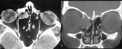
The physical examination revealed the presence of right eye medial canthus bulging, not painful upon palpation. Anterior rhinoscopy showed mild hypertrophy of lower nasal conchae with pale mucosa. Nasal endoscopic exam showed slight bulging of agger nasi region. We ordered Computed Tomography (CT scan) of nasal fossa and paranasal sinuses that demonstrated rounded image, with soft part content in the anterior ethmoid region and right frontal sinus, with displacement of papyraceous lamina and ocular globe, in addition to signs of bone remodeling suggesting clinical picture compatible with frontoethmoid mucoceles (Figure 1).
We performed anterior ethmoidectomy and endonasal frontal sinusotomy on the right, via endoscopic approach, with drainage and marsupialization of the mucoceles.
Postoperative endoscopic exam (2 months) showed epithelialization of ethmoidal cavity, nasofrontal recess and frontal sinus.
Case 2
M.A.L, Caucasian female patient, complaining of nasal obstruction and left frontal headache with displacement of the eye ipsilaterally for 6 months. The patient did not report history of previous nasal surgery or head trauma and did not present any eye disorders. The physical examination detected left eye proptosis with significant displacement towards the lateral-inferior side of it. Anterior rhinoscopy presented bulging of middle meatus on the left. We ordered nasal fossa and paranasal sinuses CT scan that demonstrated the presence of rounded, expansive image with soft part content (tumor) occupying the nasal fossa, ethmoidal cells, frontal sinus and internal portion of the orbit on the left, with compression of ocular globe and medial rectus muscle, compatible with frontoethmoid mucoceles.
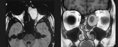
Magnetic resonance imaging (MRI) demonstrated frontoethmoid expansive process on the left of benign aspect, suggestive of mucoceles (Figure 2).
We conducted drainage and marsupialization of the lesion (with thick mucous inside it), evidencing single large cavity, involving the anterior and frontal ethmoidal sinus with exposure of periorbit on the left. The patient presented good postoperative evolution and control of nasal endoscopic exam showed epithelialized cavity without signs of recurrence.
DISCUSSION
Clinical aspects of mucoceles affecting the frontal sinus were primarily described by Langenbeck, in 1819 and Berthon proposed surgical drainage of this type of lesion in 1880. The term mucoceles was introduced by Rollet in 1896 and the first histopathological description was made by Onodi in 1901.
Mucoceles is a cystic lesion of the epithelial recover layer that affects the paranasal sinuses, contains thick mucus inside, has slow growth and expansive characteristics 1, 2. Its etiology has not been fully defined yet, but it is believed that it is caused by obstruction of drainage ostium of affected paranasal sinus owing to chronic processes of rhinosinusitis (infectious or allergic), nasosinusal polyposis, craniofacial trauma, previous surgery, benign tumors (osteomas, bone fibrous dysplasia), or malignant neoplasms (primary or metastatic) 1,3,8-10. It occurs more frequently in frontal and ethmoidal sinuses, but sphenoid and maxillary sinuses may also be affected; in the maxillary sinus, previous history of Caldwell-Luc surgery is almost always present 3,9,11. Maxillary sinus mucoceles are rare, represent less than 10% of all paranasal sinuses mucoceles and are more prevalent in Asian-descendents 11. This disease has equivalent incidence in men and women and normally affects people on their 3rd and 4th decades of life. Mucoceles tends to expand, remodel and reabsorb bone walls of affected paranasal sinus, changing their integrity and occasionally affecting the neighboring structures, such as the orbit and intracranial cavity 2, 8. Pathophysiology of the mechanism of bone reabsorption produced by mucoceles is still obscure. It is believed that osteolysis is produced by reduction of vascularization of the bone due to the mechanism of compression and/or by the action of inflammatory mediators abundantly present in the mucous of this affection, such as cytokines (IL1, IL6), vascular adhesion molecules, prostaglandins 4, 8.
The clinical picture varies according to the involved region, which may cause facial pain, headache, nasal obstruction, diplopia, reduction of visual acuity, displacement of ocular globe, facial edema, cerebral abscess, pneumoencephaloceles and meningitis 1,2,12,13. If there is acute infection of mucoceles, leading to mucopyoceles, there is higher likelihood of complications (orbital or intracranial) 10, 12. In our service, the patients' complaint involved nasal obstruction, frontal headache and displacement/bulging of orbit as a result of case progression.
The diagnosis is based on imaging exams 12. Even though simple x-ray may show opacification, bone erosion or expansion of mucoceles, CT scan is the preferred exam because it evidences bone involvement, assesses intracranial and/or orbital extension and supports surgical planning. In patient 1, the CT scan showed rounded image with soft part content occupying the anterior ethmoid cell region and frontal sinus on the right, slightly displacing the papyraceous lamina and consequently the ocular globe, with signs of bone remodeling. Patient 2 presented similar CT scan image. MRI is ordered when CT scan does not elucidate the case, especially in cases in which there is suspicion of neoplastic process. The disadvantage of MRI is its inability to assess bone anatomy. In case 2, we detected frontoethmoid expansion process on the left of benign aspect.
Differential diagnosis of mucoceles includes encephaloceles, cholesterol granuloma, epidermoid cyst, meningioma, chordoma, neurofibroma, salivary adenoma, paraganglioma, nasoangiofibroma, and malignant neoplasms 3.
Treatment of mucoceles is surgical and the access routes may be either external or endonasal 6,7,11,14. External approach is made through frontoethmoidectomy (Lynch's procedure) or by osteoplastic flaps with or without frontal sinus obliteration and total excision of mucosa 7,13. For many years, these techniques were the only surgical alternative to treat frontoethmoidal mucoceles. They are aggressive procedures with high morbidity and currently they are reserved for extreme cases with significant intracranial or orbital extension 1, 2. The current tendency is to conduct functional, little invasive and low morbidity procedure with nasosinusal endoscopic surgery, with marsupialization and abundant drainage of the lesion, preserving the epithelium 1,2,5,7,10. Recent studies demonstrated that mucoceles does not affect the characteristics of the respiratory mucosa and that marsupialization and consequent improvement of local ventilation is possible to reverse epithelial metaplasia into normal respiratory epithelium or at least bring it close to normal 5. In patient 1, we conducted ethmoidectomy and frontal sinusotomy on the right via endoscopy and drainage and marsupialization of the mucoceles, similarly to patient 2.
Outpatient follow-up with nasal endoscopy is essential to ensure disease control. Image exams (CT scan and MRI) may be useful, especially when there is suspicion of lesion recurrence.
CONCLUSION
Mucoceles are benign lesions of expansive characteristic that may cause severe complications at orbital and intracranial levels and for this reason they should be diagnosed and treated early. Marsupialization with drainage through nasosinusal approach proved to be a safe and efficient procedure in therapeutic approaches of frontoethmoidal mucoceles.
REFERENCES
1. Chiarini L, Nocini P.F, Bedogni A, Consolo U, Giannetti L, Merli G.A. Intracranial spread of a giant frontal mucocele: case report. British Journal of Oral & Maxillofacial Surgery 2000; 38: 637-40.
2. Hurley DB, Javer AR, Kuhn FA, Citardi MJ. The endoscopic management of chronic frontal sinusitis associated with frontal sinus posterior table erosion. Am.J.Otolaryngol 2000; 14: 113-20.
3. Lloyd G, Lund VJ, Savy L, Howard D. Radiology in focus. The Journal of Laryngology and Otology 2000; 114: 233-36.
4. Benninger MS, Marks S. The endoscopic management of sphenoid and ethmoid mucoceles with orbital and intranasal extension. Rhinology 1995; 33: 157-61.
5. Har-El G, Dimaio T. Histologic and physiologic studies of marsupialized sinus mucoceles: report of two cases. The Journal of Otolaryngology 2000; 29: 195-8.
6. Rubin JS, Lund VJ, Salmon B. Frontoethmoidectomy in the treatment of mucoceles. Arch Otolalaryngol Head Neck Surg 1986; 112: 434-6.
7. Ulualp SO, Carlson TK, Toohill RJ. Osteoplastic flap versus modified endoscopic Lothrop procedure in patients with frontal sinus disease. Am J Rhinology 2000; 14: 21-6.
8. Lund VJ, Henderson B, Song Y. Involvement of cytokines and vascular adhesion receptors in the pathology of fronto-ethmoidal mucoceles. Acta Otolaryngol 1998; 113: 540-5.
9. Stiernberg CM, Bailey BJ, Calhoun KH, Quinn FB. Management of invasive frontoethmoidal sinus mucoceles. Arch Otolaryngol Head Neck Surg 1986; 112: 1060-3.
10. Bussaba NY, Salman S.D. Maxillary sinus mucoceles: clinical presentation and long-term results of endoscopic surgical treatment. The Laryngoscope 1999; 109: 1446-9.
11. Busaba NY, Kieff D. Endoscopic sinus surgery for inflammatory maxillary sinus disease. The Laryngoscope 2002; 12: 1378-83.
12. Lund VJ, Rolfe ME. Ophthalmic considerations in fronto-ethmoidal mucoceles. The Journal of Laryngology and Otology 1989; 103: 667-9.
13. Benninger MS, Steven M. The endoscopic management of sphenoid and ethmoid mucoceles with orbital and intranasal extension. Rhinology 1995; 33: 157-61.
14. Gady H. Endoscopic management of 108 sinus mucoceles. The Laryngoscope 2001; 111: 2131-4
Figure 1. Nasal fossa and paranasal sinuses CT scan at axial and coronal sections evidencing right frontoethmoidal region.
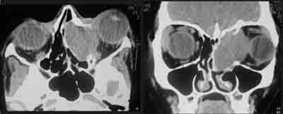
Figure 2. Nasal fossa and paranasal sinuses CT scan at axial and coronal sections evidencing image of left frontoethmoidal region with displacement of ocular globe on the left (proptosis).
Figure 3. Nasal fossa and paranasal sinuses CT scan at axial and coronal sections with contrast evidencing images of left frontoethmoidal region with displacement of ocular globe on the left (proptosis).
Preceptor, Medical Residence Program in Otorhinolaryngology, Instituto CEMA. Master in Otorhinolaryngology, UNIFESP/ EPM.
Resident Physicians in Otorhinolaryngology, Instituto CEMA.
Otorhinolaryngologists, Instituto CEMA. Master studies in Otorhinolaryngology under course, Hospital do Servidor Publico Estadual.
Preceptor, Medical Residence Program in Otorhinolaryngology, Instituto CEMA. Otorhinolaryngologist, HSPM-SP.
General Coordinator, Medical Residence in Otorhinolaryngology, Instituto CEMA. Master and Ph.D., UNIFESP/EPM.
Address correspondence to: Centro de Estudos A/C Sra. Leila. Rua do Oratório 1369 Mooca São Paulo SP 03117-000. Tel (55 11) 6602.4034 - Fax: 55 11-6602.4098 - E-mail: centrodeestudos@cemahospital.br
Affiliation: Instituto CEMA, Sao Paulo.
Print: ![]()