

Year: 2004 Vol. 70 Ed. 6 - (20º)
Relato de Caso
Pages: 829 to 832
 PDF PT
  PDF EN
Fibrous dysplasia of the temporal bone: case report and review of the literature
Author(s): Vanier S. Júnior , Eduardo C. Andrade1, Ana L. S. Didoni1, José C. Jorge , Nelson S. Filho , Fabiana R. Yoshimoto3
Keywords: Key words: fibrous dysplasia, temporal bone, hearing loss.
Abstract:
Summary
Fibrous dysplasia is a disease characterized by the progressive replacement of normal bone elements by fibrous tissue. It's an uncommon benign disorder of unknown etiology. There are two primary categories of the disease: monostotic fibrous dysplasia that involves a single bone and polyostotic fibrous dysplasia that involves multiple bones. Although the craniofacial skeleton is a common site of the disease, the temporal bone rarely becomes involved. The most common presenting symptom is deafness and diagnosis is based on radiological images. When fibrous dysplasia is accompanied by significant clinical symptoms, surgery is recommended. The follow up of this patient is very important to make an early diagnosis of recurrences. In this article we report a case of fibrous dysplasia of the temporal bone and the review of literature.
![]()
INTRODUCTION
Fibrous dysplasia is an uncommon disorder of unknown etiology. It represents a bone developmental disorder, specially a defect in osteoblastic differentiation and maturation 1. Bone abnormalities represent a remarkable point in the disease, whereas endocrinopathies, abnormal skin pigmentation and mucosa membrane alterations may also be present 2. There are two types of primary disease: monostotic fibrous dysplasia, which involves only one bone and amounts to 70% of the cases, and polyostotic fibrous dysplasia, which presents involvement of multiple bones. Approximately 3% of the patients have the so-called McCune-Albright syndrome, in which bone involvement is followed by skin lesions and endocrine pathologies 1. Even though the craniofacial skeleton is a frequent site of the disease, temporal bone affection is rare 3. It tends to develop at early childhood with predominance of male gender (2:1) and preference for Caucasians 1,4.
There is special interest for Otorhinolaryngology owing to the fact that fibrous dysplasia may involve craniofacial bones, causing deformities and dysfunctions, such as hearing loss, retroauricular bulging, and complications (cholesteatoma and facial nerve damage) 1, 5. We reported a case of temporal bone monostotic fibrous dysplasia and review the pathology and its involvement in the region.
CASE REPORT
Male 17-year-old patient, leukodermal, came to our center of Otorhinolaryngology with clinical picture of unilateral hearing loss on the left for 3 years and one episode of otorrhea. He did not report otalgia, otorrhagia, tinnitus or vertigo. He did not report associated diseases or any family history. Otoscopy showed narrowing of left external auditory canal, with no visualization of the tympanic membrane. Otoscopy on the right was normal.
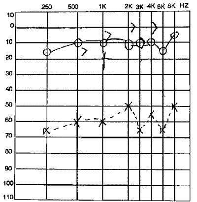
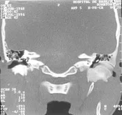
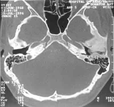
Pure tone audiometry revealed moderate unilateral conductive hearing loss on the left. Normal audiometry on the right (Figure 1). Temporal bone computed tomography (CT scan) demonstrated bone growth with temporal bone sclerosis and narrowing of external auditory canal on the left, compatible with picture of temporal bone fibrous dysplasia on the left and normal right temporal bone (Figures 2 and 3). The patient was submitted to mastoidectomy, we collected fragments of the damage and histopathology analysis revealed result compatible with bone fibrous dysplasia.
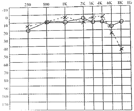
The patient presented reversion of symptoms after the surgery with significant clinical improvement. Postoperative audiometry showed normal responses for frequencies 250 to 4000Hz and mild hearing loss in frequencies 6 and 8KHz on the left (Figure 4). Patient has been followed up for 3 years with no signs of clinical or audiological abnormalities.
DISCUSSION
During the past 4 decades, fibrous dysplasia has become a skeletal affection 2 characterized by slow and progressive bone replacement by abnormal isomorphic proliferative fibrous tissue, intertwined by immature or normal bone trabecular arrangement 1, 3. The cause of fibrous dysplasia is unknown, even though many different etiologies have been suggested. The disease tends to develop in the pre-adolescence years, and is predominant in male patients (2:1). It presents slow progression and the monostotic form frequently ceases after the end of puberty 1, 4.
The incidence of craniofacial involvement is 10 to 25% in the monostotic form of the disease. In this region, the most affected areas are mandible and maxilla, and the temporal bone is affected in only 18% of the cases 5, 6. In general, only one temporal bone is affected, but in the polyostotic form of the disease, both temporal bones may be involved 2.
The most common clinical manifestation of temporal bone involvement is narrowing of external auditory canal leading to progressive conductive hearing loss. Other symptoms include retroauricular bulging, otorrhea, otalgia and tinnitus. Sensorineural hearing loss may occur when there is involvement of ear capsule 3, 4. Cholesteatoma (usually located in the external auditory canal) occurs in about 40% of the cases, and it is the most frequent complication of the disease. The involvement of the facial nerve is seen in 10% of the patients 1,4.
Radiological findings reflect morphology of the disease and vary according to number of calcifications and fibrosis. High resolution CT scan provides further details in the extension of the temporal bone fibrous dysplasia compared to conventional radiological imaging, considered the best imaging test to study temporal bone changes. CT scan is very useful to assess stenosis of external auditory canal, involvement of middle ear structures, associated presence of cholesteatoma, and extension to the facial nerve1,3,6.
Biochemical abnormalities may be noticed, but they are inconstant. Serum calcium, phosphorous, alkaline phosphatase may be normal or slightly affected 4. Malignant transformation has not been reported for temporal bone fibrous dysplasia, but it has already been reported in other disease sites. In 2/3 of the malignant cases, there is transformation to osteosarcoma, being that the mean time to transformation is 13.5 years 3.
The most important differential diagnoses with temporal bone fibrous dysplasia include Paget's disease, hyperparathyroidism, local reaction to meningioma, osteoma, eosinophilic granuloma, osteochondroma, and sarcomatous neoplasm 7,8.
It is not always possible to make the diagnosis of temporal monostotic fibrous dysplasia with isolated clinical and radiological data. Diagnostic confirmation may sometimes require the result of bone histological analysis 4.
Currently, there is no conservative treatment to control fibrous dysplasia. The simple presence of the lesion does not justify surgical intervention 8. If followed by significant clinical symptoms the surgery is recommended. The indication for surgery of temporal bone fibrous dysplasia includes bone invasion of external auditory canal (enough to produce conductive hearing loss), recurrent infections and secondary cholesteatoma in the external auditory canal. Surgical procedure aims at achieving 3 objectives: restoration of the function, prevention of complications, and esthetical improvement. Radiotherapy should be avoided owing to high incidence of malignant transformation (44%)1.
Prognosis is good in most cases, depending on disease severity. Clinical assessment associated with periodical CT scan may be useful in following up the patient to assess disease progression and the need for further surgical interventions 5.
CLOSING REMARKS
Fibrous dysplasia is an uncommon benign pathology of unknown etiology. The Otorhinolaryngological interest lies in the fact that this disease may affect facial and head bones, causing deformities and dysfunctions. Temporal bone fibrous dysplasia is rare, but this diagnosis should always be remembered in patients with hearing loss and external auditory canal stenosis. Moreover, the patient should always be informed about the possibility of disease recurrence, requiring periodical follow-up.
REFERENCES
1. Papadakis CE, Skoulakis CE, Prokopakis EP, Nikolidakis AA, Bizakis JG, Velegrakis GA, Helidonis ES. Fibrous dysplasia of the temporal bone: report of a case and a review of its characteristics. Ear Nose Throat J 2000; 79(1):52-7.
2. Nager GT, Holliday MJ. Fibrous dysplasia of the temporal bone: update with case reports. Ann Otol Rhinol Laryngol 1984; 93:630-3.
3. Reddy KTV, Vinayak BC, Jefferis AF, Grieve DV. Fibrous dysplasia of the temporal bone. Ann Otol Rhinol Laryngol 1994:103(1):74-6.
4. Donnelly MJ, McShane DP, Burns H. Monostotic fibrous dysplasia of the temporal bone with associated lymphadenopathy. Ear Nose Throat J 1994; 73(5):328-30.
5. Morrissey DD, Talbot JM, Schleuning AJ2nd. Fibrous dysplasia of the temporal bone: reversal of sensorineural hearing loss after decompression of the internal auditory canal. Laryngoscope 1997; 107(10):1336-40.
6. Chinski A, Beider B, Cohen D. Fibrous dysplasia of the temporal bone. Int J Pediatr Otorhinolaryngol 1999; 47(3):275-81.
7. Brown EW, Megerian CA, McKenna MJ, Weber A. Fibrous dysplasia of the temporal bone: imaging findings. AJR Am J Roentgenol 1995; 164(3):679-82.
8. Nagger GT, Kennedy DW, Kopstein E. Fibrous dysplasia: a review of the disease and its manifestations in the temporal bone. Ann Otol Rhino Laryngol Suppl 1982; 92:1-52.
Figure 1. Audiogram showing conductive hearing loss on the left.
Figure 2. Coronal section CT showing extension of the lesion into the left temporal bone.
Figure 3. Axial CT with narrowing of the left external auditory canal.
Figure 4. Postoperative audiogram with improvement of hearing loss.
Print: ![]()