

Year: 2004 Vol. 70 Ed. 6 - (19º)
Relato de Caso
Pages: 824 to 828
 PDF PT
  PDF EN
Laryngeal Chondrosarcoma: Case Report and Review of Literature
Author(s): Giordania Gomes Campos , Luzia Abhrão El Hadj , Marcelo Lodi de Araújo², Paulo Pires de Mello², Luiz Fernando Pires de Mello
Keywords: Key words: chondrosarcoma, condroma, larynx, cartilaginous tumors.
Abstract:
Summary
Cartilaginous tumors of the larynx are extremely rare neoplasma that account for approximately one per cent of all tumors of this organ. Less than 0,1% correspond to chondrosarcomas. Chondroma and "low-grade" chondrosarcoma are the most common, 70-75% of these tumors arise on the endolaryngeal surface of the posterior lamina of the cricoide cartilage. The diagnosis of laryngeal chondrosarcoma is likely to be missed because of its infrequent occurrence and its indolent pattern of growth. The clinical presentation is varied and directly dependent on the size and location of the tumor: stridor, hoarseness, dyspnea or a neck mass are common present signs. The objective of this study is to show an unusual case of laryngeal chondrosarcoma originating from thiroyd cartilage, discussing its clinical presentation, diagnoses, treatment and prognoses.
![]()
INTRODUCTION
Laryngeal cartilaginous tumors, even though rare, may occasionally be detected in the daily practice of Otorhinolaryngologists, requiring greater attention from the examiner to develop clinical suspicion.
Chondrosarcoma is a malignant neoplasm of slow growth resultant from the proliferation of hyaline cartilage. It is rarely found in the head and neck region, and it is normally detected in the pelvis, femur, ribs, humerus, scapula, fibula, sacrum and sternum. There is evidence that only 10 to 12% of chondrosarcoma are located in the head and neck region. In the larynx, the most common location is the cricoid cartilage in its posterior-lateral region. It is exceptionally described in the lower margin of the laryngeal aspect of the thyroid cartilage or in arytenoid cartilages.
It may be found at any age, but it is normally detected at adult age and there is male predominance of 3:1.
CASE REPORT:
MFS, 48 years of age, male, retired, born in Paraíba and resident in Rio de Janeiro for many years, came to the Ambulatory of Bronchoesophagology and Head and Neck Surgery, Hospital Geral de Bonsucesso - RJ, presenting right neck mass, of slow and progressive growth for 4 years, progressing with dysphonia for 3 years.
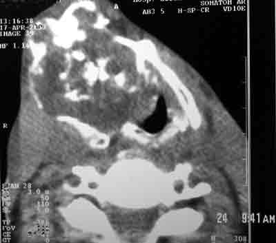
The ENT examination evidenced tumor formation in the neck on the right, fixed to the larynx and measuring approximately 6cm, of hard consistency and non-painful upon palpation (Fig. 1). Laryngoscopy showed bulging of paraglottic space on the right that prevented visualization of glottis. We ordered Computed Tomography (CT scan) of the neck and it showed suggestive image of thyroid cartilage chondrosarcoma (Figure 2), indicating surgical exeresis.
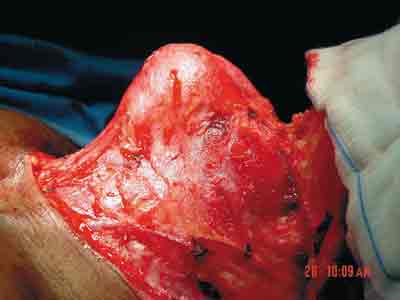
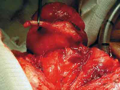
Surgery was conducted under general anesthesia and the patient was initially submitted to tracheotomy. Surgical approach was conducted by neck incision, anterior bimastoid arch form, deep down to platysma muscle, whose flap was displaced. Exposure of pre-laryngeal muscles, placement of muscles to the midline level, and detection of globous lesion, petrous consistency, white, measuring approximately 6cm, that comprised the right portion of the thyroid cartilage (Figures 3 and 4). Displacement of internal perichondrium of the affected hemilarynx, excision of the thyroid lamina containing the tumor with section of thyro-hyoid ligament close to the lesion (Figures 5 and 6); fixation of right hemilarynx with pre-laryngeal muscle, placement of drainage, closing by planes, skin suture and external compressive dressing.
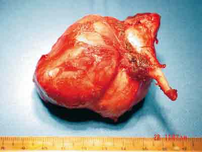
The frozen analysis was impaired by calcified cartilaginous tumor (Figure 7). The clinical pathology showed macroscopy result of: bosselated nodular formation, well delimited, apparently encapsulated, firm and elastic, weighting 70g and measuring 6.5x5.5x5.4 cm, encompassing the major horn of thyroid cartilage, measuring 1.7cm in length. The sections showed that the lesion was white, bright, with many intertwined calcifications areas. Microscopy: low grade chondrosarcoma (Grade I) and surgical limits represented by the lesion itself.
Postoperative care was uneventful and on the 8th day the patient was discharged with reasonable vocal quality, occluded tracheotomy, good oral feeding and satisfactory airways.
Currently, he is being followed up as an outpatient and has no signs of lesion recurrence.
DISCUSSION
Laryngeal chondrosarcomas are rare tumors and there are only about 250-300 cases described in the world literature 1.
Its onset may be favored by laryngeal trauma 2.
The clinical presentation is varied, which may range from no symptoms in small lesions to manifestations of progressive dyspnea, stridor, dysphagia, dysphonia, but it rarely causes pain. Cases of idiopathic paralysis of vocal folds have been described in the literature as a consequence of laryngeal chondrosarcoma 2,3. The onset of chondrosarcoma in two patients after radiotherapy for primary treatment of vocal fold squamous cell carcinoma has been reported, which can occur up to 16 years after exposure 3,4.
In the larynx, chondrosarcoma presents some typical characteristics: slow growth, rare metastases, and low tendency to recurrence 5, but it may happen in the long-term and is normally controllable.
Neck palpation may reveal hard mass, fixing the cricoid cartilage that is mobilized during swallowing.
Endoscopic exam should be carefully conducted because it is a lesion normally located in the subglottic region, and we can observe regular submucous mass, posterior, rarely larger than 2-3cm that is very hardened and may hinder the biopsy.
Neck radiology in profile shows the larynx projected forward. CT scan typically shows hypodense, well-circumscribed mass with agglomerated calcifications 6, in addition to showing extensive tumor in the laryngeal framework. Magnetic resonance imaging (MRI) may demonstrate lesion in the larynx and it has superior advantage of resolution with use of contrast for the tumor and paralaryngeal tissues 7.
The histopathology revealed lobulated mass of homogenous structure formed by hyaline cartilage lobes. The presence of giant cartilaginous cells with nuclear pleomorphism and hyperchromatism, formed by multiple nodules that are interconnected and have varied sizes, is characteristic of chondrosarcoma. Intercellular space may be of chondroid or myxomatous origin. The presence of mitosis is not necessary for the diagnosis, and calcifications and ossifications present bone erosion and not osteoid production common in malignant cells.
In general, chondrosarcoma causes local destruction due to mass effect before it invades neighboring tissues.
Differential diagnosis is made with chondromyxoid fibroma, osteosarcoma, fibrosarcoma, chondroma and chordoma.
Chondrosarcomas are divided into 3 histology grades (I, II and III) according to grade of cell differentiation. These cell differentiation grades are classified according to rate of mitosis, cellularity and nuclear size.
Low-grade chondrosarcomas (I), which may look like benign neoplasms in many situations 10, are characterized by small and dense nuclei, predominantly chondroid intercellular space, frequent calcifications and absent mitotic activity. In grade II, there are large-proportioned and moderate size nuclei, with small rate of mitosis and myxoid intercellular space, in general. In grade III, there are 2 or more mitosis in 10 fields.
Fortunately, most chondrosarcomas are indolent and normally detected in grades I and II. In a 5-year analysis of patients with chondrosarcoma grades I, II and III, survival was 90%, 81% and 43%, respectively. No metastases were found in tumors grade I, but 70% of grade III tumors were metastatic. Other prognostic factor is related to tumor size, and those that are larger than 10cm are significantly more aggressive than smaller tumors. If there are metastases, the most frequent sites are the lungs and skeleton. Level of resectability of the tumor also contributes to prognosis.
Laryngeal chondrosarcomas are less aggressive than those located in other regions of the body; neck or distant metastases are rare (8.5%), local recurrences are not uncommon but they tend to be not aggressive and normally late, and clinical follow-up is recommended for at least 10 years.
Treatment is preferably surgical exeresis with preservation of laryngeal structure and function 8, 9, 10. Total laryngectomy is indicated in cases of high-grade malignancy, when total exeresis of the lesion is not feasible, or in recurrent lesions 5,11.
Chondrosarcomas are little radiosensitive and radiotherapy is reserved for cases of recurrence, in extensive forms, for high grades or irresectable lesions 13.
Chemotherapy is the palliative management for aggressive tumors with local invasion.
CLOSING REMARKS:
Laryngeal chondrosarcomas are a rare entity and making the diagnosis is a challenge to Otorhinolaryngologists. Its occurrence should be always considered when there are symptoms of laryngeal affection, neck masses and idiopathic paralysis of vocal folds.
It is important to make early diagnosis of this neoplasm to allow early treatment and preservation of laryngeal structure and functions.
REFERENCES
1- De Stefani A, Fadda Gl, Cavalot A, Nazionale G, Mola P. Chondrosarcoma of the larynx: Case report and review of the literature - Tumori 2000 - Jan-Feb; (1):79-81.
2- Leonetti ED, Collins SL, Jablokow V, Lewy R. Laryngeal chondrosarcoma as a lat-appearing cause of "idiopathic" vocal cord paralysis - Otolaryngol Head Neck Surgery - 1987 - Oct; 97(4):391-5.
3- Glaubiger DL, Casler JD, Garret WC, You HS, Lillis-Hearne PK. Chondrosarcoma of the larynx after radiation treatment for vocal cord cancer - Cancer 1991 - Oct 15; 68(8):1828-31.
4- Campos ED, Conde PN, Dana JJC, Alvarez MF, Castelle JMT. Advanced stage laryngeal chondrosarcoma - Ann Otorrinolaryngol Ibero - Am 2002; 29(5):473-81.
5- Wang SJ, Borges A, Lufkin RB, Se Carz JA, Wang MB. Chondroid tumors of larynx: computed tomography finding - Am J Otolaryngol - 1999 - Nov - Dec; 20(6):379-82
6- Mishell JH, Shild JA, Mafee MF. Chondrosarcoma of the larynx: Diagnosis with magnetic resonance imaging and computed tomography. Arch Otolaryngol Head Neck Surg - 1990 - Nov; 116(5):1935-39.
7- Nicolai P, Ferlito A, Sasaki CT; Kirchmmer JA; Laryngeal chondrosarcoma: incidence, pathology, biological behavior and treatment. Ann Otol Rhinol Laryngol - 1990 - Jul; 99(Pt 1):515-23.
8- Lemarchand V, Bequignon A, Chanel S, Moreaus S, Valdazo A. Chondrosarcomas and low-grade chondrosarcomas of the larynx: a case report - Ann Otolaryngol Chir Cervicofac - 2002 Sep; 119(4):252-6.
9- Cantrell RW, Reibel JF, Jahrsdoerfer RA, Johns ME. Conservative surgical treatment of chondrosarcoma of the larynx - Ann Otol Rhinol Laryngol - 1980;(6 Pt 1):567-71.
10- Hicks JN, Walker EE, Moor EE. Diagnosis and conservative surgical management of chondrosarcoma of larynx - Ann Otol Rhinol Laryngol - 1982 - Jul-Aug;91(4 Pt 1)389-91.
11- Kambic V, Zargi M, Gale N. Laryngeal Chondrosarcoma: is conservative surgery adequate treatment? - J Laryngol Otol - 1989 - Oct; 103(10):970-2
12- Saleh HM, Guichard C, Russier M, Kemeny JL, Gilain L. Laryngeal chondrosarcoma: a report of five cases - Eur Arch Otorhinolaryngol - 2002 - Apr;259(4):211-6.
13- Dailiana T, Nomikos P, Kapranos N, Thanos L, Papathanasiou M, Alexoupoulou E, Papaioannou G, Kelekis D A. Chondrosarcoma of the larynx: treatment with radiotherapy - Skeletal Radiol - 2002 Sep; 31(9):547-9.
Fig. 1: Preoperative neck mass.
Fig. 2: Preoperative CT: areas of calcification at the level of the cartilage. Right side thyroid with lateral displacement of larynx.
Fig. 3: Intraoperative view.
Fig. 4: Intraoperative view.
Fig. 5: Removal of lesion close to thyroid cartilage.
Fig. 6: Surgical specimen.
¹ Resident Physician, Service of Bronchoesophagology and Head and Neck Surgery, Hospital Geral de Bonsucesso (HGB) - Rio de Janeiro - RJ
² Staff member, Service of Bronchoesophagology and Head and Neck Surgery, HGB
³ Head of Service of Bronchoesophagology and Head and Neck Surgery, HGB
Study conducted at the Service of Bronchoesophagology and Head and Neck Surgery, Hospital Geral de Bonsucesso - Rio de Janeiro - RJ.
Address correspondence to: A/C Drª Giordania Gomes Campos - Rua José Higino, nº 30 - Tijuca - Rio de Janeiro - RJ - Cep: 20520-200
Tel: (55 21) 9241-2246 - E-mail: giordaniagc@ig.com.br


Print: ![]()