

Year: 2004 Vol. 70 Ed. 6 - (10º)
Artigo Original
Pages: 773 to 776
 PDF PT
  PDF EN
Neck dissection in the treatment of squamous cell carcinoma of the lip
Author(s): Ali Amar ,2, Sergio A. Franzi1, Otávio A. Curioni1, Abrão Rapoport1, Onivaldo Cervante
Keywords: Key words: squamous cell cancer, lip, metastasis, neck dissection.
Abstract:
Summary
Squamous cell carcinoma of the lip usually is diagnosed at an early stage and has low incidence of neck metastases. Aim: To assess the incidence and location of lymph node metastases in squamous cell carcinoma of the lip. Study Design: Retrospective study, case series. Material and Method: A file review of 78 patients treated between 1990 and 2001. It was evaluated the relation between the primary tumor size, degree of differentiation, comissure involvement and the presence and location of lymph node metastasis. Results: Lymph node metastasis were detected in 7% of lesions 3 cm and in 41% of cases > 3 cm (p=0.002). Ten patients had metastases, all of them had metastases in level I and only 2 cases had metastases in other levels too. After elective treatment of the neck, metastasis was found in level I only. Conclusion: Metastasis are uncommon in lesions less than 3 cm. Lymph node metastasis usually occur at level I and suprahyoid neck dissection can be indicated for elective treatment of the neck.
![]()
Introduction
Lip cancer is the most frequent tumor of the mouth and is related to sun exposure, particularly in Caucasians. In the city of Sao Paulo, lip cancer accounts for 2.4/100.000 and 0.6/100.000 incidence among men and women, respectively, corresponding to 0.6% and 0.2% of all malignant lesions diagnosed1.
The most common histological type is squamous cell carcinoma, which frequently affects the lower lip. During treatment, regional lymph node involvement should always be investigated. Although nodal metastases are not so frequent in initial tumors, they are related to significantly poorer diagnosis, especially in cases of neck recurrence of non-treated neck2. The specialist must assess the risk of occult metastases and the indication of elective neck dissection (as a prophylactic procedure); also, extensive dissection should be performed if palpable lymph nodes are present. Suprahyoid dissection (levels I, II and III) is considered adequate for elective treatment of mouth tumors, but it may not be sufficient in the presence of metastases or it may be too aggressive when elective treatment involves both sides of neck in an elderly patient3,4.
The present study assessed the incidence and location of metastases of squamous cell carcinoma of the lip taking neck treatment into consideration.
Material and Method
The medical charts of 78 patients with squamous cell carcinoma of the lip, with no previous treatment, admitted to the Head and Neck Surgery and Otorhinolaryngology Department of Hospital Heliópolis, between January 1990 and December 2001, were analyzed. There were 62 men and 16 women. The average age was 60 years (20 to 93 years) and the mean age was 61 years (Q25%-75%= 53 to 71 years). The primary site was the lower lip, presented by 75 patients, whereas tumors of the upper lip were found in 3. Relative to T staging, 40 were staged T1, 23 T2, 10 T3 and 5 T4. The average time of complaint was 12 months (1 to 84 months). Surgery was performed in 67 cases, among which 6 received complementary radiotherapy. Neck dissection was initially carried out in 18 patients, out of which 9 underwent bilateral dissection (27 dissections). Concerning the type of dissection, 5 were radical modified (levels I to V), 18 were supraomohyoid (levels I, II and III) and 4 were suprahyoid (level I) dissections. Radiotherapy alone was employed in 3 cases and 8 patients were lost for follow-up after the initial evaluation. Post-treatment follow-up lasted on average 17 months (0 to 143 months).
The incidence of lymph node metastases was assessed in accordance with primary tumor size, histological grading and lip commissure involvement. Moreover, metastases' locations were evaluated taking into account 8 nodal levels, according to the American Head and Neck Society classification3. Only histologically-confirmed metastases (pN) and cervical recurrences were considered in this analysis; untreated patients were excluded. Patients were restaged in accordance with 2002 UICC TNM Classification. The statistical analysis employed Fisher test and Mann-Whitney test.
Results
Palpable lymph nodes were found in 14 patients, among which 8 underwent cervical dissection and 6 did not return for follow-up. Out of 64 patients without palpable lymph nodes, 10 were submitted to elective neck dissection. The histological results confirmed the presence of metastases in 4/8 patients who underwent therapeutic dissection, and in 6/10 patients submitted to elective dissection.
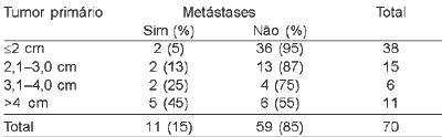
Out of 10 subjects with postoperative histological confirmation of disease dissemination, all presented level I metastases - 5/10 level Ia and 6/10 level Ib. One patient presented levels I, II and IV, and 1 had levels I and III metastases. Patients submitted to elective neck dissection presented level I metastases only. There were 5 cases of cervical recurrence, out of which 4 underwent previous cervical dissection. A patient with untreated neck was rescued with neck dissection, whose metastasis was identified as of level Ib. Incidence of lymph nodes metastases was 7% for 3cm tumors and 41% for >3cm tumors, p=0.002, suggesting close relation between primary tumor size and risk of metastases (Table 1).
Lip commissure was involved in 12 (15%) of the patients, and this involvement was related to 11-fold higher risk of lymph node metastases (CI 95%=2.4 to 54 and p=0.003). Tumors that reached lip commissure had average diameter of 5.8 cm, while other tumors were on average 2.2 cm (p=0.000004). Differentiation grade was identified in 70 cases. Metastases were found in 8/50 patients with grade I tumors, 2/18 grade II and 1/2 grade III.
Discussion
Incomplete follow-up of patients limited the study results, once most of them were at an initial stage of disease and were not submitted to neck dissection. Considering that regional recurrences of lip carcinomas may appear within 2-3 years after treatment of primary tumor, the incidence of metastases may be underestimated. Squamous cell lip carcinoma presents low incidence of lymph node metastases in lesions smaller than 3 cm, thus not justifying elective neck prophylactic dissection.
Tumor extension and thickness should strongly be considered. Infiltrating lesions greater than 6 mm, as well as those less differentiated, are liable to disseminate5. Therefore, elective dissection may be chosen for lesions 3 cm, taking into account tumor thickness or differentiation, particularly when patients' follow-up is difficult. Patients with local recurrences have greater risks of lymph node metastases, for which neck treatment is indicated3. Although involvement of the lip commissure is related to higher probability of metastases, this factor tends to be related rather to tumor size than to its location. Only 1.2%-1.5% of the tumors originate from the commissure, whose involvement usually occurs due to tumor growth6,7. In this study, involvement of lip commissure was observed in 15% of the cases and was related to larger primary tumor. Sample size was not enough for multivariate analysis and, consequently, this is a questionable issue. However, other authors suggested that lip commissure involvement was not associated with presence of lymph nodes6.
In general, metastases occur in level I cases, while levels III, IV and V are rarely affected; thus, less extensive neck dissection is feasible3,8. The central region usually drains to submentalis lymph nodes (Ia), while the side thirds drain to submandibular lymph nodes (Ib)9. In N0 cases, neck dissection should be considered only at level I (suprahyoid dissection)10. Taking into account that submandibular lymph nodes are located on the lower border of mandible, the gland could be preserved during dissection. Among patients with clinically palpable lymph nodes, the false-positive rate is 50%. Even though intraoperative evaluation with frozen section examination of suspected lymph nodes is inaccurate, this method may help identifying the cases in which neck dissection should extend to levels II and III11. Investigation of sentinel lymph node allows better intraoperative evaluation, which is frequently found as level I in lip tumors12. Despite the presence of confirmed metastases, supraomohyoid dissection (levels I, II and III) is indicated for neck treatment, once metastases in other levels are not common8. Although most cases show good evolution, lymph node metastases identify a subgroup of patients with poorer prognosis, particularly concerning neck recurrences. Thus, elective neck treatment should always be considered when the risk of metastases is significant. Nearly 25% of patients were older than 70 years, for whom benefits and risks from elective dissection should be carefully considered, especially when bilateral dissection is required. The suprahyoid dissection is strongly indicated in those cases due to its lower morbidity.
Conclusions
Lymph node metastases seldom occur in tumors 3 cm. Level I dissection (suprahyoid) is indicated for elective neck treatment of lip squamous cell carcinomas. This type of dissection should be extensive to levels II and III (supraomohyoid) for patients presenting positive level I lymph nodes. For levels IV or V, neck dissection should be performed when there is evidence of metastases beyond level I.
References
1. Mirra AP, Latorre MR, Veneziano DB. Incidência de Câncer no Município de São Paulo, Brasil: 1997-1998. Brasília, DF: Ministério da Saúde, 2001. Available at http\\www.fsp.usp.br/rcsp/rcsp1.pdf, accessed on 30/04/2004.
2. Santos LRM, Cernea CR, Kowalski LP et al. Squamous cell carcinoma of the lower lip: A retrospective study of 58 patients. Sao Paulo Med J 1996; 114: 1117-26.
3. Robbins KT, Atkinson JD, Byers RM, Cohen JI, Lavertu P, Pellitteri P. The use and misuse of neck dissection for head and neck cancer. J Am Coll Surg 2001; 193: 791-802.
4. Spiro JD, Spiro RH, Shah JP et al. Critical assessment of supraomohyoid neck dissection. Am J Surg 1988; 156: 286-9.
5. Rodolico V, Baarresi E, Di Lorenzo R et al. Lymph node metastasis in lower lip squamous cell carcinoma in relation to tumor size, histologic variables and p27kip1 protein expression. Oral Oncol 2004; 40: 92-8.
6. Zitsch RP, Lee BW, Smith RB. Cervical lymph node metastases and squamous cell carcinoma of the lip. Head Neck 1999; 21: 447-53.
7. McCombe D, MacGill K, Ainslie J, Beresford J, Matthews J. Squamous cell carcinoma of the lip: A retrospective review of the Peter MacCallum Cancer Institute experience 1979-88. Aust N Z J Surg 2000; 70: 358-61.
8. Vartanian JG, Carvalho AL, Araujo Filho MJ, Hattori Jr M, Magrin J, Kowalski LP. Predictive factors and distribution of lymph node metastasis in lip cancer patients and their implications on the treatment of the neck. Oral Oncol 2004; 40: 223-7.
9. Rouvière H. Anatomie des lymphatiques de l'homme. Paris: Masson et C Editeurs; 1932.
10. Koç C, Akyol UM, Çelikkanat S, Çekic A, Özdem C. Role of suprahyoid neck dissection in the treatment of squamous cell carcinoma of the lower lip. Ann Otol Rhinol Laryngol 1997; 106: 787-9.
11. Rassekh CH, Johnson JT, Myers EN. Accuracy of intraoperative staging of the N0 neck in squamous cell carcinoma. Laryngoscope 1995; 105: 1334-6.
12. Altinyollar H, Berberoglu U, Çelen O. Lymphatic mapping and sentinel lymph node biopsy in squamous cell carcinoma of the lower lip. Eur J Surg Oncol 2002; 28: 72-4.
Table 1. Size of primary tumor and post-treatment lymph node metastases.
Print: ![]()