

Year: 2004 Vol. 70 Ed. 5 - (10º)
Artigo Original
Pages: 645 to 649
 PDF PT
 Audiological profile of patients with Goldenhar syndrome
Author(s):
Karina Costa Brosco1,
Neivo Luiz Zorzetto2,
Antonio Richieri da Costa3
Keywords: Goldenhar's syndrome, oculoauriculovertebral dysplasia, audiology.
Abstract:
The Goldenhar's syndrome is a rare congenital anomaly, of which the etiology is yet unknown, and characterized by a classical triad of ocular, auricular and vertebral abnormalities. This study used 30 individuals presenting Goldenhar's syndrome, that were regularly enrolled in the Craniofacial Anomaly Rehabilitation Hospital (HRAC), of both sexes, varying from 8 to 34 years old. The goal was to characterize the audiologic profile to the individuals presenting this syndrome, thus, assuring a better treatment and orientation for them, and also the establishment of the frequency of contra-lateral auditory commitment in individual with the classical unilateral involvement. The auditory assessment of this study showed ATL, tympanometry, EOA-T and BERA. According to the results, we concluded that 35% (N=10) of the individuals presented, as a characteristic of the audiologic profile, loss of hearing type sensorioneural, mixed with varied levels from moderate to deep (7 uni and 3 bilateral); 13% (n=4) presented conductive loss (bilaterally) with levels mild to severe and 3% (n=1) presented a unilateral deep type of sensorioneural loss. This study showed 40% n=12) with normal bilateral hearing and in 10% (n=3) it wasn't possible to establish the characteristic of the audiologic profile, since only the BERA was used as an assessment tool for establishing the electro-physiologic threshold. Of the twelve (12) individuals presenting unilateral malformations of the left ear, only two (02) presented contra-lateral auditory commitment of the ear, one type mixed with a severe level and one conductive of a moderate level. Considering the sex variable, the mayor predominance of the Goldenhar's syndrome was for females (57%) compared to the males (43%), but these results weren't statistically significant, as well as for the unaffected anatomical side, that was predominantly the right side.
![]()
INTRODUCTION
Goldenhar syndrome is a rare congenital anomaly, of unknown etiology, characterized by ocular, auricular and vertebral abnormalities, frequently associated with other visceral or facial congenital malformations (Laredo et al. 1985).
The syndrome was described for the first time by Von Arlt in 1845 and it was recognized as a clinical entity by Maurice Goldenhar in 1952, who described it in a child, as reported by Salvitti et al. (1978). It was also named first arch syndrome and Gorlin syndrome. Currently, it is frequently known as oculoauriculovertebral dysplasia, the name given by Gorlin et al. (1963) and Sugar (1966). Baum et al., in 1973, listed 114 cases of oculoauriculovertebral dysplasia. The pioneer case in Brazil was described by Paiva in 1971. Oculoauriculovertebral dysplasia is a complex congenital symptomatic manifestation of unknown etiology in which the main abnormalities are limited to the eyes (dermoid and/or epibulbar lipodermoid), external ear (auricular appendices, blind fistulae) and vertebral spine (hemivertebra, vertebral fusions and other malformations). According to Laredo et al. (1985) it is also associated with other visceral or facial congenital malformations that involve the structures derived from the 1st and 2nd branchial arches.
The following authors have already studied this syndrome: Gorlin et al. (1990), Gorlin and Pindborg (1964), Poswillo (1973), Bluestone et al. (1983), Smith (1985), Musarella and Young (1986), Lisboa et al. (1987), Meireles and Tomita (1987), Rollnick et al. (1987), Gorlin et al. (1963), Kaye et al. (1989), Grabb (1965), Pashayan et al. (1970), Rollnick and Kaye (1983), Rollnick et al. (1987), Alves et al. (1991), Bertazzo et al. (1991), Johnson et al. (1994), Casey et al. (1996), Manfre et al. (1997), Mocellin et al. (1998), Lopes Filho (1998), Schaefer et al. (1998), Kirkham (1970), Converse et al. (1979), Kaban et al. (1981), Phelps et al. (1983), Seltzer et al. (1981), Opitz and Faith (1969), who described the possible etiologies and clinical manifestations of the syndrome but did not define the audiological profile of these patients. This is the purpose of the present study: to analyze regular patients from Hospital de Reabilitação de Anomalias Craniofaciais da USP-Bauru (HRAC-USP-Bauru) and to define prevalence of contralateral auditory behavior in cases of classical unilateral involvement.
MATERIAL AND METHODS
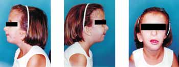
This study was conducted with 30 subjects clinically diagnosed with Goldenhar syndrome (Figure 1); 13 were male and 17 were female, aged 8 to 34 years, regularly enrolled in the clinical genetic center, HRAC-USP- Bauru. Audiological assessment comprised:
- Pure tone Audiometry;
- Acoustic immittance measures;
- Brainstem evoked response audiometry (BERA);
- Transient otoacoustic emissions (TOAE).
Out of 30 tested cases, 16 were assessed using all audiological assessment procedures. The cases with pinna and unilateral external acoustic canal agenesia, owing to the impossibility of introducing the probe for acoustic immittance measures and TOAE were submitted only to pure tone audiometry and BERA. Similarly, we did not perform tympanometry and TOAE in cases that had external auditory canal stenosis (n=4).
Pure tone audiometry and the classification of hearing loss followed the standards devised by Santos and Russo (1991). We used audiometer Midimate 622-Madsen Electronics.
Acoustic immittance measures shown in tympanograms were classified and analyzed according to the data reported by Jerger (1970).
BERA was analyzed according to the standards proposed by Jewett and Willistom (1971). The device used was BERA model-Hirnstamm-Audiometer Brain Stem Audiometer. Otoacoustic emissions were recorded with ILO 88 version 92 brand OTODYNAMICS, coupled to computer IBM-APTIVA.
The results obtained were statistically treated by applying chi-square test, with significance level of 5%.
RESULTS
Clinical assessment
As to gender, there was no statistically significant difference in the occurrence of the syndrome, and there were 13 (43%) male and 17 (57%) female subjects.
Physical examination revealed 12 subjects with unilateral external ear malformation, one with bilateral malformation and 17 without malformation.
Out of 13 cases with external ear malformation, 10 presented pinna and external acoustic canal agenesia, 5 on the right and 5 on the left; three had external acoustic canal stenosis, two on the right and one on the left, and one case presented pinna microtia and external acoustic canal stenosis on the right.
Out of 12 subjects with unilateral external ear malformation, ten did not present contralateral auditory impairment and two presented auditory affections, with one moderate conductive loss and another severe mixed loss, both on the left side.
Audiological assessment
After total audiological assessment of the sample (30 cases), we detected that 12 subjects presented bilateral normal hearing, 9 had unilateral hearing loss and 9 had bilateral hearing loss. The 12 cases with normal hearing were submitted to genetic tests that confirmed the diagnosis of the syndrome.
Out of 12 subjects with unilateral affections, seven presented mixed loss, 5 on the right and 2 on the left, and one had sensorineural loss on the right. This group had only one subject who underwent electrophysiological threshold investigation with BERA. Still concerning the 9 cases of unilateral loss, we noticed that four presented moderate hearing loss, two had severe loss, and two had profound loss. Out of 9 cases with bilateral auditory affections, four presented bilateral conductive loss, two had bilateral mixed loss, and one had mixed loss on the right and sensorineural mild loss in high frequencies on the left. In this group, 2 subjects underwent BERA.
In the group with bilateral hearing loss, a subject presented bilateral mild loss, two had bilateral moderate loss, and one had mild loss on the right and moderate loss on the left, one had moderate loss on the right and mild loss on the left, and one had moderate loss on the right and severe loss on the left.
Out of three subjects who did not understand pure tone audiometry conditioning and underwent BERA to define electrophysiological thresholds, one of them had unilateral abnormal thresholds (absence of response on the right) and two had bilateral affections [60dBHL (RE)/absence of response (LE) and 80dBHL (RE)/60dBHL (LE)].
DISCUSSION
Goldenhar syndrome or oculoauriculovertebral dysplasia is a rare congenital anomaly of unknown etiology; different hypotheses have been suggested, either hereditary or embryopathic (Salvitti et al. 1978; Laredo et al. 1985, Meirelles and Tomita, 1987). Even though they have been described as new cases with dominant autosomal heritage, authors such as Smith (1985), Mussarela and Young (1986), Kaye (1992) and Gorlin (1995) thought that most of them were sporadic. Conversely, Johnson et al. (1994) and Gorlin et al. (1990) referred that anomalies of the 1st and 2nd branchial arches have been observed in children born from mothers exposed to thalidomide, primidone, retinoic acid, and diabetic mothers. Our results recorded that out of 30 subjects with Goldenhar syndrome, 57% (13 cases) were female and 43% (17 cases) were male, not agreeing with the observations made by Smith (1985), Rollnick et al. (1987), Gorlin et al. (1990) and Schaefer et al. (1998), who identified predominance of male gender in 3:2 ratio.
Out of the total sample (30 cases), 13 cases presented bilateral external ear malformation, 12 had unilateral affection and 17 cases had no malformation.
As to external ear malformation, they corresponded to what is mentioned in the literature, that is, we found 10 cases of pinna and external auditory canal malformations, three cases of stenosis of external auditory canal and one with pinna microtia and external canal stenosis. Pinna agenesia was predominant in the study and it corresponds to the most severe expression of malformations of type III, as provided by Altmann (1965) classification, mentioned by Paparella and Shumrikck (1973). To Bluestone et al. (1983) and Gorlin et al. (1995) agenesia or anotia is an external ear abnormality that can be found in oculoauriculovertebral dysplasia and range from a poorly defined mass of tissue on the bone, displaced anterior and inferiorly, to slightly deformed ear.
The predominantly unilateral damage found in our results corresponds to literature data. Anatomical conditions are frequently asymmetrical, with one side of the face more severely affected than the other, as referred by Charles et al. (1983), and the right side was more affected than the left side in studies conducted by Smith (1985), Rollnick et al. (1987) and Gorlin et al. (1990), which was also observed in the present study.
Audiological assessment showed 12 subjects with bilateral normal hearing, a fact reported by the studied literature. In the battery of tests conducted in these cases, hearing thresholds, speech recognition index (SRI) and speech reception thresholds (SRT) had results within the normal range, as determined by Santos and Russo (1991). We found type A tympanometry characterizing normal tympanic-ossicle system in most subjects, type B curve in only one case on the left, characterizing conductive abnormality type AD in three cases, one bilaterally and the other just on the left ear, suggesting laxity of tympanic-ossicle complex. In cases with type A and Ad tympanometry curves, we found presence of TOAE, that is, integrity of cochlear mechanism observed by reproducibility greater than 50% and amplitude of response greater than 3dBSPL over noise, according to studies conducted by Gattaz et al. (1994) and the criteria used by CPA (Centro de Pesquisas Audiológicas -FOB-USP). Type A tympanometry subjects presented absent TOAE, with reproducibility values below 50%, which could be justified by the conductive problem present. BERA showed normal responses for interpeak (I-III, III-V, I-V) intervals and absolute latency values (I, III, V), which let us suggest the presence of neural integrity of auditory central nervous system up to the brainstem. In the anatomically integral side, subjects presented pure tone thresholds and SRT and SRI values within the normal range. Tympanometry showed that most subjects presented type A curve, and only one case presented type Ar curve and another one had type C curve. In TOAE they all had responses present, except for one case, which could be interpreted as absence of response recording on this side. Interpeak measures (I-III; III-V; I-V) and absolute latency times (I-III-V) were within the normal range, suggesting neural integrity of auditory central nervous system up to the brainstem. On the contralateral side, the six cases of pinna and external auditory canal agenesia and the only case with external canal atresia did not undergo tympanometry nor TOAE, and the mean of pure tone thresholds in these cases was abnormal, presenting varied level of hearing loss. Values of SRT were compatible with pure tone averages and SRI values were compatible with audiological configuration. In one subject we conducted only speech detection threshold because of hearing loss level (profound) and the results were compatible. In one case only it was possible to conduct tympanometry in the contralateral ear, presenting type B curve and absence of TOAE, justified by middle ear affection. In BERA, two subjects presented absence of responses and it was not possible to assess time of absolute or interpeak latencies. Four subjects presented increased absolute latency time (I-III-V) and normal range interpeak intervals (I-III; III-V; I-V) suggesting conductive abnormalities.
Three subjects were submitted to BERA to investigate electrophysiological thresholds and neural integrity because they could not be conditioned for audiometry. Results ranged from 20 dB HL to absence of response. In cases with pinna and external auditory canal agenesia it was not possible to conduct tympanometry and TOAE because it was impossible to introduce the probe into the canal. In cases of type B tympanometric curve in the contralateral ear we found reproducibility below 50% and response amplitude of 3dB SPL in TOAE, justified by middle ear affections. In two subjects, BERA showed absence of interpeak interval response (I-III, III-V, I-V) and increased absolute latency times (I, III and V). In one case, interpeak intervals (I-III, III-V, I-V) were within the normal range, but absolute latency values (I, III, V) were increased, suggesting conductive affection compatible with the tympanometry.
Out of 18 subjects with hearing loss, concerning type and level, most of them presented mixed hearing loss (n=10), suggesting middle and external ear affection as well as internal ear abnormalities, and 8 had unilateral and two had bilateral loss, ranging in level from mild to profound; four presented bilateral conductive mild to severe losses, and one presented profound sensorineural loss on the right; three were submitted to BERA because they did not appropriately respond to pure tone audiometry. Out of these 3 cases, one had unilateral auditory affection with absence of response at 100dB HL and two had bilateral affections raging from 60dB HL to absence of response.
The present study detected that type and level of hearing loss in most of the subjects with Goldenhar syndrome were different from those reported by the studied literature, which referred higher incidence of conductive losses and few cases of sensorineural losses. Our sample, in turn, showed that in most cases there were mixed losses: seven mixed losses, one sensorineural case and no unilateral conductive loss. In cases of bilateral hearing loss, four were conductive, two were mixed and one was sensorineural and no cases were purely sensorineural cases.
The discussion of cochlear functional integrity by TOAE in subjects with conductive affections and decreased auditory thresholds is a difficult process, which may even prevent recording of TOAE. In the analyzed cases, the results found were worse than 50% in relation to the curves, indicating absence of responses. In subjects with decrease of hearing thresholds associated with type A and type Ar tympanometry curve, we detected absence of TOAE, which could suggest cochlear mechanism affection. BERA did not detect subjects with neural conduction affection up to the brainstem (I-III, III-V and I-V within the normal range), even among those that did not understand the audiometry instructions. Absolute latency times I-III-V were compatible with hearing affections found in audiometry, tympanometry and TOAE.
Our results are in accordance with information reported in the studied literature concerning predominance of conductive hearing loss caused by external and/or middle ear malformations, impairing normal sound conduction, and some rare cases of inner ear affection. However, most of the subjects studied here presented external ear malformation and mixed hearing loss. It is, therefore, fair to infer that there may be some type of middle and/or inner ear malformation, a fact that was not the assessment object of the employed methodology.
REFERENCES
1. Altmann F. Malformations of the auricle and the external auditory meatus. Arch Otolaryng 1951; 54:115.
2. Alves MAS, Real SV, Souza SR. Síndrome de Goldenhar (displasia óculo-aurículo-vertebral): relato de um caso. Rev Bras Oftalmol 1991; 50:60-2.
3. Baum JL, Feingold M. Ocular aspects of Goldenhar s syndrome. Am J Ophthalmol 1973; 75:250-7
4. Bertazzo JRV, Queiroz Neto LS, Queiroz Filho LS. Síndrome de Goldenhar-Gorlin. Arq Inst Penido Burnier. 1991: 33:50-2
5. Bluestone CD, Stool SE, Arjona SK. Congenital malformations of the mouth and pharynx. In: Pediatric otolaryngology. Philadelphia: WB Saunders; 1983. p.917.
6. Casey HD, Braddock SR, Haskins RC, Carey JC, Morales Junior L. Frontonasal malformation and the oculoauriculovertebral spectrum: the oculoauriculofrontonasal syndrome. Cleft Palate Craniofac J 1996; 33:519-23.
7. Converse JM, McCarthy JG, Coccaro PJ, Wood-Smith D. Clinical aspects of craniofacial microssomia. In: Symposium on diagnosis and treatment of craniofacial anomalies. St. Louis: C.V. Mosby & Co.; 1979.
8. Grabb WC. The first and second branchial arch syndrome. Plast Reconstr Surg 1965; 36: 485-508.
9. Gorlin RJ, Pindborg JJ, editors. Syndromes of the head and neck. New York: McGraw-Hill; 1964.
10. Gorlin RJ, Cohen MM, Levin LS. Branchial arch and oro-acral disorders. In: Syndromes of the head and neck. 3rd. New York: Oxford University Press; 1990. p.641-8.
11. Gorlin RJ, Kenneth LJ, Jacobsen U, Goldschmidt E. Oculoauriculovertebral dysplasia. J. Pediat 1963; 63: 991-9.
12. Jerger J. Clinical experience with impedance audiometry. Arch Otolaringol 1970; 92: 311-24.
13. Jewett DI, Williston JS. Auditory evoked far fields averaged from the scalp of humans. Brain 1971; 4: 681-96.
14. Johnson K, Fairhurst J, Clarke NM. Oculoauriculovertebral spectrum: new manifestations. Pediatr Radiol 1994; 25: 146-8.
15. Kaban LB, Mulliken JB, Murray JE. Three-dimensional approach to analysis and treatment of hemifacial microssomia. Cleft Palate J 1981; 18: 92-9.
16. Kaye CI, Rollnick BR, Hauck WW, Martin AO, Richtsmeier JT, Nagatoshi K. Microtia and associated anomalies: statistical analysis. Am J Med Genet 1989; 34: 574-8.
17. Kirkam TH. Goldenhar's syndrome with inner ear defects. J Laryngol Otol 1970; 84: 855-6.
18. Laredo FJ, Braga JMB, Kasinski SK, Caballero JMP. Síndrome de Goldenhar (displasia óculo-aurículo-vertebral). Folha Med Bras 1985; 91: 361-4.
19. Lisboa RC, Mendez HMM, Paskulin GA. Síndrome de Goldenhar e variantes: relato de sete pacientes. Rev AMRIGS 1987; 31: 265-9.
20. Lopes Filho OC. Imitância acústica: aplicações clínicas. In: Tratado de Fonoaudiologia. São Paulo: Roca; 1998. p.171-82.
21. Manfre L, Genuardi P, Tortorice M, Lagalla R. Absence of the common crus in Goldenhar syndrome. AJNR Am J Neuroradiol 1997; 18: 773-5.
22. Meirelles R, Tomita S. Síndrome de Goldenhar com surdez neurossensorial. Folha Med Bras 1987; 95:105-9.
23. Mocellin M, Capasso R, Catani GSA, Gasperin AC, Vizzoto Júnior AO. Síndrome de Goldenhar (displasia óculo-aurículo-vertebral). Relato de caso e revisão da literatura. Rev Bras Otorrinolaringol 1998; 64: 77-9.
24. Mussarela MA, Young ID. A patient with median cleft face anomaly and bilateral Goldenhar anomaly. Am J Med Genet Suppl 1986; 2: 135-41.
25. Opitz JM, Faith GC. Visceral anomalies with Goldenhar syndrome. Birth Defects Orig Artic Ser 1969; 5: 104-5.
26. Paiva C. Goldenhar's syndrome (oculo-auriculo-vertebral dysplasia): a propos of a case. Rev Brás Oftalmol 1971; 30: 139-45.
27. Paparella M, Shumrick DA (editor). Otolaryngology. Philadelphia: Sauders; 1973. v.2. p. 3-23.
28. Pashayan H, Pinsky L, Fraser FC. Hemifacial microssomia oculo-auriculo-vertebral dysplasia: a patient with overlapping features. J Med Genet 1970; 7: 185-8.
29. Phelps PD, Lloyd GA, Poswillo D. The ear deformities in craniofacial microssomia and oculo-auriculo-vertebral dysplasia. J Laryngol Otol 1983; 97: 995-1005.
30. Poswillo D. The pathogenesis or the first and second branchial arch syndrome. Oral Surg Oral Med Oral Pathol 1973; 35: 302-28.
31. Rollnick BR, Kaye CI. Hemifacial microssomia and variants: pedigree data. Am J Med Genet 1983; 15: 233-53.
32. Rollnick BR, Kaye CI, Nagotoshi K, Hauck W, Martin AO. Oculoauriculovertebral dysplasia and variants: phenotypic characteristics of 204 patients. Am J Med Genet 1987; 26: 361-75.
33. Salvitti C, Azulay RD, Heringer ML, Almeida FLA. Oculo-auriculo-vertebral dysplasia: presentation of a case and attempt at organizing the symptomatology. Rev Ass Med Bras 1978; 24: 160-2.
34. Santos TMM, Russo ICP. A prática da audiologia clínica. 3a ed. São Paulo: Cortez; 1991.
35. Setzer ES, Ruiz Castañeda N, Severn C, Ryden S, Frias JL. Etiologic heterogeneity in the oculoauriculovertebral syndrome. J Pediatr 1981; 98: 89-90.
36. Schaefer GB, Olney HA, Kolodziej P. Oculo-auriculo-vertebral spectrum. Ear Nose Throat J 1998; 77: 17-8.
37. Smith DW. Síndromes de malformações congênitas: aspectos genéticos, embriológicos e clínicos. 3a ed. São Paulo: Manole; 1985.
38. Von Arlt (1945). Salvitti C, Azulay RD, Heringer ML, Almeida FLA. Oculo-auriculo-vertebral dysplasia: presentation of a case and attempt at organizing the symptomatology. Rev Ass Med Bras 1978; 24: 160-2.
Print: ![]()