

Year: 2004 Vol. 70 Ed. 4 - (3º)
Artigo Original
Pages: 457 to 462
 PDF PT
 Glottal closure in diagnosis of minor structural alterations in children
Author(s):
Noemi De Biase1,
Paulo Pontes2,
Vanessa Pedrosa Vieira3,
Simone De Biase4
Keywords: Key words: dysphonia, vocal fold, glottal closure.
Abstract:
Introduction: The glottal closure varies during phonation, even in subjects who bears no vocal complaints and no alterations on medical examination, according to age, sex, vocal register, fundamental frequency, tension and lesions. There has been noticed complete or incomplete junction of the vocal fold free boarder; when incomplete there are formation of chinks presenting different formats. Objective: Our point is to find in the glottal coaptation mode, during sustained phonation of the vowel / /, in children having minor structural alterations, components that allow us to set them apart from subjects having vocal nodule or from subjects presenting no vocal complaints. Material and Methods: We have used a retrospective study of children's data assisted from 1996 to 2001, composed of children's larynx images that presented diagnosis of minor structural alterations, vocal nodule and also of children not showing any vocal complaints. From these images there has been analyzed the glottal configuration during phonation of the vowel / / and there has been realized statistical analysis to compare the three groups. Results: The triangular chinks are found in the three groups, while the spindle chink only occurred in minor structural alterations. Conclusion: The use of glottal coaptation mode in children as a diagnosis criterion to set the minimal structural alteration apart from the vocal nodule and regular larynx is important when we observe spindle chink, a situation found only in the minimal structural alterations. The triangular chinks were not meaningful to differentiate minimal structural alterations from vocal nodule and from regular larynx.
![]()
INTRODUCTION
Vocal nodule is a benign lesion caused by abusive use of voice and continuous friction usually in the mid-third region of the vocal folds; microscopically, thickening of the epithelium and the basal membrane is verified1. When laryngoscopy is used, a nodular bilateral lesion of varied sizes and asymmetrical aspect is revealed.
Minor structural alteration (MSA) is a congenital variation of the laryngeal anatomy, which is clinically expressed exclusively by phonation, and therefore is not considered a lesion2. Hirano's studies3,4, which have established the relation of mucosa tunica structures in vocal production, showed evidence of how small architectural variations can modify the vocal folds' vibration, interfering in voice quality. MSA are considered histological architecture deviations from vocal fold mucous membrane and involve the following conditions: vocal sulcus, epidermoid cysts, laryngeal microweb, mucosal bridges and vasculodysgenesis - in general they do not appear alone in the larynx. Vocal impact caused by these changes is strictly related to individual vocal demand and, in some cases, with size and number of alterations present in each larynx.
Minor structural alterations are not always evident by laryngoscopy, as they often present discreet signs, although vocal repercussion may be considerably significant. Stroboscopy can be useful in assessing MSA larynx, once reduction or lack of mucosa wave vibration2,5 can be observed. Also, this device can visualize hidden sulcus. In a study carried out in 1985 with 157 patients, Bouchayer et al.5 found out that only 10% of cysts were revealed by the exam and 55% were cases suspected by indirect signs obtained by stroboscopy or the presence of enlarged vessels converging into one single spot. Other cases were identified during surgery only.
Apparently, incidence of cysts has increased in latest years, not only due to technological advances, but also to improved knowledge for their recognition2,5-8. Similar situation is observed in the identification of other minor structural alterations, as they are associated conditions. Moreover, information on the vocal fold surface's vessels distribution has shown to be significant in diagnosing laryngeal benign lesions6,9, while the presence of vasculodysgenesis is a strong sign of minor structural alterations.
The aspect of minor structural alterations varies according to the changes observed. Conditions such as epidermoid cysts, pocket sulcus and stria sulcus with edema of the lower lip may lead to contralateral reactions, taking the appearance of a bilateral nodule, even if it is a unilateral lesion. In this case, the lesion resembles vocal nodules and leads to difficult diagnosis.
Accurate diagnosis of laryngeal alterations is relevant as there are a variety of treatment approaches and prognosis9-11. In cases of vocal nodules, no previous alterations of the vocal fold architecture are observed before the trauma, for which vocal therapy is indicated - or surgery, in specific cases -, leading to good prognosis. On the other hand, for minor structural alterations, improvement of balance and voice quality is sought, although vocal therapy leads to poor results and the need for surgical procedure is frequent.
Glottal configuration during phonation varies in accordance with age, gender, vocal register, fundamental frequency, tension and lesions, even in subjects without vocal complaints or clinical alterations12-16. Complete or incomplete coaptation of the vocal fold free border was verified; in cases of incomplete coaptation, formation of chinks of different shapes was observed. Recent studies have demonstrated that glottal configuration is related to individual's higher or lower predisposition to develop vocal granuloma or nodule17-18, as the impact zone in the vocal fold varies during adduction. In this context, presence of mid-posterior triangular chink has favored diagnosis of vocal nodule18.
Glottal configuration is often determined by glottis proportion, which in turn is a parameter for the relation between length of the membranous and the cartilaginous portions of the vocal fold. In children without vocal complaints, presence of posterior or mid-posterior triangular chink is associated with low values of glottis proportions19-21. Children and young women's larynges present lower values of glottic proportion as compared to male adults, indicating greater susceptibility of women and children to lesions caused by trauma of the vocal fold's mid-third region19. Posterior or mid-posterior triangular chinks may follow minor structural alterations in children, differently from adults, in which this condition is usually verified in case of vocal nodule18.
OBJECTIVE
The purpose of this study was to find elements in the glottal closure mode that could allow us to differ children with minor structural alterations, during sustained phonation of the vowel / /, from subjects with vocal nodule or those without vocal complaints.
MATERIAL AND METHODS
Retrospective data study of children under the age of 10 years assisted in the period between 1996 and 2001, at the Larynx Institute of Sao Paulo. The sample included images of children's larynges, distributed by three diagnoses: minor structural alterations; vocal nodules; and no vocal complaints.
Diagnosis of minor structural alteration was established through direct microlaryngoscopy during surgical procedure indicated for treatment of dysphonia. Criterion to enter the MSA group was the presence of minor structural alteration diagnosed by videonasofibroscopy and confirmed by microlaryngoscopy during surgical procedure. The vocal nodule group consisted of children presenting the following conditions: bilateral front-to-front nodular lesion in the mid-third of the vocal folds' membranous portion; mid-posterior triangular chink; abusive and under pressure use of voice; children submitted to microsurgery for laryngeal nodule removal, confirmed by surgical procedure or vocal therapy, with voice improvement and significant reduction of nodules.
For the group of children with normal larynx, images of routine videolaryngoscopy of subjects without vocal complaints or alterations/lesions in the vocal folds were selected. Among the examined children, 18 images of larynges with minor structural alterations, 11 images of larynges with vocal nodules, and 20 images of regular larynges were investigated. The videonasolaryngoscopy images were analyzed in the first medical visit; children's ages in the MSA group ranged from 3 to 11 years, mean age of 7.7 years, among which 14 were boys and 4 were girls. In the vocal nodule group, ages ranged from 3 to 12 years, mean age of 6.9 years - 8 boys and 3 girls. The control group included children aged 5 to 11 years, mean age of 8.2 years - 10 boys and 10 girls.
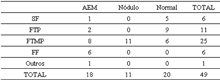
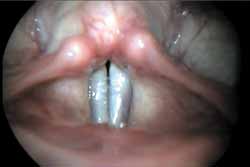
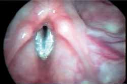
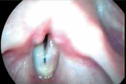
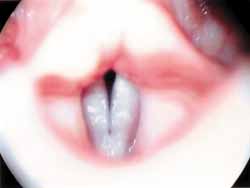
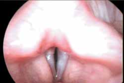
Laryngoscopy images were randomly edited in tapes and had no diagnostic identification. Two otorhinolaryngologists who were not involved in tape edition carried out images' evaluation. Glottal configuration during phonation of vowel / / was analyzed and classified into complete closure or without chink (SF) and incomplete closure or with chink (CF), identified according to site and shape, as follows: posterior triangular chink (FTP), triangular configuration and limited to intercartilaginous area (figure 1a), mid-posterior triangular chink (FTMP) of the same shape and length extending beyond the vocal process of arytenoids - i.e, reaching the vocal folds' membranous portion (figure 1b and 1d), which can be followed by anterior chink (figure 1c), secondary to nodular lesion of the mid-third area, and spindle chink (FF) (figure 1e). Other types of chinks, such as parallel, irregular, and double-spindle chinks were included in the group denominated "Others". A statistical analysis was conducted for group comparison, including the following tests:
- Test for two proportions (independent frequencies)
- Test for K proportions (independent frequencies)
- Chi-square test of adherence (dependent frequencies)
As only accurately diagnosed cases with long follow-up periods and consistent to admission criteria could be selected, the sample was small.
RESULTS
Incidence of each type of glottal closure observed under each specific pathological condition, as well as the control group, are shown in Table 1. Incidence of glottal closure may also be checked in Graph 1, where cases of posterior chinks - either posterior triangular or mid-posterior chinks - were grouped.
DISCUSSION
Study and evaluation of the larynx under certain physiological conditions and with high-quality imaging were possible due to technological improvement, which has enabled more accurate clinical diagnoses of vocal alterations and identification of small lesions, which were formerly not accessed by Garcia's mirror examination . However, there are still some difficulties that are inherent to the lesions and resultant from laryngoscopy use. Frequently, diagnosis is only determined or confirmed through surgical investigation or clinical analysis exam, particularly in MSA cases2,5,7,8,11. Clinical diagnosis is fundamental as to choose treatment among vocal therapy, surgery or clinical therapy. In this sense, any alteration is scrutinized during clinical examination; in case of a nodular-like lesion, determining whether it is a vocal nodule or a minor structural alteration is imperative. Videolaryngoscopy images of the lesion not always yield to a diagnostic decision and, therefore, other parameters are taken into consideration. One parameter includes the presence of enlarged vessels traveling transversally to the vocal fold, with sudden reductions or with a twisted or spring aspect9. Other minor structural alterations associated with vessels are called vasculodysgenesis, which are relevant in differential diagnosis of vocal nodule - considering its lamina propria is not altered and vessels are similar to those observed in larynges of patients with no vocal complaints or lesions9.
Glottal closure during phonation is always observed for diagnostic determination, as glottis configuration is strongly related to predisposition to certain benign lesions of larynx associated with the impact zone during adduction of vocal folds9,17,18. Presence of FTMP, with or without anterior opening, is a sign of important trauma at the triangle vertex during phonation, that is, the vocal nodule's site. This chink is observed in the presence of vocal nodule and may help eliminating suspected minor structural alteration, as reported in a study with adult population18.
Technological improvement has enabled more comprehensive evaluation of children's larynx and, although this kind of study is less frequent than with adults, they are relevant for characterization and understanding of the physiology and alterations typically observed in this age range20,21. Anatomical aspects of the larynx vary in consistence with gender and age2,4. One of the most outstanding difference is larynx size, which differs in adults according to gender2,4. The relative difference between membranous and intercartilaginous regions is subtle and is reflected by glottis proportion, which changes the impact zone during phonation. This aspect explains the differences observed between the incidence of vocal nodule and granuloma in both gender17,18.
At early age, there are no outstanding anatomical differences related to gender, while the membranous portion is relatively small19, leading to low values of glottis proportion, even when compared to values found in female adults15. Such anatomical configuration leads to frequent posterior opening of the larynx during phonation, resulting in 75% of glottal closure observed in our control group (Table 1), which is in accordance with the values reported by Crespo21. This fact explains the high incidence of vocal nodules in children, consistent with vocal behavior. However, when diagnostically confirmed cases of minor structural alteration are observed - once all cases refer to patients submitted to microsurgery of the larynx and therefore examined by direct microlaryngoscopy - it is evident that posterior openings are still very frequent. In fact, more than half of the cases of minor structural alterations maintained a non-lesion infantile larynx pattern - FTP or FTMP (Graph 1). Consequently, observation of the triangular chink during laryngoscopy does not seem to help in differentiating vocal nodule from minor structural alteration, as it occurs with adults, although FTMP is considered an aspect of a vocal nodule. However, FF was observed nearly in one-third of individuals presenting minor structural alterations (Table 1; Graph1); this chink does not cause trauma at the vocal nodule development site, but gives rise to a concavity in this region reducing the friction force during phonation. The presence of this chink is statistically significant in minor structural alterations cases as it determines diagnosis, while it is considered to be the result of histological changes involving fibrous proteins present in the vocal fold lamina propria, particularly collagenous fibers. In this sense, FF findings may express the occurrence of modifications during formation of lamina propria components observed in minor structural alterations, or even that a compensatory tension resulting from minor structural alterations can lead to posterior closure during phonation.
Concerning FTP, its higher incidence among regular larynx subjects is expected compared to the other analyzed groups, as in this condition there are no alterations in the mid-third region and no efforts to phonation.
CONCLUSIONS
The use of glottal closure mode during phonation in children as a diagnostic criterion to differ minor structural alteration of the vocal nodule from regular larynx is relevant when a spindle chink is found, as this condition is only observed in minor structural alterations; although posterior or mid-posterior triangular chinks are indicative of vocal nodule, they were not significant to differentiate minor structural alteration from regular larynx.
REFERENCES
1. Gray SD. Basement membrane zone injury in vocal - nodules. In: Gauffin J, Hammarberg B. Vocal fold physiology. Stockolm, Sweden: Singular; 1991:21-7.
2. Pontes P, Behlau M, Gonçalves MI. Alterações Estruturais Mínimas Da Laringe (AEM). Considerações básicas. Acta Awho 1994; 13 (1):2-6.
3. Hirano M., Yoshida T, Hirade Y, Sanada T. Improved Surgical Technique For Epidermoid Cysts Of The Vocal Fold. Ann Otol Rhinol Laryngol 1989; 98: 791-5.
4. Hirano M. Phonosurgical Anatomy of The Larynx. In: Ford CN, Bless DM. Phonosurgery: Assessment And Surgical Management Of Voice Disorders. New York: Raven Press; 1991. p.25-41.
5. Bouchayer M, Cornut G, Witzig E, Loire R, Roch JB, Bastian RW. Epidermoid Cysts, Sulci, And Mucosal Bridges of the true vocal cord: a report of 157 cases. Laryngoscope 1985; 1087-93.
6. Bouchayer M, Cornut G. Microsurgery for Benign Lesions of The Vocal Folds. Ear Nose Throat J 1988; 67: 446-66.
7. Monday LA, Cornut G, Bouchayer M, Roch JB. Epidermoid Cysts of Vocal Cords. Ann Otol Rhinol Laryngol 1983; 92: 124-7
8. Pontes P, Gonçalves MI, Behlau M. Vocal fold Cover Minor Structural Alterations: Diagnostic Errors. Phonoscope 1999; 2(4): 175-85.
9. Pontes PAL, De Biase NG, Behlau M. Vascular Characteristics Of The Vocal Fold Cover In Control Larynges and Larynges with Benign Lesions. Phonoscope 1999; 2(3): 129-35.
10. Forrest LA. Treatment of Benign Cysts and Tumors of The Larynx. Otolaryngol Head Neck Surg 1995; 3: 149-54.
11. Sataloff R, Spiegel J. Endoscopic Microsurgery. In: Gould WJ,. Sataloff RT, Spiegel JR. Voice surgery. St Louis, Mosby; 1993. p. 227-68.
12. Bless D, Hirano M, Feder, RJ. Videostroboscopic evaluation of the larynx. Ear Nose Throat J 1987; 66: 289-98.
13. Pinho S, Pontes P. Disfonias funcionais: avaliação otorrinolaringológica dirigida à fonoterapia. Acta Awho 1991; 10:34-7.
14. Pinho SMR. As fendas glóticas e a terapia fonoaudiológica. In: Ferreira LP. Um pouco de nós sobre voz. Carapicuíba, São Paulo: Pró-fono; 1993. p. 51-60.
15. Pontes P, Behlau M, Kyrillos L. Configuration et rapport glottic: um essai pour comprendre la fente glottique postérieure. Rev Laryngol 1994; 115(4): 261-6.
16. Murry T, Xu JJ, Woodson GE. Glottal configuration associated with fundamental frequency and vocal register. J Voice 1998; 12(1): 44-9.
17. Pontes P, De Biase N, Kyrillos L, Pontes A. Importance of glottic configuration in the development of posterior laryngeal granuloma. Ann Otol Rhinol Laryngol 2001; 110(8): 765-9.
18. Pontes P, Kyrillos L, Behlau M, De Biase N, Pontes A. Vocal Nodules and Laryngeal Morphology. J Voice 2002; 16(3): 408-14.
19. Behlau M, Pontes P. Exame Laringológico. In: Behlau M, Pontes P. Avaliação E Tratamento Das Disfonias. São Paulo, SP: Lovise, 1995. p 143-66.
20. Hirano H, Kurita S, Nakashima, T. Growth development and aging of human vocal folds. In: Bless D, Abbs, JH. Vocal Fold Physiology. San Diego: College-Hill; 1983. p.22-43.
21. Crespo NA. Coaptação glótica, proporção glótica e ângulo de abertura das pregas vocais em crianças. Tese de doutorado: Universidade Federal de São Paulo - Escola Paulista de Medicina. São Paulo, 1995.Table 1. Distribution of subjects studied, according to type of glottal configuration.
SF: without chink; FTP: posterior triangular chink; FTMP: mid-posterior triangular chink with or without anterior opening; FF: spindle chink; MSA: minor structural alteration.Graph 1. Distribution of subjects with no minor structural alteration, vocal nodule, and the control group, in percentage, according to type of glottal configuration.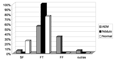
MSA, Nodule, Normal
SF: without chink, FT: posterior triangular and mid-posterior chinks, FF: spindle chink.Figure 1. Telelaryngoscopy during sustained phonation of vowel / / showing chinks.
a.Posterior triangular chink in minor structural alteration
b. Mid-posterior triangular chink in minor structural alteration
c. Mid-posterior triangular chink with anterior opening in minor structural alteration
d. Mid-posterior triangular chink in vocal nodule
e. Spindle chink in minor structural alteration
Print: ![]()