

Year: 2004 Vol. 70 Ed. 4 - (2º)
Artigo Original
Pages: 450 to 455
 PDF PT
 Unilateral vocal fold paralysis: association and correlation between maximum phonation time, position and displacement angle
Author(s):
Luciane M. Steffen1,
Maristela B. Moschetti2,
Nédio Steffen3,
Eliana M. Hanayama4
Keywords: Key words: larynx, vocal cord paralysis, voice, glottis.
Abstract:
Introduction: Vocal fold paralysis (VFP) is due to an injury of the vagus nerve or one of its branches and may cause dysfunctions in the glottic competence. The Maximum Phonation Time (MPT) is a test usually applied on dysphonic patients to assess glottic efficiency, mainly in patients with VFP and a decreased phonation time. The clinical classification of the VFP as median, paramedian, intermedian, abduction or cadaveric is controversial. Objectives: To check association and correlation between Maximum Phonation Time (MPT) with position and with the displacement angle of the paralyzed vocal fold (PVF), to measure the distal angle of the PVF in different positions from median line, correlating it with the clinical classification. Methods: Records of 86 PVF individuals were reviewed, videoendoscopic exams were analyzed and a computer program measured the distal angle of the PVF. Results: The MPTs for each position of paralyzed vocal fold have statistical significance only for /z/ in the median position. There is a relationship between the MPT of /i/, /u/ with PVF distal angle. Correlation and association of the displacement angle with clinical position demonstrate statistical significance when the PVF is in abduction. Conclusion: By the present study it was impossible to classify positions of the paralyzed vocal fold using either MPT or the displacement angle measurement.
![]()
INTRODUCTION
The injuries that impair the laryngeal nerves may occur at any point of the vagus nerve path, from medulla oblonga to the larynx ends and it may cause paresis or uni or bilateral1 vocal fold paralysis.
Under such conditions, the larynx capability of closing the vocal folds to produce phonation, cough, swallowing and some other activities that require glottic closing may be impaired2, 3..
Dysphonia, dysphasia, fatigue or problems to breathe or cough are clinical symptoms whose level of severity can be shown according to the level of glottic incompetence3.
Maximum Phonation Time (MPT) is obtained through a simple test, which is usually applied to dysphonic patients3, 4, aiming to assess glottic efficiency5, 6. To men we expected normal values of 20 seconds and to women, 15 seconds 4, 6,7. Therefore, these values can be a bit higher: 25 to 35 to men and 15 to 25 to women 7. The used vowels are /a/, /i/ and /u/ and the consonants /s/and /z7.
The importance of MPT is highlighted in the assessment of VFP5 cases, where values are lower 3-6, about four to five seconds8. As a result, it decreases efficiency of expiration air during phonation, and it requires new air reload every one or two seconds of emissions, causing fatigue and hyper ventilation8.
PVF positions have been vastly discussed and invariably, the studies that approach this topic relate that to laryngoscopic test findings9-16. There is controversy about the different positions of PVF. According to the position taken, they can be classified in median, paramedian, intermediary, abduction or cadaveric17. This is a classic description, usually used in the medical practice. However, there are reports10 that state it is impossible to standardize them, given that we should take into consideration the examiner's subjectivity. Further studies are suggested to determine precisely the PVF position, as well as the terminology used18.
Just few studies show quantitative and objective factors which are related and/or associated with VFP and are relevant in the decision of type of treatment, either surgical or therapeutic.
The present study aimed at measuring the glottic configuration in different positions of the paralyzed vocal folds and correlate them, as well as to associate Maximum Phonation Time with the position and displacement angle.
MATERIAL AND METHOD
Sample
Two hundred and two subjects were diagnosed in the clinic Dr Nédio Steffen with unilateral vocal fold paralysis from January 1990 to November 2002.
Exclusion criteria: post-surgical patients with vocal fold medialization, paralyzed falsetto, incomplete medical records and videolaryngoscopic images which did not allow visualization of the vocal folds.
There were 86 adults, 62 (72%) were men and 24 were women. Ages ranged from 18 to 88 years, median age of 53 years.
Data Assessment
Medical charts were reviewed and videolaryngoscopic tests analyzed.
MPT of the assessed subjects were registered by a speech pathologist in the medical charts. Digi chronometry was used to do that.
The selected vowels were a/, /i/, /u/ and the consonants /s/ and /z/.
The ratio s/z was the other phonation time measure used. It is MPT of consonant /s/ divided by the maximum time of sustained production of consonant /z/.
Laryngeal endoscopic test was carried out with rigid fibrolaryngoscope, with the following equipment: rigid telescope, with 70° degree angulation (MACHIDA< LY-C30; 150 watts of halogen light or xenon stroboscopic light with 300 watts power (KAY); Panasonic camera (model GP-KS 162HD); videorecorder Panasonic 4 heads (model AG-1730), and magnetic tape (videorecorder JVC and TDK).
In case the patient showed hyper reflexes, the examiner used a flexible fiberscope, which was introduced into the nasal fossa, previously anesthetized.
The test was conducted by an ENT specialist and taped in video, VHS system. The larynx was assessed during vowel /e/ emission, sustained by the patient as long as he could in a comfortable tone and normal loudness.
At this time, the image was sent to the computer through a video capture board.
Adobe Photoshop, version 6.0 was used to calculate glottis chink. In order to measure the angle, a line was drawn from the anterior commissure up to the vocal apophysis of the healthy vocal fold. After that, starting from the same place, another line was drawn up to the maximum opening of the paralyzed vocal fold.
All collected data were included in the analysis.
The statistical analysis was carried out with Pearson's coefficient test. Maximum phonation time was compared between the different paralyzed vocal fold position groups and by the analyses of variance based on Duncan test. In the bi-variance analysis, the level of significance (alpha) adopted was 0.05. In the multiple variance analysis the alpha level was = 0.010. Data were processed and analyzed with the help of Statistical Package for the Social Sciences Program, version10.0 (SPSS).
RESULTS
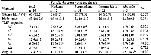
The described epidemiological data and the opening angle of the paralyzed vocal folds conducted through videoendoscopy in 86 patients with vocal fold unilateral paralysis are described in Table 1.
Table 1 shows that the findings of the correlation between the different PVF positions as well as age were: Median: 45.4 years (+/- 13.3); Paramedian: 53.1 (+/- 18.0); Intermediary: 56.6 (+/- 18.2); Abduction: 63. 3 (+/- 9.9), and were not statically significant.
The association and correlation between the measurements of MPT of the variables /a/, /i/, /u/, /s/, /z/ and the relation between the different positions taken by the PVF, can be seen in Table 1, where non-coinciding index-letters represent statistically significant difference in Duncan multiple comparison test.
The MPT of the 86 subjects with VFP in relation to the different positions were on average: /a/: 7.1 + 4.0; /i/: 7.3+ 4.7; /u/: 6.9+ 4.5; /s/: 12.4 +6.3; z: 6.7 +4.9 and the ratio s/z was 2.0 + 2.0.
MPT to each position taken by the PVF are shown in Table 1, where there was statistically significant relation to /z/ in the median position (13.3 + 8.5, P<0.001) and MPT in different positions is progressively reduced once the positions are more abducted.
The values of the s/z ratio (Table 1) are higher than 1.3 in the paramedian, intermediary and abduction positions, the median position s/z amounted to about 0.9+ 0.4.
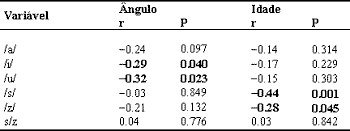
The association and correlation between the displacement angles of PVF are summed up in Table 2. MPT /i/, /u/ related with displacement angle of PVF (/i/: P= 0.040 and u/: P= 0.023), whereas for vowel /a/ and consonants /s/ and z/, there was no relation. In table 2, the correlation coefficients between MPT and age show that /s/ had statistically significant difference (P=0.001).
When correlating angle with position, it is seen in Table 1 that there was statistically significant difference only in abduction position (22.3+ 6.2). The paramedian and median positions showed very similar angle values (11.1+7.6 and 10.7+3.9, respectively).
DISCUSSION
The topic of PVF positions has been broadly debated and the different positions taken by the VFP9-16 are reported in the literature.
The glottic configurations adopted in the unilateral vocal fold paralyzes are influenced by several factors which interact simultaneously and in a complex way; in addition to that, there is the examiner's opinion. 19
It is considered to be too subjective 20 to only relate the vocal function with the glottis clinical exam in the horizontal position. This position does not show significant correlation with some of the vocal functions measurements, and it suggests that a combination of factors (such as age, pulmonary capacity, co-morbidity) can be predictors of the vocal20 function.
The MPT are short at VFP3-6. Colton and Casper 8 referred on average between four to five seconds, whereas in the current study there was an average of seven seconds to vowels. However, despite the fact that MPT depends on a number of factors, such as the performance effort of the subject, gender, age, height, emotional status, neurovegetative conditions, airflow and also his/her respiratory function, regardless of laringeal5, 21 aspects, MPT can be improved by means of manual compression of the larynx, leading to better glottic closure21, or surgery for medialization of paralyzed vocal fold 18.
The study shows in Table 1 that the results of the correlation between different positions of VFP and age are parallel variables, even though there was no statistical significance. The older the subject, the more enhanced is the abducted position. Such finding can be biased and even lead to future field research. However, it is important not to forget that anatomical and physiological changes also occur in the vocal folds with aging15. Tanaka22 noticed vocal fold bulging in subjects over 60 years of age showing no laryngeal pathology. Evidences of muscular atrophy and changes in the mucosa vibration due to changes in the superficial and intermediate layers were also highly reported in men.
The coefficient of correlation between MPT and age in Table 2 shows that /s/ had statistically significant difference and values of /z/ close to /s/.
The theory on consonant production explains that they have a friction source base, differently from vowels that have greater constriction in the vocal tract and reduction of resonance tube. The fricative consonants are produced when air goes through large month constriction, producing turbulence. They may be either voiced or voiceless. The /s/ sound is continuous and counts on the air friction through the vocal tract path. Friction duration is greater in voiceless sounds , e.g.in /s/, rather than in voiced sounds 23.
Based on the above theory and considering the anatomical and physiology changes to the vocal folds during aging15, 22, and since fricative sounds depend more on frictional source and not so much on the glottic one, as in the vowel case, it was expected in the present study that vowels would have greater MPT related to age. However, statistic significance was obtained for fricative consonants, especially /s/.
The elderly vocal production is not directly related only to the laryngeal changes, but also to a number of other factors. The MPT of /s/ is significant to age once this sound depends more on other sources than the larynx. MPT to each position taken by the PVF are shown in Table 1, and there was statistic significance only to /z/ in the median position.
There was correlation between VFP displacement angle for /i/, /u/, but not for vowel /a/ and consonant /s/ and /z/. Once the angle is measured from the image obtained during sustained emission of /e/ or /i/, there were no results shown to variables /a/, /s/ and /z/ which are not used during the laryngoscopic test.
MPT value lower than 10 seconds during vowel /a/emission is considered to be pathological7, 24. In the present study, the average MPT to /a/ is the only variable which shows values lower than 10 seconds, regardless of PVF position.
Production of /s/ is a measure of the expiratory control and /z/ sound uses the laryngeal component. Usually the voiceless fricative /s/ and the voiced fricative /z/ are sustained by a normal speaker for a similar period of time or /z/ is sustained for a bit longer. Therefore, there is a s/z ratio which ranges from 1 to 1.3 8,25,26.
The s/z ratio provides interesting data about the phonation dynamics and it has been considered a very reliable tool to assess glottic efficiency26. Values higher than 1.3 show lack of glottic coaptation 8, 25,26.
In the presence of glottic closing disorder, the duration of the sustained vocalization of /z/ changes. In Table 1, glottic inability is observed on s/z ratio, where values are higher than 1.3 in the paramedian, intermediary and abduction positions. These findings are similar to those in the literarture3-6. In median position, the s/z ratio values where about 0.9 +/- 0.4 8,25,26 and considered normal, which suggests that they are close to those individuals that have efficient glottic coaptation. It is common to see symptomatic unilateral vocal fold paralyzes when VFP is located in the median position. These are compatible findings.
The median and paramedian positions show very close angle values. Pinho and Pontes27 referred to the prevalence of the median position as 45.6% (21/46), whereas in this study the paramedian position was found in 59.3% (51/86), and only 11.6% (10/86) were present in the median position. Such close angle measurements, paradoxically larger than in the median position, represent the difficult to set an immediate, precise and subjective clinical diagnosis of median or paramedian position in unilateral vocal fold paralysis cases. A number of publications relate that the modification of the glottic configuration does not prove to have correspondence with the standard clinical classification of paralyzed vocal fold 10, 11,18. There is no consensus so far about the criteria used to characterize them.
Koufman, in order to define the VFP, had considered the distance between the vocal apophysis during phonation12. According to the study, the glottic gap to the paramedian position was inferior to 1.5mm, to the posterior closing commissure, intermediary: 1.5 to 2.5mm, with posterior closing or opening commissure and in abduction superior to 2.5mm. According to the author, the median position is confused with the paramedian and three positions are enough: paramedian, intermediary and lateral. Johns and Rood14 understand that only two classifications would be enough: paramedian and intermediary. We have not found any study that measured the angle of these different positions.
Even though there was no statistical significance, the intermediary and abduction positions pointed out to higher angle values.
In table 1, the angle is associated and correlated with the position, therefore there was statistically significance only in the abduction position. We point out that even though this numeric data is significant it can be a bias, given that the abduction position sample only represented three subjects. According to Koufman12, during abduction position, the glottic chink distance between the vocal process is higher than 2.5 mm. Therefore, it is very difficult to determine these measurements at the very precise moment of laryngoscopy. There are similarities between those measurements and illustrations of PVF positions published in textbooks and anatomy atlas.
CONCLUSIONS
The study of patients with Unilateral Vocal Fold Paralysis led to the conclusion that:
1. The association and correlation between the Maximum Phonation Time measurements of variables /a/, /i/, /u/, /s/, /z/ with the different positions taken by PVF showed statistical significance only to /z/ in the median position.
2. The association and correlation between the measurements of s/z ratio with the different positions taken by PVF showed values higher than 1.3 in the paramedian, intermediary and abduction positions.
3. The association and correlation between MPT and PVF displacement angle were related to vowels /i/ and /u/.
4. There was no association and correlation between angle measurements and the subjective clinical position of the paralyzed vocal fold.
REFERENCES
1. Tsuji DH. Paralisia das cordas vocais. In: Costa SS, Cruz OLM, Oliveira JAA. Otorrinolaringologia, princípios e prática. Porto Alegre: Artes Médicas; 1994; p.468-473.
2. Greene M, Mathieson L. The voice and its disorders. San Diego, California: Singular Publishing Group; 1991. p.293-307.
3. Crary MA, Glowaski AL. Vocal fold mobility. In: Brown WS, Vinson BP, Crary MA. Organic voice disorders assessment and treatment. San Diego, London: Singular Publishing Group; 1996. p.301-21.
4. Kent RD, Kent J, Rosenbek J. Maximum performance tests of speech productions. Journal of Speech and Hearing Disorders 1987; 52: 367-87.
5. Isshiki N, Okamura H, Morimoto M. Maximum phonation time and air flow rate during phonation: simples test for vocal function. Ann Otol Rhinol Laryngol 1967; 76: 998-1007.
6. Zemlin WR. Princípios de anatomia e fisiologia em fonoaudiologia. 4ª ed. Porto Alegre: Artmed; 2000.
7. Behlau M, Pontes P. Avaliação e tratamento das disfonias. São Paulo: Lovise; 1995.
8. Colton RH, Casper JK. Compreendendo os problemas de voz. Porto Alegre: Artes Médicas, 1996.
9. Dedo HH. The paralyzed larynx: an eletromyographic study in dogs and humans. Laryngoscope 1970; 80: 1455-517.
10. Woodson GE. Configuration of the glottis in laryngeal paralysis. I: Clinical study. Laryngoscope 1993; 103: 1227-33.
11. Woodson GE. Configuration of the glottis in laryngeal paralysis. II: Animal experiments. Laryngoscope 1993; 103: 1235-1241.
12. Koufman JA, Walker FO, Joharji GM. The cricothyroid muscle does not influence vocal fold position in laryngeal paralysis. Laryngoscope 1995; 105: 368-72.
13. Becker W, Naumann HH, Pfaltz CR. Larynx, hypopharynx and, trachea. In: Ear, nose, and throat diseases. 2nd. ed. New York: Georg Thieme; 1994. p.398-433.
14. Johns ME, Rood SR. Vocal cord Paralysis: diagnosis and management (a self instructional package). Washington: American Academy of Ophthalmology and Otolaryngology/Committee on Continuing Education in Otolaryngology, 1978.
15. Mamede RCM, Filho FVM, Entschev BM. A lei de Wagner-Grossman é aplicável nas paralisias da laringe? Rev Bras de Otorrinolaringologia 2000; 66(1): 52-57.
16. Hirano m, Noose I, Shin T et al. Electromyography for laryngeal paralysis. In: Hirano M, Kirchner JA, Bless DM, editors. Neurolaryngology: recent advances. Boston: Little Brown; 1987. p.232-48.
17. Furukawa M, Furukawa MK, Ooishi K. Statistical analysis of malignant tumors detected as the cause of vocal cord paralysis. ORL J Otorhinolayngol Relat Spec 1994; 56: 161-5.
18. Benninger MS, Crumley RL, Ford CN, Gould WJ. Evaluation and treatment of the unilateral paralyzed vocal fold. Otolaryngology - Head and Neck Surgery 1994; 497-508.
19. Riad MA, Kotby, MN. Mechanism of glottic in a model of unilateral vocal fold palsy. Acta otolaryngol 1995; 115: 311-3.
20. Inagi K, Khidr AA, Ford CN, Bless D. Correlation between vocal functions and glottal measurements in patients with unilateral vocal fold paralysis. Laryngoscope 1997; 107: 782-91.
21. Blaugrund SM, Taira T, Dren AE, Lin P, Isshiki N, Gould WJ. Effects of lateral manual compression upon glottic incompetence: objective evaluation. Annals of Otology, Rhinology and Laryngology 1990; 99: 248-55.
22. Tanaka S, Hirano M, Chijiwa K. Some aspects of vocal fold bowing. Ann Otol Rhinol Laryngol 1994; 103: 357-62.
23. Russo I, Behlau M. Percepção da fala: análise acústica. São Paulo: Ed Lovise; 1993.
24. Hirano M, Koile Y, Von Lenden H. Maximum phonation time and air usage during phonation. Folia Phoniatr 1968; 20: 185-201.
25. Boone DR, MacFarlane SC. A voz e a terapia vocal. Porto Alegre: Artes Médicas; 1994.
26. Eckel FC, Boone DR. The S/Z ratio as an indicator of laryngeal pathology. J Speech Hear Disord 1991; 46: 147-9.
27. Pinho SMR, Pontes PAL, Gadelha ME; Biasi N. Vestibular vocal behavior during phonation in unilateral vocal fold paralysis. Journal of Voice 1999; 13: 36-42.Table 1. Comparison of demographic variables, maximum phonation time and vocal fold displacement angle according to the diagnosis of paralyzed unilateral vocal fold.
M: male, TMF: maximum phonation time, P: statistical significance. Data are presented as mean and standard deviation or frequency (%).? Analysis of variance, non-coinciding index-letters represent statistically significant differences in the Duncan multiple comparison test .Table 2. Correlation coefficients between maximum phonation time and vocal fold displacement angle and aging in patients with unilateral vocal fold paralysis.
r: Pearson, coefficient of correlation, P: statistical significance.
Print: ![]()