

Year: 2004 Vol. 70 Ed. 3 - (20º)
Relato de Caso
Pages: 419 to 422
 PDF PT
 Wegener's granulomatosis and subglottic stenosis: case description
Author(s):
Fernanda Guidolin 1,
Carlos Eduardo Magro 2,
Francisco Bezerra Neto 3,
Thelma L. Skare 4
Keywords: Wegener granulomatosis, subglottic stenosis, Anca-c
Abstract:
We describe a patient with Wegener Granulomatosis being treated with steroids and cyclofosfamide that developed progressive dyspnea and hoarseness despite of treatment. Investigation showed a subglotic stenosis. The dyspnea was relieved by tracheotomy. We discuss the differential diagnosis as well as a number of treatment modalities in patients with Wegener granulomatosis and subglottic stenosis.
![]()
INTRODUCTION
Wegener's granulomatosis (WG) is an inflammatory multi-systemic disease characterized by small and middle vessel vasculitis with formation of granulomas and tissue necrosis 1.
Among the most common malformations, this disease has involvement of upper and lower airways and kidneys 1. However, many times WG can present only complications directed purely to the upper airways 2. Therefore, it is essential that specialists in this area, who are not in constant contact with rheumatology, be aware of these possibilities to make early diagnosis of this pathology and also to provide the necessary support to possible complications.
LITERATURE REVIEW AND DIFFERENTIAL DIAGNOSIS
WG is a necrotizing vasculitis that is relatively rare. The diagnosis of the pathology is made by the criteria proposed by the American College of Rheumatology, which are shown in Table 1. The recognition may be difficult in cases in which involvement is limited to the upper airways. In these situations, ANCA-c (antineutrophil cytoplasmic antibody) can support diagnosis. The antigen for which this antibody is directed is a lissosomic enzyme - proteinase 3 - PR3 and its presence is detected in 90% of classical Wegener's granulomatosis cases, even though in less typical situations, positivity can be lower 1.
Tracheal stenosis is present in 17% of the cases of WG 1, being that this complication is more common in female 4 young 5 patients. A study conducted by Rottem et al.5 considered that tracheal stenosis is the manifestation that allows clinical distinction between WG in adults and in children.
Subglottic stenosis should be considered in all patients with Wegener's granulomatosis that have increase in dyspnea, cough and vocal affections. The presence of audible wheezing can help detection, but it is not always present. This complication can present a wide range of clinical manifestations that are variable and range from asymptomatic form to cases in which severe dyspnea is life threatening.
The cases of tracheal stenosis in patients with Wegener's granulomatosis are described invariably as subglottic cases 6, and they tend to show circumferential narrowing 7. The reason for this preference in location is unknown, but this is the narrowest part of the trachea and it certainly produces symptoms quite easily, which are quickly identified. Moreover, it is believed that this portion of the trachea has the poorest tracheal blood supply, and consequently, it shows more difficulties to recover after any damage 6. Another possibility to explain the preference is that this region is exposed to larger amounts of inhaled antigens 6.
The cases of patients that presented only tracheal stenosis and positive ANCA-c were described as holders of the simple disease form 2, 8, 9. However, it is always important to perform careful differential diagnosis. Other causes of subglottic stenosis include sarcoidosis, recurrent polychondritis, trauma after intubation, amyloidosis, tuberculosis, lymphoma, and there are also idiopathic cases 10.
In patients with WG, subglottic stenosis presents some interesting characteristics. It is quite common to manifest as an isolated form, without other manifestations of disease activity 11. In most situations, it responds to treatment of vasculitis, which is made with corticoid and cyclophosphamide 4, 11. Moreover, it is relatively common to have onset of complication during the treatment with these drugs 11.
Treatment of stenosis is made with dilations, resections with carbon dioxide laser, use of intralesion injections of corticoid and laryngotracheal reconstructions 4.
In a study conducted by the National Institute of Health (NIH), with 128 patients with WG diagnosed in 24 years, they found a percentage of subglottic stenosis of 16% in the patients (25 patients). In this study, about 13% of the cases were irreversible (that is, they did not respond to treatment). Only 3% were reversible. About 13 out of 25 patients required tracheostomy at some point in treatment. In the same series, all subjects submitted to carbon dioxide laser treatment to resect the lesion developed recurrent forms of fibrosis, requiring alternative ways to treat it more definitely, showing that this option should be reserved as second best 4.
Table 1. Diagnostic Criteria by American College of Rheumatology for Wegener' Granulomatosis, 199012
1. Nasal or oral inflammation (oral ulcers, painful or not, or nasal bleeding discharge);
2. Abnormal chest X-ray (nodules, fixed or cavity infiltrates);
3. Abnormal urinary sediment (micro hematuria or hematic cylinder);
4. Granulomatous inflammation in biopsy (histology should show granulomatous inflammation on the arterial wall, perivascular or extravascular region of arteries or arterioles).
The patients should have at least 2 out of 4 criteria present. The presence of 2 or more criteria conveys 88.2% sensitivity and 92.0% specificity.
CASE REPORT
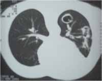
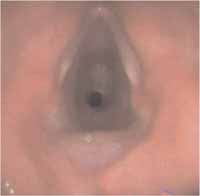
Female 31-year-old patient, with diagnosis of Wegener's granulomatosis since February 2001, presenting lung cavitations associated with recurrent infections of upper airways (Figure 1) and proliferative glomerulonephritis compatible with ANCA-related small vessels angeitis, detected at renal biopsy. At the time of diagnosis, she had positive ANCA-c, titer of 1:20. The initial picture was complicated by renal failure requiring dialyses to treat it, which was made with peritoneal dialysis. She was treated with oral cyclophosphamide (dose of 2mg/Kg/day) and high doses of corticoid (1mg/Kg/day prednisone) with remission of renal activity and improvement of respiratory condition. Dialysis was suspended and she remained with creatinine depuration of about 50ml/min. After disease control, we started to gradually suspend prednisone and maintained cyclophosphamide. Six months later, when the corticoid dose was about 20mg/day, patient had a flare up and presented progressive dyspnea, without response to bronchodilators. She complained of associated hoarseness. The exam revealed stridor in the cervical region, intercoastal retraction in inspiration and symmetric reduction of vesicular murmur. Chest x-ray showed tracheal narrowing, which was confirmed by videolaryngoscopy with fibrotic subglottic stenosis of 50% (Figure 2). She was submitted to tracheostomy to improve dyspnea and normalization of lung auscultation immediately after the procedure. After tracheostomy, the patient developed bronchial infection and was treated with antibiotics. She is currently waiting for definite tracheoplasty.
Figure 1. Chest CT scan showing cavitated nodules.
Figure 2. Videobronchoscopy showing circumferential subglottic stenosis.
DISCUSSION
In the case described, WG presented in a progressive and quickly form severely affecting vital organs such as the lungs and kidneys. Despite the severe initial progression, the appropriate therapy led to prompt remission, and the patient, who was already treated with dialysis, experienced recovery of renal function. When the patient presented remission of vasculitis, she had dyspnea, which was initially mistaken by recurrence of the lung underlying disease. Thanks to the exams, it was clarified that it was subglottic stenosis.
This occurrence of stenosis in the quiescence period of vasculitis, as demonstrated by the literature, justified the lack of response to underlying therapy. In this situation, an increase in corticoid dose and cytostatic drugs can hinder the progression of the patient because it favors infections. A possible explanation for this imbalance is the fact that stenosis represents a scarring fibrotic process referring to cure of previous vasculitic lesion.
CONCLUSION
Subglottic stenosis in WG is a rare complication of a relatively common disease. However, its knowledge can be essential to allow not only recognition of the disease, but also appropriate management of these patients, who are affected by severe and potentially fatal disease.
REFERENCES
1. Valente RM, Hall S, O´Duffy JD, Conn DL. Vasculitis and related disorders. In Kelley WN, Harris Jr ED, Ruddy S, Sledge CB. (Eds), Textbook of Rheumatology, 5th Ed. Philadelphia: WB Saunders; 1997. p.1079-122.
2. De Vries N, Gans ROB, Donker AJM, Goldschmeding R, Hoorntje SJ, Snow GB. Autoantibodies against constituents of neutrophils in diagnosis and treatment of isolated subglottic stenosis. Arch Otolaryngology Head Neck Surg 1992; 118:1120-3.
3. Daum TE, Specks U, Colby TV et al. Tracheobronchial involvement in Wegener's granulomatosis. Am J Resp Crit Care Med 1995; 151:522-6.
4. Lebovicz RS, Hoffman GS, Leavitt RY et al. The management of subglottic stenosis in patients with Wegener Granulomatosis. Laryngoscope 1992; 102: 1341-5.
5. Rottern M, Fauci AS, Hallahan CW et al. Wegener granulomatosis in children and adolescents: clinical presentation and outcome. J Pediatr 1993; 112: 26-31.
6. Ross CN, Tam FWK, Winter RDJ, Pusey CD, Rees AJ. Anti neutrophil cytoplasmic antibodies and subglottic stenosis (letter). Lancet 1990; i (335): 1231-2.
7. Yumoyo E, Sacki K, Kadota Y. subglottic stenosis in Wegener's granulomatosis limited to the head and neck region. Ear, Nose &Throat J 1997; 76 (5):571-4.
8. Gaughan RK, Sesanto LW, McDonald T. Use of anticytoplasmic autoantibodies in the diagnosis of Wegener's Granulomatosis with subglottic stenosis. Laryngoscope 1990; 100: 561-3.
9. Hoare TJ, Evans PHR. Anti-neutrophil cytoplasmic antibody assay in diagnosis of recurrent subglottic stenosis. (letter) Lancet 1988; ii: 1360.
10. Hoare TJ, Jayne D, Evans PHR, Croft CB,Howard DJ. Wegener's granulomatosis, subglottic stenosis and antineutrophil cytoplasm antibodies. J Laryng Otol 1989; 103: 1187-91.
11. Langford CA, Sneller MC, Hallahan CW et al. Clinical features and therapeutic management of subglottic stenosis in patients with Wegener's granulomatosis. Arthritis & Rheum 1996; 39 (10): 1754-60.
12. Leavitt RY, Fauci AS, Bloch DA, Michel BA, Hunder GG, Arend WP, et al. The American College of Rheumatology 1990 Criteria for the Classification of Wegener's Granulomatosis. Arthritis Rheum 1990; 33: 1101-17.
1 Resident Physician in Rheumatology, HUEC.
2 Resident Physician in Rheumatology, HUEC.
3 Preceptor, Service of Residence in Rheumatology, HUEC.
4 Professor of Rheumatology, Medical School, Faculdade Evangélica do PR.
Service of Rheumatology, Hospital Universitário Evangélico de Curitiba (HUEC)-PR.
Address correspondence to: Thelma L Skare Rua João Alencar Guimarães, 796 Curitiba PR 803110-420
Tel (55 41) 274-1659 E-mail: tskare@onda.com.br
Print: ![]()