

Year: 2004 Vol. 70 Ed. 3 - (5º)
Artigo Original
Pages: 316 to 322
 PDF PT
 Comparative study between two tonsillectomy techniques: Ultracision harmonic scalpel and traditional dissection with cold scalpel
Author(s):
Fernando A. Ramos 1,
Roberto D. P. Ferreira 2,
Rubens H. da Silva 2,
Eloísa P. do Prado 2,
Renato J. Corso 2,
José Antonio Pinto 3
Keywords: tonsillectomy, Ultracision, surgical time, post operative pain.
Abstract:
Tonsillectomy is the most commom surgical procedure in Otolaringology. The modifications and the technique surgical evolutions aim to simplifly and reduce the complications. The UltacisionÒ harmonic scalpel begin to be used in 1999 in tonsillectomies with good outcomes. Aim: To compair the time of the procedure, bleeding and the need of trans operative hemostasis, post operative pain, healing aspect of the tonsilar fossa and complications in patients submitted to tonsillectomy with cold and ultrassonic scalpel. Study design: Transversal cohort. Material and Method: Twenty six patients underwent tonsillectomy: 13 using the traditional thecnique with cold instruments and 13 using the ultrassonic one. They were evaluated with a standart protocol. The post operative pain were graduated through the horizontal visual scale analogue. Results: Surgical time were shorter with the ultrassonic thecnique compared to the traditional cold instruments. The amount of stitch at tonsilar fossa was lesser than the traditional thecnique. There were no post operative statistical difference in pain and in the aspect of the tonsilar fossa. Conclusion: The ultrassonic scalpel is an excellent choice in surgeries where surgical time and transoperatory bleeding are important.
![]()
INTRODUCTION
The surgery to remove the palatine tonsils is one of the most ancient in the world, with reports dating as back as 3 AD, made by Cornelius Caesus.1
Many tonsillectomy techniques have been described, among which cold dissection, guillotine dissection, bipolar dissection, laser dissection and bipolar scissor dissection 2.
Tonsillectomy remains as the most common surgery in Otorhinolaryngology, and all those technological advances have aimed at lower morbidity in the procedures.
Currently, the indications for tonsillectomy are well defined and well divided into absolute and relative. The absolute indications are palatine tonsil hypertrophy with upper respiratory obstruction, sleep obstructive apnea/hypoapnea syndrome, suspicion of malignancy, hemorrhagic tonsillitis, failure to thrive, language abnormalities, difficulty to eat, poor dental occlusion and orofacial growth abnormalities. The relative indications are: recurrent tonsillitis, tonsillitis associated with systemic diseases (carriers of group A beta-hemolytic Streptococcus), peritonsillar abscess, and halitosis by caseous tonsillitis 3-6.
Ultracision scalpel or harmonic scalpel is indicated for soft tissue incisions, when we intend to control hemorrhage and minimize thermal lesions, complementing or replacing electrocautery, laser or even cold scalpel.
Procedures in the areas of Gynecology, Urology, Thoracic Surgery, and Laparoscopy have already used harmonic scalpel for some years 2.
Its use was successfully introduced in Otorhinolaryngology in 1999 for microscopic tonsillectomy. The scalpel has vibration mechanical energy of about 55.5 kHz and acts to promote cut and coagulation and depends on level of power (1 to 5), type of technique, tissue fraction, and pressure of the blade.
The dissecting hook is the standard for tonsillectomies. In general, the instrument has a blunt tip that provides better coagulation and slow cut. The amount of energy provided to tissues and the effects on them depend on different factors, such as level of selected power, characteristics of the blade, tissue tension, type of tissue, pathology and surgical technique 7. The greater the power, the greater the vibration, and consequently, the larger the cutting area 2.
The use of Ultracision scalpel is an innovation in surgical treatment of palatine tonsils. The present study demonstrated its use, advantages and disadvantages, comparing it to traditional dissection with cold scalpel, whose main parameters were surgical time, transoperative bleeding and hemostasis, pain pattern and aspect of postoperative tonsillar site, and trans and postoperative events.
MATERIAL AND METHOD
We operated on 26 patients randomly divided into 2 groups: 13 were submitted to the coagulation hook blade, Ultracision (Group 1) and 13 were submitted to dissection technique with cold blade scalpel and detacher aspirator (Group 2). Minimum and maximum ages were, respectively, 3 to 20 years. The age of patients in group 1 ranged from 4 to 16 years (mean age of 6.53 years) and in group 2 it ranged from 3 to 20 years (mean age of 7.5 years). Six of the 13 patients operated by both techniques were male and 7 were female (Table 1). Tonsil hypertrophy with upper respiratory obstruction and sleep apnea syndrome were the inclusion criteria. The exclusion criteria were recurrent tonsillitis, previous peritonsillar abscess, and chronic caseous tonsillitis. All patients were assessed and included in the study, none of them had to be excluded.
The surgeries were performed in the following hospitals: Hospital Geral e Maternidade Tereza Ramos and Hospital Nossa Senhora dos Prazeres, both in the city of Lages, Santa Catarina. The study was approved by the medical ethics committee of both hospitals. Ultracision or harmonic scalpel, produced by Johnson & Johnson, was offered by Ethicon Endo-Surgery for the study. The scalpel has vibration mechanical energy with frequency of about 55.5kHz and acts by cutting and coagulating, depending on level of power (1 to 5), type of technique, fraction of tissue, and pressure of blade. The instrument was sterilized and used only for one patient since it comprises a titanium blade with non-removable sheath. The 5mm instruments are available with three types of blades: dissecting hook, sharp dissecting hook, and curved blade. All blades are 14cm long2, 7. We adopted tonsillectomies with dissecting hook as the standard practice.
All surgeries were conducted by the same surgeon, and there was a period of specific training (learning curve) to develop the technique with Ultracision. The surgeon conducted a series of 10 surgeries with the technique at Hospital e Maternidade São Camilo, in Sao Paulo, together with the team of Núcleo de Otorrinolaringologia e Cirurgia de Cabeça e Pescoço de Sao Paulo, and the technicians responsible for maintaining the device, trained by the manufacturer.
The following surgical parameters were analyzed through previously defined protocol: surgical time, trans and postoperative bleeding, hemostasis, pain pattern and aspect of postoperative tonsil site, medication and trans and postoperative events (Figure 1).
All patients received orotracheal intubation under inhaling general anesthesia. The duration of each surgery was calculated as of the onset of the first incision, regardless of the technique, up to the moment in which hemostasis was satisfactory, through suture or simple compression of the site with gauze soaked in sterile solution. After dissection of the left tonsil, we conducted adenoidectomy with Beckmann's curette, if so indicated. Orotracheal tube was placed on the other side. After exposure of right palatine tonsil, we started incision by counting again surgical time for this side. Hemostasis was conducted, whenever necessary, by compressing gauze for a period of approximately 2 minutes and with simple Categut 2.0 and 2cm needle.
In order to compare the techniques, we applied a previously defined protocol to all children operated by both techniques. We assessed through the protocol surgical time and hemostasis on both sides, considering the need to suture each site. The statistical analysis was considered as total time, that is, the sum of times from both sides. If there were events during the surgery, they were reported separately.
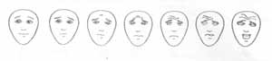
The first postoperative assessment was conducted in the post-anesthetic recovery room, in which we analyzed the presence of bleeding and pain. Second postoperative assessment was made about 10 hours after the surgery, upon hospital discharge. We assessed pain intensity using the Visual Analog Scale (VAS) and the aspect of tonsil sites. Horizontal VAS consisted of linear drawings of 7 faces, 1 neutral face and 6 faces that expressed increasing pain sensation. The faces were organized in increasing order from 1 to 7, from the left to the right - no pain (score 1) on the left and maximum pain (score 7) on the right. The subjective score was equal to the numbered scale, ranged from 1 to 7 (Figure 2).
Hospital discharge was preceded by instructions given to parents about administration of scales, diet, use of standardized drugs and relative rest.
Patients were instructed to come back on 4th or 5th postoperative day for the first assessment, and the second visit was scheduled for day 14th after surgery. Postoperative follow-up of all patients was made by the same surgeon. In each visit, VAS was analyzed and ENT examination was conducted, always questioning the presence of unexpected events.
Visual analog scale (VAS)
First of all, we tested preoperative understanding of pain in VAS. We asked the patients and/or accompanying people which face demonstrated the most and the least pain.
Next, VAS was presented to the patient upon hospital discharge, and he/she was asked to show the point in the pain scale that better represented his/her painful sensation in the oropharynx, and we checked whether the patient and the accompanying person had understood by assessing pain intensity (Figures 2 and 3).
They were instructed to fill out the scale daily, right after getting up, without administration of analgesics for the first 10 days.
The patients were assessed on day 4 or 5 postoperative after the surgery and they took their VAS from the period and came back at least one time for the assessment.
Postoperative evolution
The aspect of tonsillar site was assessed according to the protocol, through isolated analysis of right and left sites and considering the following aspects: presence of edema, fibrin, clot, dry tonsillar site, and focal or diffuse bleeding. The first assessment of the region was made upon hospital discharge.
In each new visit, we applied the protocol through which we could assess VAS score and conducted physical ENT examination, assessing the aspect of the site. We also assessed whether there were any episodes of nausea and/or vomiting in the meantime, reflex otalgia or headache and we questioned them about the use of standardized analgesics and their efficacy.
As to postoperative analgesics, we standardized Paracetamol, one drop per Kg of weight (solution of 100 mg/ml) if below 40 kg, or one 750mg-pill if over 40 Kg, up to QID. In case of fever (temperature equal or greater than 37.8o C), we instructed them to use similar drugs and administration routes.
All patients were instructed to follow postoperative care (avoid physical exercise, exposure to heat and warm showers) required to prevent external factors from affecting postoperative evolution. We introduced liquid, paste, warm or cold diet until the first assessment.Figure 1. Postoperative assessment protocol.
Figure 2. Horizontal visual analog scale.
Figure 3. Visual analog scale used on the 4th postoperative day by a child submitted to tonsillectomy with harmonic scalpel (Ultracision).
RESULTS
Out of 26 patients included in the study, only one operated with Ultracision was submitted to isolated tonsillectomy. All the others underwent adenotonsillectomy.
Power 2 was used in 77% of the patients operated with harmonic scalpel. For all the others, we used power 3.
The mean time of surgery for the technique with Ultracision (Group 1) was 14.92 minutes, with standard deviation of 3.38, and mean time of surgery with cold scalpel (group 2) was 25.46 minutes, standard deviation of 3.82. There was statistically significant difference between the surgical times by t Students' analysis with p<0.05 (Table 2).
There was bleeding during surgery in only 15.4% of the patients in group 1, which required hemostasis with simple categut 2.0. All patients in Group 2 required hemostasis with the same thread.
As to postoperative assessment during post-anesthetic recovery, there were no bleeding episodes. Pain was present in 30.8% of the patients in group 1 and in 7.7% of patients in group 2.
All patients were discharged 12 hours after the surgery. The assessment upon hospital discharge showed the following results concerning tonsillar site aspect: edema was present in 92.3% of patients in group 1 and in 61.5% in group 2; 100% of the patients in group 1 and 23% of the patients in group 2 had fibrin; no patients in both groups had bleeding in this period; one patient (7.7%) in group I and 5 (38.5%) in group 2 had clots in the tonsillar sites. Only one patient in group 1 had some complication event, which was an episode of vomiting (Table 3).
The results of the assessment on postoperative days 4 and 5 were the following: 69.24% of the patients in group 1 and 30.8% of the patients in group 2 presented edema. All patients in group 1 and 46.1% of the patients in group 2 had fibrin in the period. Only one patient (7.7%) in group 1 referred bleeding, but none in group 2. One patient in each group presented clots on the sites. Five patients in group 1 (38.5%) and 2 patients in group 2 (15.4%) referred some type of event (reflexive otalgia, nausea and vomiting) (Table 4).
As to assessment on postoperative days 9, 10 and 11, 38% of the patients had mild edema in group 1, and 7.7% presented edema in group 2; 69% of the patients in group 1 and 46% of the patients in group 2 had fibrin on the site. No patients referred bleeding. One patient in group 1 presented clots on the sites. Three patients (23%) in group 1 referred some kind of event (otalgia). No complication events were reported by group 2.Table 1. Demographical data.
Note: no statistically significant difference with t Student's test and significance level of 5%.
Table 2. Mean and standard deviation surgical time for both techniques.
Note: there was statistically significant difference with t Student's test with significance level of 5%.
Table 3. Mean and standard deviation of pain after hospital discharge.
Note: p=0.08
Table 4. Mean and standard deviation of pain on day 4 and 5.
Note: There was no statistically significant difference with t Student's test with significance level of 5%.
DISCUSSION
Discussions on the advantages and disadvantages of different tonsillectomy techniques are frequent 11, 12.
In a sample of 26 patients submitted to tonsillectomy using harmonic scalpel technique, D'Ávila reported that duration of surgical act for each tonsil was 14 minutes. There was excellent control of transoperative bleeding in almost all vessels of the tonsillar region. Early and late postoperative care did not show any complications. There was no bleeding. Postoperative pain was a symptom not frequently reported by this group of patients 13.
Sood et al, in a study with 158 palatine tonsillectomies in 59 patients using harmonic scalpel, found a statistical difference concerning duration of surgical procedure and volume of transoperative bleeding in comparison to dissection using cold scalpel 2.
Our study also showed a significant difference between the techniques concerning surgical time. The mean time of surgery by harmonic scalpel was 14.92 minutes and the mean time by traditional dissection technique with cold scalpel was 25.46 minutes.
There was no statistically significant difference concerning age and gender of operated patients in each group and pain, a fact that could have been a discrepancy, since the studies show different pain patterns in different age ranges 12.
The assessment of postoperative pain through the use of visual analog scale (VAS) is a widely used method to assess intensity and subjective pain sensation 8. VAS can be vertical or horizontal, according to the line of orientation. Comparative studies of vertical and horizontal orientation of VAS showed controversial results 9. In 1998, Breivik et al. demonstrated similar sensitivity between vertical and horizontal VAS for pain assessment after oral cavity surgery.
Pain assessment in small children is widely discussed concerning reliability of factors such as expression and language. VAS can show pain assessment in patients aged younger than 7 years. Children below this age range completely understand the mechanism. Facial pain scale applied in this study can be safely used in younger children concerning pain understanding and conception 10.
Patients with Alzheimer's disease have difficulties of expression and language that are similar to young children. In a study conducted with Alzheimer's patients with mild to moderate disease it was observed that this assessment model was a safe tool to assess intensity and affection of pain. In patients that completely understand the purpose of facial pain scale, pain can be easily assessed. The same can be said about children in which the scale is applied 10.
As to the Sood et al. study, the authors stated that they were amazed to see absence of early postoperative pain, since most of the patients could eat within the first hours after the procedure. However, pain in subsequent days was not statistically different from that caused by cold scalpel procedure 2. The absence of marked early postoperative pain as well as shorter surgical duration justified the use of the device, since its operational cost is much higher than that of cold instrument dissection, but earlier hospital discharge minimizes hospital costs 2.
In the present study, early postoperative pain (post-anesthetic recovery) was present in group 1 (30.8% of the cases) and was greater than in group 2 (7.7%).
Power 2 was used in most of the patients submitted to tonsillectomy with harmonic scalpel (77%), since the lower the power, the higher the coagulation potential and the smaller the cut. Surgery conducted at this power proved to be more efficient than dissection of palatine tonsil, since it allowed greater safety in relation to hemostasis both trans and postoperatively. Out of patients that needed suture for hemostasis during surgery, half of them were being operated in power 3.
The shorter surgical duration with harmonic scalpel technique can be related with the fact that this technique allows cut and coagulation at the same time, being that bleeding is minimal compared to traditional technique. As a result of reduced bleeding, only 15% of the patients operated by this technique required suture of tonsils for hemostasis, saving a considerable amount of time to finalize the surgery.
CONCLUSION
Harmonic scalpel presents advantages in relation to traditional dissection technique with cold instruments, such as simultaneous cut and coagulation, optimizing surgical time. Other positive factors include:
No use of electrical current (no risk of burns);
Minimum tissue carbonization;
Easy installation and handling of device.
We observed that there has been no significant difference concerning pain standard and postoperative evolution (scarring aspect of tonsil site and postoperative complications). The shorter surgical time and the shorter use of surgical room, less need for tonsil site suture for hemostasis and good postoperative evolution demonstrate that it is a safe and effective technique, since it minimizes hospital cost, being capable of replacing traditional cold scalpel dissection techniques.
ACKNOWLEDGEMENT
We would like to thank Dr Fabio Eduardo Festugato for the conduction of the statistical analyses and Mariel Wahrhastig, Gabriela Dias and José Eduardo Passos with Johnson & Johnson, which provided Ultracision device and accessories.
REFERENCES
1. Ramos CM, Gonçalves EMR, Mendonça RR, Gualandro DM. Tonsilectomia: Técnica de dissecção X Técnica de Sluder. Rev Bras Otolaringol 2001; 67(2):229-32.
2. Sood S, Corbridge R, Powles J, Bates G, Newbegin CJR. Effectiveness of the ultrasonic harmonic scalpel for tonsillectomy. ENT - Ear, nose & Throut Journal 2001August: 514-8.
3. Kavanagh TK, Beckford SN. Adenotonsillectomy in children: Indications and Contraindications. Southern Medical Journal 1988; 81(4): 507-11.
4. Ahlqvist-Rastad J, Hultcrantz E, Svanholm H. Children with Tonsilar Obstruction: Indications for and Efficacy of Tonsillectomy. Acta Paediatr Scand 1988; 77: 831-5.
5. Suen SJ, Arnold EJ, Brooks JL. Adenotonsillectomy for treatment of Obstructive Sleep Apnea in Children. Arch Otolaryngol Head and Neck Surg 1995; 121: 525-30.
6. Almeida ER, Campos VAR - In Campos CAH, Costa HOO. Tratado de Otorrinolaringologia. 1a edição. São Paulo: Ed Rocca; vol 3: 248-52, 2002.
7. Fenton RS, Long J. Ultrasonic Tonsillectomy. The journal of Otolaryngol 2000: 29(6).
8. Breivik EK, Skoglund LA. Comparison of Present Pain Intensity Assessments on Horizontally and Vertically Oriented Visual Analogue Scales. Meth Find Exp Clin Pharmacol 1998; 20(8): 719-24.
9. Sriwantanakul K, Kelvie W, Lasagna L. The quantification of pain: An analysis of words used to describe pain and analgesia in clinical trials. Clin Pharmacol Ther 1982; 32: 143-8.
10. Scherder EJA, Bouma A. Visual Analogue Scales for Pain Assessment in Alzheimer's Disease. Gerontology 2000; 46: 47-53.
11. Monteiro E. Amigdalectomia: dissecção x sluder. Rev Bras Otolaringol 2001; 67(3): 319-24.
12. Sant'Anna GD. Dor pós-tonsilectomia: comparação entre pacientes com diferentes idades. Revista Brasil Orl 2000; 66(2): 123-7.
13. D'Avila JS. Microcirurgia de tonsilas com bisturi Ultracision. Arq Fund Otorrinolaringol 2001; 5(4).
1 Otorhinolaryngologist, Post-Graduation studies under course, Discipline of Otorhinolaryngology, Hospital das Clínicas, Medical School, University of Sao Paulo USP - Ribeirão-Preto - SP.
2 Resident Physicians, Núcleo de Otorrinolaringologia e Cirurgia de Cabeça e Pescoço de Sao Paulo.
3 Director, Núcleo de Otorrinolaringologia e Cirurgia de Cabeça e Pescoço de Sao Paulo, Head of the Service of Otorhinolaryngology, Hospital e Maternidade São Camilo - Sao Paulo.
Study conducted at Clínica Dr. Fernando Arruda Ramos, together with Núcleo de Otorrinolaringologia e Cirurgia de Cabeça e Pescoço de Sao Paulo.
Study presented at 36o Congresso Brasileiro de Otorrinolaringologia, Florianópolis - Santa Catarina - SC.
Address correspondence to: Fernando Arruda Ramos - Rua Lauro Muller, 547 2o andar, Lages Santa Catarina SC 88501130
Tel/Fax (55 49) 222-9165 - E-mail: arrudaramos@matrix.com.br


Print: ![]()