

Year: 2004 Vol. 70 Ed. 3 - (4º)
Artigo Original
Pages: 310 to 314
 PDF PT
 Oral candidiasis and hairy leukoplakia as progression markers of hiv infection in Brazilian patients
Author(s):
Ivan Dieb Miziara 1,
Adriana da Silva Lima 2,
Rodrigo Antonio Cataldo de la Cortina 2
Keywords: HIV, AIDS, oral candidiasis, hairy leukoplakia
Abstract:
Oral candidiasis (OC) and hairy leukoplakia (HL) are important markers of HIV (Human Imunodeficiency syndrome) infection progression for AIDS, mainly in locals where specific tests are inacessible. Aim: to intertwine OC and HL to CD4+ counting and to viral charge (VC) on HIV positive brazilian patients, confirming them as trustworthy clinical markers of the disease progression. Study design: Longitudinal cohort. Material and Method: we have prospectively evaluated 124 HIV+ patients not in use of antiretroviral therapy. All of them have undertaken otorrhinolaringologic examination and CD4+ and VC counting, being divided in two groups: P and A, accordingly to presence or absence of OC and HL. After six months, patients belonging to the A group were re-divided on groups P6 (presence of lesions) and A6 (absence of lesions). Again, CD4+ and VC were counted. The results were statistically evaluated. Results: on the P group, (43 patients, 28 OC, 15 HL) the CD4+ counting was smaller with greater viral charge when compared to A group (P<0.001). After 6 months, 15 of the 81 patients from the A group were excluded as they have initiated antiretroviral therapy. Eighteen patients (11 OC and 7 HL) were included on the P6 group. The others, who were free of lesions, were allocated on the A6 group. Again, CD4+ counting on the P6 group was smaller compared to the A6 group (P<0.001). The opposite occurred to the viral charge. Discussion and Conclusions: OC and HL indicate CD4+ counting below 300 cells/mm3 and increased VC, thus being trustable clinical markers of the disease progression.
![]()
INTRODUCTION
The oral lesions most strongly associated with HIV infection are oral candidiasis (erythematous and pseudomembranous), hairy leukoplakia, Kaposi' sarcoma, non-Hodgkin lymphoma, linear gingival erythema, necrotizing ulcerative gingivitis, and necrotizing ulcerative periodontitis. Approximately 60% of the subjects infected with HIV and 80% of those with AIDS present oral manifestations 1.
Oral lesions, such as oral candidiasis (OC) and hairy leukoplakia (HL) are also important indicators of prognosis of progression of the infection by human immunodeficiency virus (HIV) 2. The presence of lesions not only suggest HIV infection, but may be one of the first signs of the evolution of the HIV-infected subject to the development of acquired immunodeficiency syndrome (AIDS) 3.
Moreover, as in a vicious cycle, oral candidiasis and hairy leukoplakia increase the prevalence of immunosuppression and have been associated with immune dysfunction and reduced number of CD4+ lymphocytes. They are clinical progression markers of AIDS and they are present normally when CD4+ cell count is below 200 cell/mm3 and there is high viral load 4, 5, 6.
Such factors are important especially in developing countries such as Brazil, in which places distant from large urban center are not equipped to conduct specific laboratory tests, in order to have more precise clinical control of HIV infection progression.
Therefore, the objectives of the study were:
1. Assess the correlation between oral candidiasis and hairy leukoplakia with number of CD4+ cells and viral load (VL) in Brazilian patients infected with HIV;
2. Confirm the presence of these opportunistic infections as reliable clinical markers of progression of the disease in the Brazilian population.
MATERIAL AND METHOD
First Examination
We selected patients from the Group of Stomatology, division of Clinical Otorhinolaryngology, Hospital das Clínicas, Medical School, University of Sao Paulo, in the period between January 1998 and December 2001, comprising 124 patients with HIV positive serology, with or without diagnosis of AIDS (CDC-93 criteria).
All patients signed the informed consent term and the study protocol was submitted to the ethics committee of Clinical Otorhinolaryngology, HCFMUSP.
We included in the study only patients that were not using any retroviral therapy, including HAART (Highly Active Antiretroviral Therapy). The diagnosis of oral candidiasis and hairy leukoplakia was made by two examiners according to the criteria by EC Clearinghouse on Oral Problems Related to HIV Infection 7.
After the first visit (which we named baseline) patients were divided into two groups named P (determined by the presence of oral candidiasis and/or hairy leukoplakia) and group A (if lesions were absent).
Next, all patients were submitted to laboratory tests for CD4+ cell count and viral load level determination. CD4+ cell dosage was made by flow cytometry using the device FACS Calibulur (Becton-Dickinson, San Jose, CA). HIV RNA dosage was conducted using PCR technique (Roche Diagnostic Systems) with lower detection limit of 50 copies/ml.
We performed statistical analysis of both groups comparing CD4+ cell count and viral load level between affected and non-affected groups. Next, we conducted statistical analysis between CD4+ cell count and level of viral load between patients with oral candidiasis and hairy leukoplakia. For both analyses, we used software SPSS 10 and performed non-parametric Mann-Whitney test considering p value of 5% (p<0.05).
Second Examination
After 6 months (or any time before this period, provided that they presented oral lesions), patients in group A were submitted to a new clinical assessment in order to assess the onset of oral lesions during the time interval. We performed new laboratory tests (CD4+ cell count and viral load) in order to assess the immune status of these patients at the time of the second examination.
We excluded from the study all patients that had started treatment with HAART in the period between the first and second examination.
Therefore, we subdivided the patients into two new subgroups named A6 and P6, depending on absence or presence of oral lesions, respectively.
We conducted statistical analysis of the two groups, comparing CD4+ cell count and viral load level between the affected group P6 and the non-affected group A6. Next, we performed the statistical comparison of CD4+ cell count and viral load level between the patients with oral candidiasis and hairy leukoplakia. We used software SPSS 10 for both analyses and non-parametric Mann-Whitney, considering p value of 5% (p<0.05).
RESULTS
First Examination
Out of 124 patients that started the study, 52 were female and 72 were male subjects, mean age of 37 years (18-62). The most common transmission route was sexual intercourse (87% in males and 97% in females).
We observed that in the affected group (P) 43 patients had oral lesions, being 28 cases of oral candidiasis and 15 of hairy leukoplakia. The non-affected group (A) comprised 81 patients.
We did not find any patient that had concomitant manifestations of oral candidiasis and hairy leukoplakia.
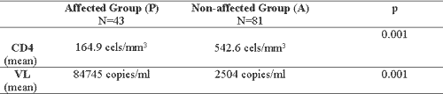
In Group (P) we detected very reduced CD4+ cell count and increased viral load levels when compared to the non-affected group, showing statistically significant difference (p<0.001), as seen in Table 1.
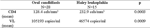
We noticed that in the affected group, the patients with oral candidiasis presented CD4+ cell count significantly lower (p=0.0003) and level of viral load significantly higher (p=0.0009) than the patients with hairy leukoplakia, as seen in Table 2.
Second Examination
We did not find any patients with concomitant oral candidiasis and hairy leukoplakia. Out of 81 patients in group A, 15 were excluded for having started HAART.
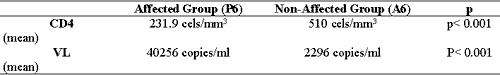
We observed that 18 patients developed oral lesions, being that 11 had oral candidiasis and 7 had hairy leukoplakia, forming group P6. The other patients (n=48) had not apparent lesion, comprising group A6. There were statistically significant differences both in CD4+ cell count and viral load, as shown in Table 3.
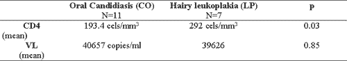
Patients with oral candidiasis in group P6 presented mean values significantly lower in CD4+ cell count than patients with hairy leukoplakia in the same group (p=0.03). However, the mean viral load level did not show statistically significant difference between both groups (p=0.85), as shown in Table 4.Table 1. Comparison of mean CD4+ cell count and mean level of viral load (VL) between the affected group (P) and the non-affected group (A) at baseline.
Table 2. Comparison between mean CD4+ cell count and mean level of viral load (VL) between patients with oral candidiasis and hairy leukoplakia at baseline.
Table 3. Comparison between mean CD4+ cell count and mean viral load level (VL) of affected patients (P6) and non-affected patients (A6) after 6 months.
Table 4. Comparison between mean CD4+ cell count and mean level of viral load (VL) in patients with oral candidiasis and hairy leukoplakia after six months.
DISCUSSION
The two most used clinical markers to assess level of immunosuppression are number of CD4+ lymphocytes and viral load level. The latter is more associated with progression of the disease to death, and it may vary during acute infections and is not associated with the risk of opportunistic infections.
Consequently, CD4+ values are the most frequently used test to assess the risk of opportunistic infections related to immunosuppression, as well as to define a case of AIDS, being extremely important to define the introduction of antiretroviral therapy, with (HAART) or without the inclusion of protease inhibitors 9.
This is an extremely important factor especially in developing countries in which lack of resources and long distance from urban centers (such as in Brazil) do not allow these tests to be conducted as a routine, when necessary. If we consider the diagnosis of hairy leukoplakia and oral candidiasis they are reasonably easy to be made by otorhinolaryngologists or experienced general practitioner and are low cost procedure.
In our study, concerning number of CD4+ cells, we observed that at baseline patients with oral lesion had on average CD4+ cell count that was significantly lower than the patients without oral lesion. This piece of data confirms that these two lesions - oral candidiasis and hairy leukoplakia can be used as an indirect inference of level of immunosuppression of the patients.
Concerning oral candidiasis, we detected that in both affected groups (P and P6), patients presented CD4+ cell count below 300 cell/?l. The strong association with oral candidiasis and reduction in number of CD4+ cells, especially in levels below 200 cell/?l, have been observed in other studies 10-14. It is important to point out that our findings are not entirely in agreement with the ones reported by Crowe et al. (1991), in which they confirmed the presence of oral candidiasis when CD4+ cell count was below 500 cells/mm3 15.
Regardless, as patients in the non-affected group (A) initially presented mean CD4+ cell count higher than 500 cell/mm3 and those that after six months progressed to OC presented significant reduction of this figure (193.4 cell/mm3), we believe that these data are precise indicators that OC is a reliable marker of evolution of the level of immunosuppression among the Brazilian population.
Some studies have also related presence of hairy leukoplakia and level of CD4+ cell below 200 cell/mm3 16, 17. However, according to Itin (1993), HL would not be clearly associated with number of CD4+ cells, but the author admitted that the lesion would be a significant marker of the progression of the disease 18.
In our study, subjects with HL, conversely, presented mean CD4+ cell counts higher than 200 cell/mm3 in both periods analyzed, in accordance with the data by Homberg et al. (1995)19.
However, since patients in the non-affected group (A) presented at baseline mean CD4+ cell count of 542.6, and six months later those in this group (and presented HL and joined the P6 group) had mean cell count of 292 cell/mm3, we believe that the disease is also a marker, even though not so precise, of the level of immunosuppression degree of these patients.
The greater precision reached by OC as marker of immunosuppression level is emphasized when directly compared with patients with HL: we detected reduction in number of CD4+ cells higher in patients with OC both at baseline and after six months (CD4+ < 200 cell/mm3). Patients with HL presented on average level of CD4+ count between 200-292 cell/mm3 at both situations.
Our findings are in accordance with those reported by Kolokotronis et al. (1994), who observed presence of more episodes of oral candidiasis in patients with lower CD4+ counts than those with hairy leukoplakia 20.
As to viral load at baseline, we detected a large number of patients with oral lesion (84,745 copies/ml on average), when compared to non-affected subjects (2,504 copies/ml on average), with statistical significance. Values were higher (105,193 copies/ml on average) in patients with OC in relation to patients with HL (46,574 copies/ml) being that this was a statistically significant difference.
Our study is partially in agreement with the one by Patton (1999), in which HIV-positive patients with hairy leukoplakia presented viral load higher or about 20,000 copies/ml regardless of CD4+ level 21, and also the one by Campo et al. (2002), to whom oral candidiasis was strongly related to viral load above 10,000 copies/ml 6.
According to those authors, patients with hairy leukoplakia normally present mean viral load higher than patients with oral candidiasis 6, 21. Our results were exactly the opposite. The explanation to this finding is possibly the fact that patients with OC were in worse immunosuppression status, favoring viral replication.
After 6 months, the patients that started to present oral lesions (OC and HL) showed significant increase in viral load, going from 2,504 copies/ml on average to 40,256 copies/ml on average.
However, differently from the studied groups at baseline, there was no statistically significant difference between mean viral load of patients with OC and HL, which is an intriguing fact. The possible explanation is the small number of cases (only 18 patients).
It was concluded that CD4+ cell count when there is infecting viral load was strongly related to onset of OC and HL, differently from what other authors stated. Patton (1999), for example, stated that HL was not clearly related with CD4+ cell count but rather with viral replication, even though the author believes that HL is also a disease progression marker 21.
CONCLUSION
Based on our results, we concluded that:
1) Presence of oral lesions, that is, oral candidiasis and hairy leukoplakia in patients infected by HIV is a strong indication that CD4+ cell count of these patients is at lower levels than 300 cell/mm3.
2) The presence of OC and HL in HIV infected patients is related to increase in viral load, and therefore,
3) The presence of OC and HL is a reliable clinical marker of progression of the disease in the Brazilian population.
REFERENCES
1. Robinson PG. The oral manifestations of HIV infection. Int J STD AIDS 1997; 8(11):668-74.
2. Patton LL, McKaig RG, Rogers D, Strauss RP, Eron JJ Jr. The role oral manifestations of HIV and body signs in suspicion of possible HIV infection. Oral Surg Oral Med Oral Pathol Oral Radiol Endod 1998; 85(4):416.
3. Ditchtel WJ. Oral manifestations of human immunodeficiency virus infection. Otolaryngol Clinics of North America 1992; 25(6):1211-25.
4. Ravina A, Ficarra G, Chiodo M, Mazzetti M, Romagnani S. Relationship of circulating CD4+ T-Lymphocytes and p24 antigenemia to the risk of developing AIDS in HIV-infected subjects with oral leukoplakia. Journal of Oral Pathology & Medicine 1996; 25(3):108-11.
5. Margiotta V, Campisi G, Mancuso S, Accurso V, Abbadessa V. HIV infection: oral lesions, CD4+ cells count and viral load in an Italian study population. Journal of Oral Pathology & Medicine 1999; 28(4):173-7.
6. Campo J, Del Romero J, Castilha J, Garcia S, Rodriguez C, Bascones A. Oral candidiasis as a clinical marker related to viral load, CD4 lymphocyte count and CD4 lymphocyte percentage in HIV-infected patients. Journal Oral Pathology & Medicine 2002; 31(1):5-10.
7. Centers for disease control and prevention (CDC). 1993 revised classification and expanded surveillance case definition for AIDS among adolescents and adults. MMWR 1992; 41(17):1-19.
8. Williams DM. Classification and diagnostic criteria for oral lesions in HIV infection. J Oral Pathol Med 1993; 22:289-91.
9. Jung AC, Paauw DS. Diagnosing HIV-related disease: Using the CD4 count as a guide. J Gen Intern Med 1998; 13(2): 131-6.
10. Klein RS et al. Oral Candidiasis in high-risk patients as the initial manifestation of the acquired immunodeficiency syndrome. N Engl J Med 1984; 311:354-8.
11. Moniaci D, Greco D, Flecchia G, Raiteri R, Sinicco A. Epidemiology: clinical features and prognostic value of HIV-1 related oral lesion. J Oral Pathol Med 1990; 477-81.
12. Katz MH et al. Progression to AIDS in HIV-infected homosexual and bisexual men with hairy leukoplakia and oral candidiasis. AIDS 1992; 6:95-100.
13. Barr CE et al. HIV-associated oral lesions: immunologic, virologic and salivary parameters. J Oral Pathol Med 1992; 21:295-8.
14. Phair S et al. The risk of pneumocystis carinii pneumonia among men infected with human immunodeficiency virus type-1. N Engl J Med 1990; 322:161-5.
15. Crowe SM, Carlin JB, Stewart KI, Lucas CR, Hoy JF. Predictive value of CD4 lymphocyte numbers for the development of opportunistic infections and malignancies in HIV-infected persons. J Acquired Immune Defic Sybd. 1991; 4:770-6.
16. Fahey JL, Taylor JMG, Detels R et al. The prognostic value of cellular and serologic markers in infection with human immunodeficiency virus type 1. M Engl J Med 1990; 322:166-72.
17. Glick M, Muzyka BC, Lurie D, Salkin LM. Oral Manifestations associated with HIV-related disease as markers for immune suppression and AIDS. Oral Surg Oral Med Oral Pathol 1994; 77: 344-9.
18. Itin PH, Lautenschlager S, Fluckiger R, Ruffi T. Oral manifestations in HIV-infected patients: Diagnosis and management. J Am Acad Dermatol 1993; 29:749-60.
19. Holmberg SD, Buchbinder SP, Conley LJ et al. The spectrum of medical conditions and symptoms before acquired immunodeficiency syndrome in homosexual and bisexual men infected with the human immunodeficiency virus. Am J Epidemiol. 1995; 141:395-404.
20. Kolotronis A et al. Immunologic status in patients infected with HIV with oral candidiasis and hairy leukoplakia. Oral Surgery Oral Medicine Oral Pathology 1994; 78(1): 41-6.
21. Patton LL et al. Oral hairy leukoplakia and oral candidiasis as predictors of HIV viral load. AIDS 1999; 13(15): 2174-6.
1Collaborating Professor, Discipline of Otorhinolaryngology, Medical School, USP.
2 Ph.D. studies in Otorhinolaryngology under course, Medical School, USP.
Address correspondence to: Rua Cristiano Viana, 450 -121 Jd. América São Paulo SP 05411-000
Tel: (55 11) 3085-1703 - E-mail: miz@uol.com.br
Awarded best paper in Laryngology at III Congresso Triológico de Otorrinolaringologia, Rio de Janeiro, 2003.
Print: ![]()