

Year: 2004 Vol. 70 Ed. 3 - (3º)
Artigo Original
Pages: 306 to 309
 PDF PT
 Clinical and histopathological aspects of the sinonasal lymphoma
Author(s):
Bernardo C. Araújo Filho 1,
Flavio A. Sakae 2,
Marcus M. Lessa 2,
Maura C. das Neves 2,
Richard L. Voegels 3,
Ossamu Butugan 3
Keywords: non-Hodgkin's lymphoma, sinonasal tract, histopathology
Abstract:
Non-Hodgkin's lymphoma of the sinonasal tract is relatively uncommon and is now recognized as an important cause of destructive lesions of the nose and midface, which show a progressive course. It is this rarity that may cause clinicians to dismiss them as a benign inflammatory infiltrate and may present a considerable diagnostic challenge to the pathologist. Aim: The objective of the present study was to identify epidemiological, clinical and histopathological aspects of the non-Hodgkin's lymphoma of the sinonasal tract, correlation site and natural history with histopathological types. Study design: Series review. Material and Method: The study group consisted of seven patients (4 men and 3 women) with clinical diagnosis of sinonasal non-Hodgkin lymphoma seen at the Clinic of the Division of Otorhinolaryngology, University of Sao Paulo, between 1985 and 2003. Patients age ranged from 22 to 54 years (mean: 38,2 years). The patients data obtained were analyzed regarding epidemiological, clinical, therapeutic and outcome factors. Results: The site of tumor, clinical manifestations and natural history of the sinonasal lymphomas of the B-cell phenotype and T/NK cell phenotype were different, so the histopathological diagnosis is very important. Conclusion: The diagnosis can be extremely difficult. Biopsy specimens were necessary to establish the diagnosis of each patient. Early diagnosis and efficient therapy must be developed to improve patient outcome.
![]()
INTRODUCTION
Sinonasal tract lymphomas are uncommon neoplasms that are known to cause destructive lesions of the nose and facial mid third 1. Non-Hodgkin lymphoma (NHL) amounts to 1.5% of all tumors detected in the body. About 20 to 30% of the NHL are extranodal and only 0.44% of these lymphomas are located in the sinonasal tract, especially in Asia 2-4. Its rarity can take healthcare professionals to make mistakes in clinical diagnosis, representing a real challenge to pathologists owing to its inflammatory nature 5, 6. NHL has been growing at a rate of 3.3% every year since 1970 and not only the advent of AIDS is responsible for this increase. Lesions that used to be called lethal midline granuloma, polymorphic reticulosis, lymphoproliferative angiocentric lesions, pseudolymphoma, idiopathic destructive disease, and others were used to name this entity, marked by destructive aspect and presence of polymorphic inflammatory infiltrate 1, 4. As a result of immunohistochemistry and molecular biology techniques it is possible to diagnose the real histopathological basis of this disease, which is a NHL 7, 8.
Non-Hodgkin lymphoma can present with different cell phenotypes: it may be from B cell line, T cell line and recently, natural killer cell (NK) line 4, 7. NHL manifest according to the phenotype, biological behavior, geographical destruction and different therapeutic implications.
In the present study we determined clinical and histopathological aspects of NHL of sinonasal tract, correlating tumor site and biological behavior of subtypes of NHL, based on the experience of the Division of Clinical Otorhinolaryngology, Hospital das Clinicas, Medical School, University of Sao Paulo (HCFMUSP).
MATERIAL AND METHOD
We conducted a retrospective study that included 7 patients seen in the Ambulatory of the service of Otorhinolaryngology, HCFMUSP, between 1985 and 2003. These patients were diagnosed as having sinonasal tract NHL, confirmed by clinical pathology and histochemical analysis of the lesion. We correlated epidemiological, clinical histological and evolution aspects of each patient.
RESULTS
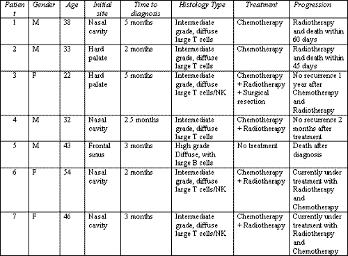
Ages ranged from 22 to 54 years, being that the mean age was 38.2 years. There was predominance of male subjects (4:3) and the interval from onset of symptoms to diagnosis was on average of 2.7 months (Chart 1).
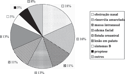
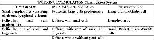
The clinical presentation was very nonspecific, insidious and progressive, mimicking common nasal symptoms (Graph 1). Symptoms such as facial edema are highly suggestive, even though they are nonspecific. The patients were staged according to the Working Formulation classification for lymphomas and diffuse intermediate grade was the most prevalent one 7, 9 (Chart 2). The advocated treatment, considering patients individually, included chemotherapy, surgical resection and radiotherapy. Five-year survival rate was very low, reflecting the high grade of malignancy of the tumor. The patients that were more recently diagnosed and submitted to chemotherapy and local-regional radiotherapy presented longer survival than patients submitted to radiotherapy only. The patients had negative HIV serology, except for one patient that developed lymphoma in an atypical and very rare site, the frontal sinus. The patients were non-smokers and did not abuse of alcoholic drinks.
Graph 1.
Chart 1. List of patients and correlation between clinical, histopathological and progression data.
Chart 2. Classification system used to classify non-Hodgkin lymphoma.
DISCUSSION
According to Cleary and Batsakis (1994)4, sinonasal non-Hodgkin lymphomas are predominantly T cell line phenotypes in Asia and in the Western world they affect mostly B cell lines. T and NK cell tumors present similar clinical and histological behavior, differentiated only in molecular aspects; thus, we consider them to be part of the same group 7, 8. In our study, most of the cases (85%) had T/NK cell line. The increase in diagnosis of lymphomas thanks to immunohistochemical techniques has changed this situation, since T cell NHL were underestimated, since the areas of necrosis were abundant, hindering the diagnosis 1, 7. Vasculitis, angiocentrism and presence of large amount of inflammatory cells rather than few tumor lymphoid cells are factors that make the diagnosis of T and NK cell NHL a real challenge 2, 5. In B cell line tumors, angiocentrism and necrosis are less frequent, being a tumor that causes little ulceration and normally has subepithelial location 1, 4, 7. The presence of markers such as CD56, CD2 and CD7 collaborates for NHL of T/NK cells to have greater vessel trophism, increasing adhesion of vessels and leading to more devastating effects 1, 10, 11.
The most common site of NHL in sinonasal tract found in our sample was nasal fossa. Devaney et al. (1999)7, Cleary et al. (1994)4 and Sheahan et al. (2001)6 emphasized in their papers the preference of T cell line tumors for the nasal fossa and its aggressiveness, frequently progressing to oro-antral fistulae and palate erosion. B cell lymphomas affect preferably the paranasal sinuses, especially maxillary and ethmoid sinuses 3, 7, 9. In one patient, the affected sinus was the frontal one, being that this patient was HIV-infected. Lymphomas preferably affect patients aged 50 years, but our patients were younger 7. NK tumors, since they have CD56 marker, affect younger subjects and are more aggressive, which explains the young age range involved 7. Intermediate grade was the predominant one, in accordance with the data reported by Fajardo-Dolci et al. (1999)8 and Devaney et al. (1999)7. The most common symptoms were nasal obstruction, rhinorrhea, facial edema and palate erosion. The almost absolute presence of T and NK lymphomas reflected in more aggressive and destructive episodes, being that the most common site was the nasal fossa. Epitheliotrophism of T/NK cell line is characteristic and it is clinically manifested with edema and facial hyperemia 7, 8. Thus, the clinical presentation of this tumor is evidenced in the most aggressive behavior of NK and T tumors.
Patients were not smokers or alcohol abusers. There is no correlation between tumors and alcohol and smoking 1, 7. There is known connection between patients with T/NK cell line NHL and infection by Epstein-Barr virus, but we did not detect this correlation in our study 12.
The combined treatment of chemotherapy and radiotherapy has undoubtedly improved survival of these patients, a fact that has been observed in the follow-up of our study and is in accordance with many authors that present larger samples 4, 7.
CLOSING REMARKS
Sinonasal non-Hodgkin lymphoma is an important entity that causes destruction of the nose and mid facial third. C and T/NK cell lines have different biological behavior, as well as site and clinical presentation, being that histopathological diagnosis is extremely important.
The diagnosis has been difficult owing to the presence of ischemic necrosis and few tumor cells in the lesion, especially in T/NK cell tumors. Thus, the appropriate biopsy favors more precise and earlier diagnosis, quickly instituting the right therapy approach and improving prognosis and survival of these patients.
REFERENCES
1. Berrettini S, Segnini G, Bruschini P, Marchetti G, Colosimo. Lethal midline granuloma syndrome: a case of Ki lymphoma. Revue de Laryngologie Otologie rhinologie 1993; 114 (1): 37-4.
2. Calderon-Garciduenas L, Delgado R, Calderon-Garciduenas A et al. Malignant neoplasms of the nasal cavity and paranasal sinuses: a series of 256 patients in Mexico City and Monterrey. Is air pollution the missing link? Otolaryngol Head Neck Surg 2000; Apr 122(4): 499-508.
3. Nakamura K, Uehara S, Omagari J et al. Primary non-Hodgkin lymphoma of the sinonasal cavities: correlation of CT evaluation with clinical outcome. Radiology 1997 Aug; 204(2):431-5.
4. Cleary KR, Batsakis JG. Sinonasal lymphomas. Ann Otol Rhinol Laryngol 1994; Nov; 103(11):911-4.
5. Quraishi MS, Bessell EM, Clark D, Jones NS, Bradley PJ. Non-Hodgkin's lymphoma of the sinonasal tract. Laryngoscope 2000 Sep; 110(9):1489-92.
6. Shehan P, Donnelly M, Murphy M. T/NK cell non-Hodgkin's Lymphoma of the sinonasal tract. The Journal of Laryngology and Otology 2001; 115: 1032-35.
7. Devaney K, Rinaldo A, Carbone A. Sinonasal Malignant Lymphomas: A distinct Clinicopathological category. Ann Otol Rhinol Laryngol 1999; 108:411-9.
8. Fajardo-Dolci G, Magana RC, Bautista EL. Huerta Sinonasal lymphoma. Otolaryngol Head Neck Surg 1999 Sep; 121(3):323-6.
9. Hanna E, Wanamaker J, Adelstein D, Tubbs R, Lavertu P. Extranodal lymphomas of the head and neck. A 20-year experience Arch Otolaryngol Head Neck Surg 1997 Dec; 123(12):1318-23.
10. Cabane J, Raphael M. Letters to the editor. Laryngoscope 1992; 102 April.
11. Babb MJ, Cruz RM, Puligandla B. Sinonasal mucosa-associated lymphoid tissue lymphoma. Arch Otolaryngol Head Neck Surg 1999 May; 125(5):585-8.
12. Cho KH, Kim CW, Lee DY, Kim DW. An Epstein Barr virus associated lymphoproliferative lesion of the skin presenting a s recurrent necrotic papulovesicles of the face. British Journal of Dermatology 1996; 134:791-6.
1 Resident Physician, Division of Clinical Otorhinolaryngology, Hospital das Clínicas, Medical School, University of Sao Paulo.
2 Physician, Post-graduate studies under course, Discipline of Otorhinolaryngology, Medical School, University of Sao Paulo.
3 Associate Professor, Discipline of Otorhinolaryngology, Medical School, University of Sao Paulo.
Study conducted at the Division of Clinical Otorhinolaryngology, Hospital das Clínicas, Medical School, University of Sao Paulo
Study presented at II Congresso de Otorrinolaringologia da USP, held on August 5 - 7, 2001 in Sao Paulo.
Address correspondence to: Bernardo Cunha Araújo Filho - Av. Dr. Ovídio Pires de Campos, 171 ap. 617 05403-010 Sao Paulo SP
Tel (55 11)8146-5405 - E-mail: bcaf @terra.com.br
Print: ![]()