

Year: 2004 Vol. 70 Ed. 2 - (17º)
Relato de Caso
Pages: 261 to 264
 PDF PT
 Bilateral peripheric facial nerve palsy in acute linfoid leukemia: a case report
Author(s):
Marcos L. Antunes 1,
Maria C. M. Soares 2,
Andy O. Vicente 3,
José R. G. Testa 4,
Yotaka Fukuda 5
Keywords: leukemia, facial nerve, palsy
Abstract:
The facial mimic is very important to the human expression and communication, which depend on the integrity of the facial nerve. So, the peripheric facial palsy (PFP) can leave esthetics, functional and psychological sequelae. The more common etiology is Bell's palsy (50 to 80%) and most of the patients show a unilateral manifestation. The simultaneous bilateral PFP is rare, and the leukemia is the neoplasia that can often that kind of manifestation. We present a clinical case of an 18-year-old patient with acute lymphoid leukemia and simultaneous bilateral facial palsy, who did not recover after the chemotherapy treatment, and died five months after the initial manifestation of the facial palsy. Important considerations were accomplished about the physiopathology of PFP in acute lymphoid leukemia, besides literature review.
![]()
INTRODUCTION
Peripheral facial palsy is a common entity that may generate significant esthetical and functional sequelae in patients. According to the literature, Bell's palsy is the most common cause, amounting to 50 to 80% of the cases 1. However, it is considered as a diagnosis of exclusion. Most of the patients affected by peripheral facial palsy present unilateral manifestation. Simultaneous bilateral affection of facial nerve is rare (less than 1% of the cases of peripheral facial palsy) 2, 3.
In a literature review with over 2,850 patients with peripheral facial palsy, there were 90 different etiologies, divided into birth, trauma, neurological causes, infections, metabolic causes, neoplasm, toxic, iatrogenic and idiopathic causes (where Bell's palsy was included, responsible for 51% of the cases). Tumors amounted to 6% of the cases 4.
Among tumors, we should bear in mind that there are tumors that directly damage the nerve, such as parotid tumors, vestibular Schwannoma, glomus tumors, among others, and neoplasms with systemic manifestations which can lead to clinical picture of unilateral or bilateral facial paralysis, such as leukemia. When we consider bilateral facial palsy, leukemia is the most frequent neoplasm with this type of manifestation 5, 6.
Leukemia has been recognized as a clinical entity for over 150 years, since the description by Virchow and Craigie and Bennett (1845). Dress, in 1945, was the first otologist that addressed otological complications of leukemia. According to Paparella et al.1, manifestations are more common in acute than chronic forms of leukemia, and mainly in acute lymphoid leukemia, in which manifestations can reach 50% of the patients, among which we can include facial palsy, sensorineural hearing loss, tympanomastoid infections, nystagmus and vertigo.
Acute lymphoid leukemia is the one that most commonly presents facial palsy. Meningeal leukemia occurs by neoplastic lymphocytes or myelanocytes of the arachnoid tissue. Proliferation of cells occurs on superficial walls of the veins and extend through the arachnoid surface up to emerging arteries, veins, arterioles and venulles that comprise the brain. Leukemic cells reduce the caliper of the vessels generating brain hypoperfusion, which can lead to meningitis, with symptoms of headache, vomiting, meningeal signs and papilledema. Owing to the fact that cranial nerves go through the leptomeninges, they can be compressed with the vessels and damaged by leukemic infiltration, resulting in neuropathy 1, 7.
The diagnosis of meningeal leukemia is made by the presence of blast cells in the cerebrospinal fluid (CSF) and increase in protein levels. However, as previously reported, normal CSF exam does not rule out the possibility of facial palsy as manifestation of reactivation of leukemia, despite being extremely rare 5, 8.
Next, we report the case of a 18-year-old patient with acute lymphoid leukemia and simultaneous bilateral peripheral facial palsy.
CASE REPORT
I.P.B., 18-year-old male patient. Seen in the emergency room of Hospital Sao Paulo complaining of weakness and asthenia, in addition to spots all over the body (petechia and ecchymosis) and swelling of some body parts (lymphadenomegalia) for over 1 month. In the clinical history, we found out he had had intermittent fever, was losing weight (6Kg in the period) and had lack of appetite.
He did not report use of chemicals, previous hospitalizations or any other diseases.
No significant family history.
Physical examination: general good health status, discolored +/+4, neck lymphadenomegalia. Reduction of vesicular murmur on right hemithorax, palpable spleen at 8cm from the left coastal margin, and palpable liver at 5cm from right coastal margin. Presence of ecchymosis on lower limbs.
Supplementary tests: Hemoglobin = 12mg/dl; Platelets = 1,4000; Leukocytes = 27,000 with lymphoid-like blat cells.
Chest x-ray: mediastinum widening.
Myelogram: 40% of lymphoid blast cells.
Conclusion: acute lymphoid leukemia.
The patient underwent the first chemotherapy session with regimen BFM5/93 (Daunorubicin, Metrothexate, Elspar and Prednisone), and was discharged one month after the 1st session, with remission of the condition.
He developed pancreatitis as a result of Elspar two days after discharge and consequent unbalanced diabetes, being hospitalized. He underwent abdomen CT scan which showed pancreas pseudocyst, endoscopically drained. At that time, he presented sudden onset of peripheral facial palsy on the left. The prognostic tests of Hilger showed 2mA difference between the normal and the paralyzed side. The physician introduced acyclovir 1.5 g/day and prednisone 60mg/day. The patient did not manage to undergo audiometry owing to his weak status and had suppuration on both ears simultaneously.
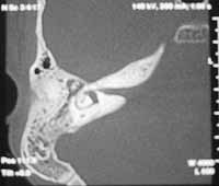
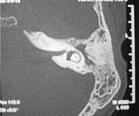
Head and temporal bone CT scan was conducted we observed leptomeningeal cyst and partial velamentum of mastoid cells and bilateral tympanic cavity, with normal ossicle chain and no apparent lesion in the facial nerve canal (Figures 1 and 2).
Three days after onset of facial paralysis on the left, he had peripheral facial paralysis on the right, classified as grade IV bilaterally. Hilger's test showed response of 8mA on the right and non-excitability on the left. CSF exam at the time revealed proteins = 34, glucose = 51, and 5,973 cells with 75% blast cells and 25% lymphocytes, confirming infiltration into the central nervous system. The patient was then submitted to intrathecal chemotherapy regimen, in addition to intravenous chemotherapy (Hyper -C -VAD) leading to clinical and laboratory improvement, but with persistence of facial palsy, which progressed to grade V in some days, plus complaints of tinnitus and vertigo.
The neurology assessed the patient and diagnosed polyradiculoneuritis secondary to leukemic infiltration (patient presented generalized motor deficit). The patient was treated with hyperimmune human immunoglobulin 400mg/kg/day for 5 days, but did not experience improvement of paralysis. Despite all treatment attempts, the patient died of sepsis 5 months after the onset of initial palsy.
DISCUSSION
Bilateral simultaneous peripheral facial palsy is rare (less than 1% of the cases) and among differential diagnoses we should include traumatic causes, syphilis, Bell, Guillain-Barre syndrome, multiple neuropathies, encephalitis, Miller Fisher syndrome, tumors, Hansen's disease, criptococcosis (AIDS), tuberculous meningitis, diabetes mellitus, sarcoidosis, lupus, Lyme's disease, mononucleosis, poliomyelitis, in addition to other diseases less frequently referred in the literature and of small frequency2, 3.
Most of the patients with leukemia and facial paralysis present leukemic cells in the CSF or activity of the disease in other organs. Facial palsy as single manifestation of leukemic reactivation is very rare 5. Cartwright et al. reported eight cases of leukemia or lymphoma preceded by Bell's palsy in a retrospective study. In four patients the palsy preceded the disease in less than one year, and in two of them, in only one month. Paralysis could occur owing to infiltration of malignant cells of the nerve before the usual signs or symptoms of leukemia or lymphoma. We wonder whether the facial nerve would be a storage site of leukemic cells, which would multiply and then lead to reactivation episodes. In this case, palsy would have to regress with regression of blast infiltration after appropriate treatment.
In our patient, paralysis occurred approximately three months after the first manifestations of the disease, which is more common to occur, that is, systemic manifestations of the disease normally precede the onset of facial palsy.
Paparella et al.1 found leukemic infiltration, hemorrhage and infection in 45 temporal bones of 25 patients with many different types of leukemia, obtained from autopsy. In many occasions, infiltration extends to the 7th and 8th cranial nerves, the internal part of the inner acoustic canal, and sometimes up to the membranous labyrinth. Among histopathological findings, leukemic infiltration and degree of infiltration were the ones that were more related with clinical manifestations when compared to hemorrhage and infection.
ENT manifestations reported in our patient were very varied, such as facial palsy, middle ear infection with persistence of effusion, and cochlear-vestibular symptoms. These findings are in agreement with the reports of Paparella1 concerning the many ENT manifestations of the disease, which are not rare.
Age is an important factor for the development of meningeal infiltration by blast cells, being more common in children and pre-school children, probably owing to the fact that there is closer relation between blood circulation and brain tissue when compared to adolescents and young adults. When there is infiltration, survival reduces considerably and the best therapy, according to the literature, would be intravenous chemotherapy at complete doses associated with intrathecal chemotherapy and meningeal radiotherapy. The most commonly used drugs for intravenous chemotherapy are cyclophosphamide, doxorubicin, vincristin, cytosine arabinoside, asparagine and prednisone. Intrathecal chemotherapy is normally conducted with metrothexate 8.
In a case report of a 14-year-old patient that had simultaneous bilateral facial palsy with an interval of 16 days from onset of unilateral to bilateral lesion, CSF did not show blast cells, but rather increase of protein levels. After the patient presented symptoms such as headache, increase of lymph nodes and otalgia, CSF analysis was repeated and they identified blast cells and in the myelogram there were 60% of lymphoblasts. The patient had remission of palsy after two months from remission through chemotherapy, but he died two months later. It demonstrates that prognosis of patients is worse when there is manifestations of leukemic infiltrate in the meninges.
In a study with 62 patients with acute myeloid leukemia, between 1975 and 1978, only six patients developed unilateral facial palsy, without ear infection or involvement of the cranial nerves; they all died within six months after its onset, average of 4 months 9.
Our patient did not present improvement of facial palsy even after many clinical treatment approaches, but owing to the complications of the treatment itself, it was not possible to reach complete remission of the disease for a satisfactory period of time. After leukemic infiltration of the central nervous system the response to treatment was not effective, and we observed increment of symptoms related to the infiltration, such as vestibular manifestations and polyradiculoneuritis. The patient died 5 months after the onset of facial palsy, period similar to that described in other case reports.
CONCLUSION
Simultaneous bilateral facial palsy is very rare and leukemia is among its differential diagnoses, since it can be an isolated manifestation of disease reactivation. Blast cells and increase of CSF protein levels are commonly found, but even in the absence of neoplasm cells, we should not rule out the possibility of meningeal infiltration leading to peripheral facial palsy.
Age is an important factor in the occurrence of meningeal infiltration and it normally indicates poor prognosis of the underlying disease. Facial palsy may be recovered after remission of blast infiltration, but toxicity of drugs can be a limiting factor to treatment and lead to facial palsy evident sequels.
Figure 1
Figure 2
REFERENCES
1. Morgan M, Nathwani D. Facial Palsy and Infection: The Unfolding Story CID 1992; 14: 263-71.
2. Keane JR. Bilateral seventh nerve palsy: Analysis of 43 cases and review of the literature. Neurology 1994; 44:1198-204.
3. Wormald PJ, Sellars SL, De Villiers JC. Bilateral facial nerve palsies: Groote Schuur Hospital experience. J Laryngol Otol 1991; 105: 625-7.
4. May M, Klein S. Differential Diagnosis of Facial Nerve Palsy. In: Management of Facial Nerve Disorders- Otolaryngologic Clinics of North America 1991; 24:613-45.
5. Juhn YJ, Inoue S. Facial Nerve Palsy as an Early Manifestation of Relapse in T-Cell Acute Lymphoblastic Leukemia. Ear Nose &Throat Journal 1996; 75:157-60.
6. Paparella MM, Berlinger NT, Oda M, Fiky FE. Otological Manifestations of Leukemia. Laryngoscope 1973; 13:1510-26.
7. Sawada H, Matsui M, Udaka F, Nishimura M, Fujita M, Kameyama K. Adult T-Cell Leukemia Initially Manifesting as Facial Diplegia. American Journal of Hematology 1989; 32:61-5.
8. Pinkel D, Woo S. Prevention and Treatment of Meningeal Leukemia in Children. Blood 1994; 84:355-66.
9. Lilleyman JS, Antoniou AG, Sugden PJ. Facial Nerve Palsy in Acute Leukemia. Scan J Haematol 1979; 22:87-390.
1 Ph.D. studies under course.
2 Specialization under course.
3 Master studies under course.
4 Professor, Discipline of Pediatric Otorhinolaryngology.
5 Full Professor.
Affiliation: Department of Otorhinolaryngology and Human Communication Disorders, Federal University of Sao Paulo-UNIFESP-EPM.
Address correspondence to: Maria Claudia M. Soares - R. Prof. Zoraide de Campos Helú, 04 Ipiranga, Sao Paulo SP 04265-020
E-mail: mcms26@aol.com
Study presented as poster at 36o Congresso Brasileiro de Otorrinolaringologia, Florianópolis, November 2002.
Print: ![]()