

Year: 2004 Vol. 70 Ed. 2 - (16º)
Relato de Caso
Pages: 255 to 259
 PDF PT
 LARYNGEAL TUBERCULOSIS AND BLASTOMYCOSIS - Report of three cases and review of literature
Author(s):
Roberta I. D. Garcia 1,
Suzana B. Cecatto 1,
Renata R. Mendonça 1,
Carlos E. M. Barcelos 1,
Rodrigo O. Santos 2 ,
Priscila B. Rapoport 3
Keywords: granulomatosis diseases, larynx, tuberculosis, blastomycosis
Abstract:
The granulomatosis diseases of the larynx have different clinical manifestations, which difficult the clinical diagnosis. When the treatment is early introduced, the result is satisfactory. The first manifestation of granulomatosis diseases can occur in larynx, and it always has to be remembered among laryngeal inflammatory diseases. We reported three cases were symptom was hoarseness, and after investigation the diagnosis was tuberculosis and blastomycosis of the larynx. The evaluation and management are discussed.
![]()
Introduction
Tuberculosis is a universal problem and 20 years ago it presented clinical pattern that was different from the current one. The modification, as well as the increase in number of described cases, can be associated with the onset of AIDS virus infections 5.
Elderly, debilitated, low social-economic population and immunosuppressed patients - especially affected by AIDS - have higher risk of developing the disease. However, extra-pulmonary forms present different epidemiology.
The most common form of extra-pulmonary affection of tuberculosis (TB) of the head and neck is neck lymphadenopathy. The disease can involve other sites such as middle ear, nasal cavity, oropharynx, nasopharynx, parotid gland, esophagus, submandibular glands, palate, larynx and thyroglossus cyst duct 2. Among laryngeal granulomatous diseases, tuberculosis is the most common one 5.
During pre-antibiotic years, laryngeal tuberculosis was considered one of the most serious and common complications of lung tuberculosis, which was normally fatal 7.
As a result of the development of anti-tuberculosis medications and early detection methods, laryngeal involvement has reduced to 1% in patients with TB and mortality rate has reduced to less than 2%. However, as a result of AIDS epidemics, there has been a global increase of TB incidence, which tends now to be more aggressive, comprising over 80% of extra-pulmonary involvement in the HIV infected population 15.
Classically speaking, laryngeal TB is related with extensive lung tuberculous lesions, whose dissemination is explained by the bronchogenic theory and is characterized as being highly contagious 15.
It is important to emphasize differential diagnosis with laryngeal carcinoma, owing to the characteristics of the clinical examination and presence of the same risk factors, such as alcohol abuse and smoking 10. The response to anti-tuberculosis treatment is excellent and quick, being that most lesions disappear within a period of 2 months.
South-American blastomycosis is systemic, progressive and uncommon mycosis, caused by Paracoccidioides brasiliensis. The disease is restricted to the population in Latin America with heterogeneous distribution 13. In Brazil, it is endemically distributed in the states of the southeast regions, Goias and Mato Grosso do Sul.(7). Laryngeal blastomycosis was described by Dennis in 1918, and since then some literature reviews have been published, but the number of cases is not very significant 3.
The purpose of the present study was to evidence the importance of differential diagnosis from granulomatous diseases of the larynx, especially tuberculosis and blastomycosis, since precise diagnosis allows appropriate therapeutics, preventing unfavorable evolution and occurrence of iatrogenia.
Case Report
Case 1: M.J.R., 45 years, female, Caucasian, worked as maid. Came to the Ambulatory of Otorhinolaryngology, Medical School, ABC (FMABC) complaining of progressive dysphonia for three weeks, without periods of aphony. She reported night dry cough for six months, daytime fever of 38o C and 8 kg weight loss for the past 2 months. She did not report dyspnea, dysphagia or odynophagia, and no changes to vocal quality throughout the day. She had contact with bacillus positive patients. She did not report smoking or alcohol abuse.
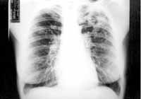
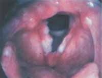
We ordered the following tests: chest x-ray (Figure 1), which evidenced cavitation on the left pulmonary apex; sputum bacillus exam, whose result was positive, and PPD with 14mm diameter (reactive factor). Direct laryngoscopy revealed depression on the free vocal fold margins (Figure 2). We diagnosed laryngeal and pulmonary tuberculosis and the patient was referred to the primary healthcare center to be treated and clinically followed up. Forty-five days after beginning of treatment she presented complete regression of laryngeal lesions and improvement of dysphonia.
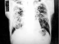
Case 2: L.B., 50 years, male, worked as builder. He came to the Ambulatory of Otorhinolaryngology, FMABC complaining of constant and progressive dysphonia for 4 months, without worsening or improving factors. He did not report dyspnea, dysphagia, odynophagia, fever, loss of weigh or periods of aphony. He reported smoking of 60 packs/year and severe alcohol abuse. General physical examination showed no significant findings. We ordered chest x-ray (Figure 3) that evidenced diffuse micronodular infiltrate and cavitation on the apex. Nasofibrolaryngoscopy: epiglottic edema, interarytenoid and both vocal folds with edema, plus right vocal fold paresis. BAAR analysis in sputum (3 samples) with positive results and negative fungi culture. After diagnosis of tuberculosis, the patient was referred to the primary healthcare center for treatment. Fifteen days after beginning of treatment with medication he presented improvement of dysphonia and nasofibrolaryngoscopy showed only interarytenoid and epiglottic edema, with normal vocal fold mobility. There was total remission of symptoms and laryngeal affections after 2 months of treatment.
Case 3: R.S.S., 43 years, male, Caucasian, agriculture worker. He came to the Ambulatory of Otorhinolaryngology complaining of constant dysphonia for 1 year. He did not report dyspnea, dysphagia, odynophagia, cough, weigh loss and fever. He smoked 10 packs/year cigarettes.
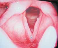
We ordered chest x-ray that was within the normal range. Direct laryngoscopy showed vegetating lesion on the posterior middle third of the left vocal fold (Figure 4). He was submitted to laryngeal biopsy and the clinical pathology exam showed chronic inflammatory process, granulomatous type, with giant-cellular reaction and presence of rounded fungi with sprouting, compatible with blastomycosis.
The patient was referred to the Service of Infectology, and there was improvement of dysphonia 20 days after beginning of medication treatment.
He is currently being followed up in the ambulatory and has been asymptomatic up to the present moment.
Discussion
Tuberculosis is the most common granulomatous laryngeal disease. It is caused by Mycobacterium tuberculosis (M. tuberculosis), highly pathogenic in debilitated patients, being inhalation route the main contamination form.
In the past, laryngeal TB normally progressed to severe pulmonary sequelae. Currently, there are patients without pulmonary symptoms or even history of pulmonary TB. Recent theories have been proposed to explain laryngeal involvement. The first theory is named bronchogenic theory, in which the larynx is infected directly by bronchial tree discharge. In the hematogenic theory, patients do not present lung impairment and dissemination is through the circulation system 15.
There is predominance of male patients (3:1) and association with smoking and alcohol abuse. Symptoms are those of chronic laryngitis, and the most common one is dysphonia, present in 100% of the patients in many studies 15. Other manifestations include odynophagia, dysphagia, otalgia, stridor, foreign body sensation, as well as cough and hemoptysis by lung affection 5.
Laryngoscopy did not show a typical form of affection, being that the lesions are sometimes not distinguishable from carcinoma or chronic laryngitis. The lesion may be nodular, exophilic, present as mucosa ulceration, as well as hyperemia or edema of the vocal folds, simulating polyps. Epiglottis and subglottis can present exuberant edema, in some cases requiring tracheostomy 14. The similarity of TB and carcinoma may hinder the diagnosis, but both diseases can co-exist, demonstrating the importance of biopsy to be performed in all suspected lesions 9.
The most commonly affected laryngeal site is the vocal fold (50 to 70%), followed by ventricular band (40 to 50%), and the remaining 10 to 15% can involve the epiglottis, aryepiglottic fold, arytenoid, posterior commissure and subglottis 1.
The diagnosis can be defined by isolation and culture of M. tuberculosis. The best material for culture is obtained from biopsy, but it is positive in only 40% of the cases 2.
Some authors consider that clinical pathology analysis is the gold standard. Mantoux or intradermal test is frequently used, and the positive result is 10mmm induration up to 48 hours in patients with normal systemic immunity, or 5mm in immunocompromised patients. This positive test is strongly indicative of active disease, but does not confirm the diagnosis when isolated. Similarly to most of the patients that present pulmonary involvement, chest x-ray can help with diagnosis. It normally presents nodule images at the lung apex and cavitations are found in half of the patients 14. Differential diagnosis includes sarcoidosis, Wegener's granulomatosis, coccidiomycosis, blastomycosis, post-intubation trauma, syphilis and, especially, neoplasm. 15
Anti-tuberculosis drugs are the same for pulmonary or extra-pulmonary affections, and only treatment duration differs. Extra-pulmonary form should be treated for at least 9 months. AIDS patients use the same therapeutic regimen, but followed by stricter control.
After diagnosis of laryngeal TB, especial attention should be given to airway permeability, and surgical interventions are reserved to cases of airway complications 2.
The response to anti-tuberculosis therapy is excellent and most lesions disappear within the period of 2 months, as observed in two described cases. It there is persistence of symptoms, it is essential to investigate the exclusion of other diagnosis.
South-American blastomycosis is a chronic disease caused by Paracoccidioides brasiliensis, asexual and dimorphic fungi. In infected tissues it presents as rounded yeasts with thick and refringent cell walls, whose diameter ranges from 5 to 30 micra. When placed as exosporulations, it provides typical microscopic aspect: ship steering wheel 12.
It affects all age ranges, predominantly between 30 and 50 years, and after puberty there is clear prevalence of male gender (15:1). The protection provided to female gender is due to estrogen, which inhibits the transformation of conidia in mycelia, a critical point of the disease pathogenesis 11. There is preference for rural workers and alcohol abuse has been presented as an important predisposing effect 13. The scarcity of cases of AIDS patients can be explained by the use of Sulphametoxazol-Trimetopim, drug used in prophylaxis of pneumonia caused by Pneumocistys carinii,(8) which acts also over Paracocidioides brasilienses.
Inhalation route is the form through which the agent penetrates the body. From a primary pulmonary focus, by hematogenic dissemination, the fungus reaches other organs and systems, causing extra-pulmonary manifestations. Skin is the most commonly affected extra-pulmonary site and the larynx is the less frequent one, as well as the genital-urinary tract, adrenal glands, bones and mucosa of the upper respiratory tract 6.
The diagnosis of laryngeal blastomycosis can be difficult, especially since we do not normally suspect of the disease. The patient may present history and symptoms such as dysphonia, productive cough, occasional hemoptysis, as well as low fever, weight loss and general malaise for months13.
The presentation form of laryngeal blastomycosis varies depending on disease staging. According to Bennett, these stages can be divided into: 1 - inflammatory process; 2 - formation of multiple abscess; 3 - ulceration; 4 - fibrosis; 5 - stenosis; 6 - neck fistulation. The importance of early diagnosis is based on the fact that the three final stages generate irreversible lesions and they can be prevented by early diagnosis and appropriate treatment 12.
In the larynx, the macroscopic characteristics are similar to those of squamous cell carcinoma, being that differential diagnosis is critical. There are reports of patients that were incorrectly treated by surgery or radiotherapy 12. Other differential diagnoses include tuberculosis, lupus, North-American blastomycosis, histoplasmosis, sarcoidosis, syphilis, Wegener's granulomatosis, actinomycosis, leishmaniasis, among other granulomatous diseases.
Definite diagnosis is based on biopsy; some times multiple biopsies are required for histology and culture confirmation with Saboraud media. Polymerase Chain Reaction (PCR) is a new promising diagnostic tool 13.
Treatment is conducted with anti-fungal drugs such as intravenous amphotericin B, ketoconazole, itraconazole or fluconazole, resulting in improvement in about 90% of the cases13.
Closing Remarks
In laryngeal inflammatory disease, we can find polymorphic clinical pictures, being that granulomatous diseases are the most important ones.
Many times, the first manifestation of these patients is at the laryngeal level, and it is far too difficult to define the diagnosis only by clinical examination. If there is suspicion, we should proceed with biopsy of the lesion to define the etiological agent. Clinical pathology analysis is also important to define association with squamous cell carcinoma and laryngeal granulomatous diseases.
The normally favorable evolution of laryngeal granulomatous lesions demonstrates that the appropriate therapy provides effective treatment, being important to have early diagnosis in order to prevent future complications.
Figure 1: Chest RX with cavitation on the left lung apex.
Figure 2: Irregularity on the free vocal fold edges.
Figure 3: Chest RX evidencing diffuse micronodular infiltrate and cavitation on the apex.
Figure 4: Damage to the posterior third of the left vocal fold.
References
1. Bailey, C.M.; Taylor, W. Tuberculous Laryngitis. Laryngoscope 1981; 91: 93-100.
2. Bailey, B.J.; Calhoun, K.H.; Deskin, R.W.; Johnson, J.T.; Kohut, R.I. et al. Head & Neck Surgery - Otolaryngology. Second edition on CD-ROM, Lippincott-Raven, 1998.
3. Bennett, M. Laryngeal Blastomycosis. Laryngoscope 1964; 74: 498-512.
4. Caporrino, J.; Cervantes, O.; Jotz, G.P.; Abrahão, M. Doenças Granulomatosas da Laringe. Acta Awho 1998; 17(1): 6-10.
5. Cummings, C.W.; Fredrickson, J.M.; Harker, L.; Krause, C.J.; Richardson, M.A.; Schuller, D.E. Otolaryngology Head & Neck Surgery. Third edition, St. Louis, Mosby-Year Book Inc; 1998: 3294-3330.
6. Dumich, P.S.; Neel, B.H. Blastomycosis of the Larynx. Laryngoscope 1983; 93: 1266-70.
7. Galietti, F.; Giorgis, G.E.; Gandolf, G.; Asteziano, A.; Miravalli, C.; Ardizzi, A.; Favato, G. Examination of 41 Cases of Laryngeal Tuberculosis Observed Between 1975-1985. Eur Respir J 1989; 2: 731-732.
8. Goldani, L.Z.; Sugar, A.M. Paracoccidioidomycosis and AIDS: An Overview. Clin Infect Dis 1995; 21: 1275-81.
9. Houghton, D.J.; Bennett, J.D.C.; Rapado, F.; Small, N. Laryngeal Tuberculosis: An Unsuspected Danger. Br J Clin Pract 1997; 51(1): 61-62.
10. Kim, M.D.; Kim, D. I.; Yune, H.Y.; Lee, B.H.; Sung, K.J.; Chung, T.S. CT Findings of Laryngeal Tuberculosis: Comparison to Laryngeal Carcinoma. J Comput Assist Tomogr 1997; 21(1): 29-34.
11. Restrepo, A. The Ecology of Paracoccidioides brasiliensis: A Puzzle Still Unsolved. Sabouraudia 1985; 23: 323-334.
12. Sanches, M.N.; Takano, C.K.; Souza, F.; Meister, H.; Okawa, M.; Paiva, V. Blastomicose de Laringe: Relato de Caso. GED 2001; 20 (2): 48-50.
13. Sant'Anna, G.D.; Mauri, M.; Arrarte, J.L. ; Camargo, H.J. Laryngeal Manifestations of Paracoccidioidomycosis (South American Blastomycosis). Arch Otolaryngol Head Neck Surg 1999; 125 (12): 1375-78.
14. Williams, R.G.; Phil, M.; Jones, T.D. Mycobacterium marches back. J Laryngol Otol 1995; 109: 5-13.
15. Yencha, M.W.; Linfesty, R.; Blackmon, A. Laryngeal Tuberculosis. Am J Otolaryngol 2000; 21: 122-126.
* Resident Physician, Discipline of Otorhinolaryngology, Medical School, ABC (FMABC).
** Assistant Professor, Discipline of Otorhinolaryngology, FMABC; Master in Otorhinolaryngology and Head and Neck Surgery, UNIFESP-EPM.
*** Faculty Professor, Discipline of Otorhinolaryngology, FMABC.
Address correspondence to: Rua Professor Filadelfo Azevedo,400
Vila Nova Conceição - Cep: 04508-010 - Sao Paulo
e-mail:robertaidg@uol.com.br
Study conducted at the Discipline of Otorhinolaryngology, Medical School, ABC, Santo Andre, Sao Paulo - 2002, and presented as a Poster at 36o Congresso Brasileiro de Otorrinolaringologia.
Print: ![]()