
Year: 2004 Vol. 70 Ed. 2 - (7º)
Artigo Original
Pages: 188 to 193
Bilateral vestibular loss after caloric irrigation: clinical aplication of vestibular rehabilitation
Author(s):
Roseli Saraiva Moreira Bittar 1,
Marco Aurélio Bottino 1,
Maria Elisabete Bovino Pedalini 2,
Jeanne da Rosa Oiticica Ramalho 3,
Camila de Giacomo Carneiro 4
Keywords: bilateral vestibular loss, vestibular rehabilitation, balance
Abstract:
Bilateral vestibular loss is a rare diagnosis among patients with dizziness and imbalance. Nevertheless, symptoms are often disabling and therapy is yet to be establish. Aim: To evaluate and describe the clinical outcome of patients with imbalance due to bilateral vestibular loss after caloric test, treated with An analog visual scale was used to evaluated clinical results. Vestibular Rehabilitation. Study design: Retrospective case report. Method: Pre and post treatment outcomes were evaluated in 8 individuals suffering from post caloric bilateral vestibular paresis whose were submitted to vestibular rehabilitation. Results: After Vestibular Rehabilitation, 7 (87,5%) of 8 patients had clinical improvement. Conclusion: Although is not expected entirely compensation for bilateral vestibular loss, the vestibular rehabilitation may be use as a therapeutic method for these patients.
![]()
INTRODUCTION
Bilateral vestibular loss can be defined as complete absence of vestibular system response to stimuli, diagnosed by eletronystagmography (ENG) or pendulum decreasing rotation test (PDRT).
It is rare in patients with complaints of vertigo and imbalance, and it amounts to 0.6% of the electronystagmography results1. Etiology includes ototoxic use, trauma, meningitis, labyrinthic infection, bilateral tumors (vestibular schwannoma), otosclerosis, endolymphatic hydrops, otological surgery, autoimmune and idiopathic disease 2, 3. Among all possible etiologies, ototoxicity is cause number one, responsible for 21% of the cases 1.
The main symptoms are oscillopsia and imbalance. The first one is the clinical expression of vestibular-ocular reflex impairment, characterized by image oscillation that is enhanced during head movement and gait. Distant objects seem to be blurred and distorted, a situation that reduces the ability to use visual clues to maintain balance. The second one results from vestibular-spinal reflex impairment and is exacerbated in dark environments and by irregular surfaces, since in the absence of vestibular afferent information, visual and proprioceptive information is used altogether. These factors justify the higher incidence of falls - 51% in bilateral vestibular loss against 30% in unilateral vestibular loss, regardless of age range 4,5.
Functional recovery of these patients can be partially reached using vestibular rehabilitation (VR), whose objective is to improve postural stability and balance, especially when walking.
VR programs include two main strategies to manage neuronal plasticity: vestibular adaptation and vestibular substitution. The distinction is important because it is related to the type of balance disorder presented by the patient. The first strategy, known as vestibular adaptation, is indicated when there is remaining vestibular function and it tries to recover any signs of residual information. It is based on changes that take place in the Central Nervous System (CNS) as a result of the vestibular system response to new sensorial information.
Vestibular adaptation is mainly based on the so-called retinal slip stimulus, which is the sliding of images on the retina after the failure of vestibular-ocular reflex, whose function is to fix the image on the retina during head movements. The presence of retinal slip evokes responses from the CNS that are capable of changing the characteristics of the reflex. This treatment strategy, therefore, aims at changing gain, phase or direction of the vestibular response 3.
The other strategy known as vestibular substitution is based on the use of alternate mechanisms to suppress the vestibular function loss. It includes sensorial substitution (heal-eye reflex), ocular motricity and prediction and anticipation 3. The patient is instructed to use proprioceptive and visual information aiming at stabilizing ocular fixation and maintaining the posture in the absence of vestibular afferent information. Visual and somatosensory clues are used together with residual vestibular information, if any, modifying the previous existing CNS programming, improving posture and gait. These compensation strategies are useful in situations in which body balance is markedly required, indicated in cases of bilateral vestibular deafferentation.
Some authors confirmed the effectiveness of VR in the management of patients with bilateral vestibular loss 4, 6, whereas others disagreed 7,8.
OBJECTIVE
The purpose of the present study was to assess and describe the clinical response of the patients with body balance disorders secondary to bilateral vestibular loss after caloric irrigation, documented by electronystagmography, and submitted to vestibular rehabilitation.
MATERIAL AND METHOD
The present study complied with all the current rules enforced by Hospital das Clinicas, Medical School, University of Sao Paulo (FMUSP), set forth by the Research Ethics Committee. It is a retrospective descriptive study of patients seen in the Division of Otoneurology, Discipline of Otorhinolaryngology, FMUSP, who presented balance disorders associated with bilateral vestibular loss after caloric irrigation.
We included patients that had balance disorders caused by bilateral vestibular loss and were submitted to treatment with VR. The diagnosis was made using electronystagmography, defined as complete absence of response to stimulation in caloric tests (44º - 30º - 18º C).
All patients underwent classical otoneurological assessment that included clinical history, ENT physical examination, cranial nerve inspection, balance and cerebellar tests, electronystagmography, according to the routine followed in the Outpatient Unit of Otoneurology, FMUSP. In some cases we conducted computed dynamic posturography (CDP) and pendulum decreasing rotation test, but they were not part of the inclusion criteria. CDP analysis considered the result of the Sensorial Integration Test using sensorial analysis.
Patients that underwent VR included substitution exercises, based on the modified protocol of Cawthorne & Cooksey, associated with complementary exercises that emphasized static and dynamic balance 9. Therapies were adopted according to individual needs and they were conducted in the outpatient unit. Patients were instructed to carry out exercises at home, with reassessments every 15 days.
The studied variables were age, gender, pre and post-treatment symptoms, and clinical diagnosis. The assessment of clinical response to treatment was made through a visual analog scale: remission (R - 100%), partial improvement (MP - between 50% and 90%), without improvement (SM - below 50%). The statistical analysis included a design to describe the cases.
RESULTS
The sample comprised 8 cases of bilateral vestibular loss after caloric irrigation, being that 4 patients were female and 4 were male subjects, aged from 33 to 87 years (mean age of 57.6 years and standard deviation of 20.1 years). Etiological diagnosis and age of the patients can be observed in Table 1.
The most frequently found symptoms were imbalance (87.5%), vertigo (87.5%), and instability (75%). History of falls was reported in 37.5% of the patients. We observed impairment of gait in two cases (25%) and oscillopsia in two cases (25%), as shown in Table 2.
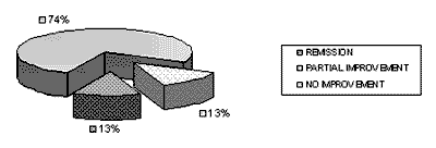
VR duration did not vary in the 8 studied cases and the mean duration of VR was 3 months (6 to 7 sessions). The results of treatment with VR are shown in Graph 1. Clinical improvement was observed in 7 (87.5%) of the 8 studied patients submitted to therapy.
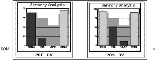
Two patients were submitted to pendulum decreasing rotation test, which showed absence of response to the rotational stimuli in case 3, and extremely reduced reflexes in case 8. Three patients were submitted to CDP, whose response in sensorial analysis of integrated sensorial test was present in the initial exam only for patient 6. After VR, there was improvement in vestibular pattern even in case 3, who did not present vestibular response in the first observation, and in visual and somatosensory patterns in the 3 patients. Data collected before and after VR can be seen in Figure 1.
DISCUSSION
Complete absence of response in caloric irrigation test or pendulum decreasing rotation test is a rare finding in patients with complaints of vertigo and imbalance 1, 10. This fact is justified by lack of literature references on the topic, the size of our sample, and the difficulty to conduct prospective clinical trials. Another restrictive factor is difficulty to compare results reported by authors since diagnostic criteria are quite different.
Oscillopsia was present in 25% of the cases and it was not a prevalent finding. This fact is in accordance with literature findings 1,7,8. However, similarly to the others, this was a retrospective study and the symptom might have not been properly investigated when the clinical history was made. The same can be said about falls, found in 37.5% of the cases, differently from the literature, which describes an incidence of falls of 70% in cases of bilateral vestibular loss in age ranges below 65 years 5.
As to etiology, the literature mentions ototoxicity and idiopathic causes as the most frequent ones 7,8,10,11. However, there was no predominance concerning etiological diagnosis in our sample, probably owing to the small size of the sample.
The results of VR showed clinical improvement in 7 patients (87.5%). Improvement was described specifically by each subject, but they all reported greater stability when performing habitual physical activities.
Since we carefully studied each patient, we describe next the clinical aspects individually observed. Case 1 presented remission of symptoms after two months of therapy and the patient managed to stop using a walking stick. Although the patient still had balance problems after the therapy, the fact that he could move without help was perceived as a significant achievement. This fact made us conclude that even though the patient does not have perfect balance according to our standards, we should consider the patients' expectations. Therefore, the expectations concerning results that we have for a child are different from that of an elderly patient, since the latter may have limited objectives, such as going out without help, for example.
Patient 2 (87 years) had no symptom improvement after rehabilitation; among the factors that interfered we can include advanced age and lack of attendance of the VR therapy sessions. In this case, a consideration is paramount: aging impairs the final treatment outcome. From a functional perspective, it reduces the number of hair cells and vestibular neurons and impairs the gain of vestibular-ocular reflex and limits the speed movements because of difficulty to fix the image on the retina. Displacement of images on the retina is higher and, consequently, maintaining a focus on the retina during head movement is more difficult. There is progressive bilateral vestibular deficit, which justifies the higher prevalence of idiopathic bilateral vestibular loss in this age range 3, 7. Another manifestation of aging is reduction in the capacity to adjust the vestibular system by the CNS, and therefore, it is more difficult to compensate and control the posture. Visual deficits are equally common and result from visual acuity deficits (glaucoma, cataract, macular degeneration), perception of depth, accommodation capability, and uniform chasing. Proprioceptive deficits complement the wide range of factors that limit the appropriate balance in elderly and result in reduction of tactile sensitivity, which is associated with reduction of muscle strength, slower responses and posture hypotension (many times maximized by the use of hypotensive drugs) that hinder recovery from bilateral vestibular loss. Owing to the higher number of risk factors, higher incidence of falls, co-morbidities and reduction of cognition, visual acuity and proprioception, elderly normally needs longer periods of therapy 3, 10. Still referring to the same patient, the second consideration is that inappropriate follow-up during VR hinders the understanding of essential issues in therapy. The insistence on key points, cognitive aspects, correction of exercises, which are rarely understood after the first explanation, in addition to active participation of the patient are essential for successful outcomes of VR.
According to our analysis, most of the patients presented partial improvement with VR, which is related with severe compromise of vestibular afferent information. Postural balance is reached only when information is sent from at least two systems and when there is substantial loss of one or more of them, limitations became greater 12. This fact is more relevant when the vestibular system is damaged, whose information is decisive in case of sensorial conflict between the other two systems. Recovery resulting from bilateral vestibular loss is slower than in cases of unilateral loss, and postural stability is never expected to be completely normal 4. Patients 3 and 4 reported gait and stairs climbing improvement without help. Patient 5 improved posture and started to cross the street without help. Patient 6 reported better performance in home chores, but still had vertigo in open-air environments, an understandable fact since there was no vertical line reference, which serves as visual support. Patient 7 reported greater physical independence, but there was still mild persistent imbalance. Patient 8 reported improvement of dizziness, but he presented difficulties to go up and downstairs without help.
Vestibular rehabilitation per se in patients with bilateral vestibular loss varies a lot according to the literature. One of the implied factors seems to be the adopted treatment strategy. Some authors reported absence of clinical improvement after therapy with VR in 50% of the patients with bilateral vestibular loss 7,8,10. In these studies, the adopted treatment strategy was based on habituation exercises, which are not the most appropriate to treat bilateral vestibular loss 4, 13. It is quite easy to realize that in these cases substitution exercises are required, so that they can stimulate the remaining visual and somatosensory pathways, in addition to the CNS prediction mechanism, since there are no vestibular stimuli to be adapted to. Other authors reported that the patients treated with adaptation and substitution exercises presented better gait and star climbing stability when compared to patients submitted to isometric exercises and conditioning 4,6,13. This aspect can also be easily understood because muscle strength does not present a significant effect on vestibular afferent information and its connections, since it does not exercise balance.
Finally, it is important to evaluate the caloric test as a prognostic tool of the result of VR. Since it is a low-frequency stimulation test, the results may fail to diagnose absence of vestibular function, and the gold standard for diagnosis is considered to be the decreasing pendulum rotation test 14.
Everyday movements have angular speed that ranges from 0.01 to 8Hz and, theoretically, a vestibular dysfunction would affect only part of this frequency interval. The most widely used test to study vestibular deficit is ENG and its main advantages are cost and the possibility of testing each labyrinth separately. Its main limitation is documenting only the horizontal semicircular canal and low frequency angular acceleration (0.002 to 0.004Hz), that is, much below the physiological range to comprise the vestibular-ocular reflex. To assess higher frequencies, the best option is the pendulum decreasing rotation test, which allows bilateral evaluation of vestibular functions in low and high frequencies angular acceleration (0.1 to 1Hz). Its main limitations are cost and difficulty to test frequencies above 2Hz, since acceleration of the body makes it difficult to stabilize the head and the distinction between slow and fast components 15.
Vestibular asymmetries can be precisely diagnosed by ENG because the affected side can be compared to the normal side. However, in bilateral vestibular loss, the pendulum rotation test becomes important to assess the vestibular-ocular reflex, since the absence of ENG response does not exclude the possibility of detecting responses in other vestibular tests, in which other frequencies are assessed 7,8,10,12. However, it does not imply that the diagnosis of bilateral vestibular loss given by the caloric irrigation test should be disregarded, since the responses, even though present in other tests, can be sometimes reduced, confirming vestibular dysfunction. Even though it is not capable of quantifying vestibular deficit, ENG is a relatively sensitive test for early diagnosis of bilateral vestibular loss, since the tendency is that in these cases the loss can be greater in low frequencies 15. We should bear in mind that ENG is a widely used tool in clinical practice and sometimes it is the only test available. To consider and evaluate its results implies the provision to the specialist of data that can help him or her plan and assess the prognosis for the patients. In fact, the two patients submitted to pendulum decreasing rotation test in our sample presented improvement of over 50% of the symptoms, even though the test showed response in only one of them. Thus, we did not detect different responses to treatment when we considered the results of pendulum test combined with ENG.
CDP is useful to quantify posture stability in different situations, measuring the contribution and the interaction of the sensorial afferent information (visual, vestibular and somatosensory). It is useful to objectively assess the functional impact of the vestibular loss and the effect on systems used to maintain balance. In bilateral vestibular loss there is oscillation of sensorial conditions, whose visual or somatosensory afferent information is absent or conflicting. The pattern is quick loss of balance and tendency to use a hip strategy in conditions 5 and 6, typically vestibular 2, 16.
The sensorial analysis of patients submitted to CDP showed absence of vestibular response and reduced visual response in two of them (patients 3 and 8), which presented visual response below that of the normal range (Figure 1). Patient 3 a 44-year-old female subject, referred oscillopsia and her main complaints was the fact that she could not go to environments with intense visual stimulation since she presented falls, which were also detected in patient 8. We can interpret the falls as overload of stimuli provided to the visual system, which takes on a key role to maintain balance before the CNS in the absence of vestibular information 17. Patients with bilateral vestibular loss tend to regain balance based on visual clues, and as time goes by they learn how to use somatosensory clues 18. For this reason, environments that present too much visual information and force the subject to make head movements, which reduce his or her visual reference, such as shopping centers or markets, cause discomfort 19.
In patient 3, post-treatment assessment showed not only improvement in vestibular participation but also visual and proprioceptive improvement, even though the latter had the expected response pattern for age (Figure 1). The improvement of vestibular pattern can be attributed to afferent information sent to other sensitive regions 20, which seems to somewhat suppresses the missing labyrinthic information. Central pre-programming is essential and even though patients present good responses to predictable stimuli, the surprise factor of unexpected movement is the cause of falls to the ground. Despite the results, the patient was very glad since she no longer had oscillopsia, no more falls, she managed to wear high heels, dance and go out at night, which were activities she did not perform before VR.
Patient 8 presented improvement of visual function, which was in deficit, and did not improve vestibular function, as observed in the previous patient. After VR, the patient was very glad even though she still had imbalance when going from one environment to another, explained by lack of visual reference. Another difficulty that persisted was going upstairs and downstairs without support.
The three analyzed patients presented the same grade of improvement, regardless of the response to CDP, and therefore, there was no different response when the results of CDP were associated with ENG.
CONCLUSION
Even though it is not expected to reach complete functional recovery of balance, Vestibular Rehabilitation in cases of bilateral vestibular loss after caloric irrigation is the appropriate management approach to be used. The outcomes can be interpreted as positive as a result of the expectations of the therapy and the patient.Table 1 - Representation of patients and etiologies of vestibular loss.
IVB = vertebral-basilar insufficiency; TCE = head trauma;
NR = not performed; PDRT = pendulum decreasing rotation test;
CDP = computed dynamic posturography.
Table 2 - Distribution of symptoms in patients with bilateral vestibular loss.
(+): symptom present; (-): symptom absent.
Graph 1- Distribution of patients according to clinical improvement presented after VR.
Figure 1- Sensorial Analysis of patient 3 before and after VR program.
somatosensory function; VIS = visual function;
VEST = vestibular function; PREF = visual preference.
Note: The dark bars are below the normal range for age.
REFERENCES
1. Vibert D, Liard P, Häusler R. Bilateral idiopathic loss of peripheral vestibular function with normal hearing. Acta Otolaryngol (Stockh) 1995; 115:611-5.
2. Herdman SJ, Sandusky AL, Hain TC, Zee DS, Tusa RJ. Characteristics of postural stability in patients with aminoglycoside toxicity. Jl Vest Res 1994; 4:71-80.
3. Herdman SJ. Reabilitação Vestibular. 2ª ed. São Paulo, Brasil: Editora Manole; 2002.
4. Herdman SJ, Schubert MC, Tusa RJ. Strategies for balance rehabilitation - fall risk and treatment. Ann New York Acad Sci; 2001, 942:394-412.
5. Herdman SJ, Blatt PJ, Schubert MC, Tusa RJ. Falls in patients with vestibular deficits. Am J Otol; 2000, 21:847-851.
6. Krebs DE, Gill-Body KM, Riley PO. Double-bind, placebo-controlled trial of rehabilitation for bilateral vestibular hypofunction: preliminary report. Otolaryngol Head Neck Surg; 1993, 109:735-41.
7. Sargent EW, Goebel JA, Hanson JM, Beck DL. Idiopathic bilateral vestibular loss. Otolaryngol Head Neck Surg; 1997, 116:157-62.
8. Telian SA, Shepard NT, Smith-Wheelock M, Hoberg M. Bilateral vestibular paresis: diagnosis and treatment. Otolaryngol Head Neck Surg; 1991, 104: 67-71.
9. Pedalini MEB, Bittar RSM. Reabilitação vestibular: uma proposta de trabalho. Pró-Fono; 1999, 11(1):140-4.
10. Gillespie MB, Minor LB. Prognosis in bilateral vestibular hypofunction. Laryngoscope; 1999, 109(1):35-41.
11. Brown KE, Whitney SL, Wrisley DM, Furman JM. Physical therapy outcomes for persons with bilateral vestibular loss. Laryngoscope; 2001, 111:1812-7.
12. Shepard NT, Telian SA. Programmatic vestibular rehabilitation. Otolaryngol Head Neck Surg; 1995, 112:173-82.
13. Herdman SJ, Blatt PJ, Schubert MC. Vestibular rehabilitation of patients with vestibular hypofunction or with benign paroxysmal positional vertigo. Curr Opinion Neurol; 2000, 13:39-43.
14. Fife TD, Tusa RJ, Furman JM, Zee DS, Frohman E, Baloh RW, et al. Assessment: vestibular testing techniques in adults and children: report of the therapeutics and technology assessment subcommittee of the American Academy of Neurology. Neurology; 2000, 55(10):1431:41.
15. Kaplan DM, Marais J, Ogawa T, Kraus M, Rutka JA, Bance ML. Does high-frequency pseudo-random rotational chair testing increase the diagnostic yield of the electronystagmography caloric test in detecting bilateral vestibular loss in the dizzy patient? Laryngoscope; 2001, 111:959-63.
16. Horak FB, Jones-Rycewicz C, Black FO, Shumway-Cook A. Effects of vestibular rehabilitation on dizziness and imbalance. Otolaryngol Head Neck Surg; 1992, 106:175-180.
17. Zee DS. Vestibular adaptation. In: Herdman SJ. Vestibular Rehabilitation. 1st ed. Philadelphia: F. A. Davis Company; 2000. pp. 77-86.
18. Bles W, De Jong JMBV, De Wit G. Compensation for vestibular defects examined by the use of a tilting room. Acta Otolaryngol (Stockh); 1983, 95:576.
19. Clendaniel RA, Tucci DL. Vestibular rehabilitation strategies in Meniere´s disease. Otolaryngol Clin North Am; 1997, 30(6):1145-58.
20. Wiest G, Demer JL, Tian J, Crane BT, Baloh RW. Vestibular function in severe bilateral vestibulopathy. J Neurol Neurosurg Psychiatry; 2001, 71:53-7.
*Assistant, Ph.D., Division of Otoneurology, HCFMUSP.
**Speech and Hearing Therapist, Responsible for the Outpatient unit of Vestibular Rehabilitation, HCFMUSP.
***Ph.D. studies under course, Discipline of Otorhinolaryngology, FMUSP.
****Intern Physician, Division of Otoneurology, HCFMUSP.
Discipline of Otorhinolaryngology, Hospital das Clinicas, Medical School, University of Sao Paulo (FMUSP)
Address correspondence to: Roseli Saraiva Moreira Bittar / Depto de ORL do HCFMUSP
R. Dr. Enéas de Carvalho Aguiar no.255 6o andar, sala 6021 - CEP:05403-000
Sao Paulo - SP - Brazil, e-mail: otoneuro@hcnet.usp.br


