

Year: 2002 Vol. 68 Ed. 6 - (21º)
Relato de Casos
Pages: 912 to 915
Aneurysmal bone cyst of mastoid
Author(s):
Sérgio K. Moussalle 1,
Débora C. Garcia 2,
Anne-Rose L. W. Baú 2,
Patrícia Ferreira 3,
Aline Lazzari 4.
Keywords: cyst bone, aneurysmatic, mastoid
Abstract:
A neurysmal bone cysts are uncommon lesions of the bone, which are osteolytic, expanding and non-neoplastic in nature. They can occur in any place of the human body, but the involviment of the temporal bone is extremely rare. The cause of these lesions remains unclear and the treatment is very difficult. The symptons depends on the part of the human body involved. The radiological findings on simple RX are inespecific, and the findings on CT scan can be suggestive. Definitive diagnosis can be done only by histological study. The first choice of treatment for these lesions has been the complete lesion resection. The incidence of recurrence is direct related with die incomplete lesion resection. Prolonged follow up evaluation is necessary to obtain good results. We present a case report of a 15-year-old young lady with a retroauricular painless enlarging mass. The pacient was submitted to radiological exams that showed a lesion suggestive of a mastoid aneurysmal bone cyst. The pacient was managed by lesion excision, and the diagnosis was confirmed by histological study. Seven years later, the pacient was found to have recurrent disease by radiographic features and clinical manifestations, and underwent a second procedure with complete lesion resection. Actually, the pacient has being followed each six month, and until now is assintomatic and recurrence free.
![]()
INTRODUCTION
Aneurysmal bone cysts are locally aggressive benign lesions that occur in any part of the skeletal structure, but are rarely found in the skull. In the temporal bone, imaging studies have shown an extracerebral heterogeneous mass that causes bone erosion. The cause remains unknown. Some reports suggested the importance of a hereditary component in the etiology of the disease. This lesion can be observed in any age range, but is more predominant between 10 and 20 years of age. The distinction between aneurysmal bone cysts and some benign or malignant bone tumors require imaging techniques and an experienced pathologist. The heterogeneous biological behavior of the aneurysmal bone cyst and its characteristic of affecting growing skeleton make it necessary in various situations to resort to surgical procedures to resect and reconstruct. Since very little is known about its etiology, diagnosis and treatment, we described a case of aneurysmal bone cyst of the mastoid, a very rare location for this type of lesion, with few cases described in the literature.
LITERATURE REVIEW
Aneurysmal bone cyst is an osteolytic expansive non-neoplastic lesion that consists of spaces filled with blood, of varied sizes, which are separated by connective tissue septa, containing bone trabeculae or osteoid tissue and osteoclastic giant cells, according to the World Health Organization. Aneurysmal bone cyst was first described by Jaffe and Lichtenstein in 1942, who studied it separated from other solitary cystic lesions, establishing the diagnostic criteria. Epidemiologically, these lesions amount to 1 to 2% of all primary bone tumors. Any part of the skeleton can be affected, but there is a predominance in long bones and the vertebrae. It rarely affects the head and neck (2.5 to 6%), and the maxilla and mandible are the most affected sites1, 2, 3, 4, 5. The involvement of temporal bone is extremely rare and there are only 10 cases reported in the literature.
Aneurysmal bone cyst usually affects young people, and in 90% of the cases, they are below the age of 20 years, but with no differences concerning gender7. Despite the fact that it is not a malignant neoplasm, aneurysmal bone cyst can grow rapidly and regress spontaneously. According to the literature review conducted by Park et al., headache was the most common symptom. Symptoms are also related to lesion site. The symptoms in the temporal bone type include hearing loss, facial nerve paresis, otorrhea and otalgia. In some cases, a palpable mass of soft consistency can be seen. In the first phase of development, there is osteolysis and elevation of the periosteum. In the second phase, there is bone destruction and in the third phase, progressive ossification. The etiology and pathophysiology are still obscure. Some authors suggested that it is a primary lesion, resulting from venous thrombosis or arterial-venous aneurysm. The most widely accepted theory is that it is a bone primary lesion that starts the formation of a bone arterial-venous fistula in which the aneurysmal bone cyst is created.
Other authors believe that the aneurysmal bone cyst is secondary to a trauma or preexisting bone pathology.
Aneurysmal bone cyst is identified in imaging exams by presenting a suggestive radiological pattern, but the alterations are nonspecific for the definite diagnosis. Head x-rays present lesions with radiological characteristics named soap bubble, radiolucent, expansive, uniloculated or multiloculated, delimited by a thin bone lining, which is also visible by computed tomography scan (CT scan). A characteristic finding in the CT scan, although inconstant, is the presence of horizontal liquid level separating the upper liquid compartment from an inferior sanguinolent liquid compartment. Considering that, definite diagnosis requires histopathology confirmation.
Even though cholesteatoma, cholesterol granuloma or histiocytosis X are frequently considered, aneurysmal bone cyst is part of the differential diagnosis of the temporal bone lesions to be evaluated by the otorhinolaryngologist. Differential diagnosis of aneurysmal bone cyst should be made with benign fibrous-bone lesions including giant cell tumors, giant cell granuloma, brown tumor of primary or secondary hyperparathyroidism (cystic fibrotic osteitis), inflammatory lesions and fibrous dysplasia. The history and radiological findings are determinant for differential diagnosis with inflammatory lesions, such as fibrous dysplasia, cholesteatoma or cholesterol granuloma. Normal calcium serum and parathyroid hormone levels exclude the possibility of brown tumor.
The preferred treatment is surgical resection. Recurrence is directly related to incomplete previous resection5. For surgically inaccessible lesions, residual lesion and recurrent disease, some authors recommend radiotherapy. It should high energy with low dose radiotherapy (500 rads) to reduce the risk of sarcoma induced by radiation, especially in children and young people. There is reference to the fact that recurrence after radiotherapy is lower than after surgery. The cryotherapy technique was reported as producing low rates of recurrence, however, the experience with this technique and the pathology in question is limited and the procedure can expose vital structures to risks, such as the facial nerve, labyrinth and the cochlea. There are reports of attempted embolization with conjugated estrogen through the external carotid artery of the suspected arterial-venous fistula in patients with assumed aneurysmal bone cyst diagnosis, but the studies with this technique are still very scarce.
The prolonged follow-up is essential for the final treatment outcome, especially in children and adolescents, considering that aneurysmal bone cyst tend to recur more frequently during the growth phase of the skeleton.
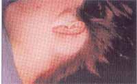
Photo 1. (External inspection): Retroauricular bulging, elastic consistency and well-defined borders.
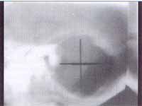
Photo 2. (mastoid X-ray): Expansive lesion in the left mastoid, causing bone erosion and destruction of intercellular septa.
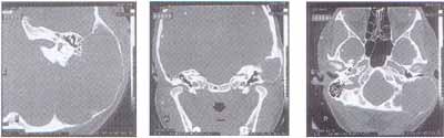
Photo 3 a, b, c. (ear CT scan): Lytic lesion, expansive, thin walls, with characteristics of aneurysmal bone cyst. There is stenosis of the external auditory canal on the same side. Tympanic cavity, ossicle chain and tympanic membrane are intact. Normal inner canal.
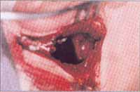
Photo 4. (second trans-operative intervention): Exeresis of giant cysts with liquid and hemorrhagic content.
CASE REPORT
History
Fifteen year-old Caucasian female patient, single, student, born and living in Porto Alegre, Brazil, had a medical visit on 15/10/93, reporting progressive retroauricular bulge on the left mastoid, developing in the past 2 years, but more markedly in the past year. She presented in the period one single episode of severe retroauricular pain. She reported headache, allergic rhinitis, renal lithiasis and back problems (did not specify).
Physical examination
Lesion on the left mastoid, expansive smooth mass of regular borders, soft consistency, depressible and painful upon palpation, with slight hyperemia. Normal otoscopy and tuning fork test.
Complementary tests
Mastoid x-ray: voluminous area of erosion on the left with destruction of intercellular septa, forming a single cavity. Did not reach the attic.
Ear CT scan: voluminous lytic expansive area with thin walls on the left mastoid with characteristics of aneurysmal bone cyst. The process caused external auditory canal stenosis on the same side. The tympanic cavity, ossicle chain and tympanic membrane were intact. Inner auditory canals were symmetrical and did not present abnormalities.
Pure tone audiometry and vocal discrimination: within normal range.
Evolution:
In October 1993, the patient was submitted to resection of the lesion, which intra-operatively was suggestive of mucocele. The macroscopic aspect of the lesion was of irregular shaped tissue, with clots adhered to it, measuring 3 x 2 x 1.7 cm. Upon sectioning, the tissue was yellow-grayish, with hemorrhagic areas and interior calcifications. The histology diagnosis was aneurysmal bone cyst. The surgery and post-operative period were uneventful. The patient returned for periodical visits (every 6 months) and remained asymptomatic and with normal ENT examination. In May 2000, the patient came to the visit complaining of pain and increase of retroauricular progressive volume ipsilateral to the previous lesion, with clinical characteristics and physical appearance similar to those presented in 1993. The findings of head CT scan emphasized on temporal bones were suggestive of recurrent lesion. In July 2000, we conducted a new surgical intervention procedure for the exeresis of the lesion of cystic aspect and with liquid content. Hemostasis was made with surgicel and gelfoam. The patient was discharged 24 hours after with antibiotic administration. Postoperatively, she presented hematoma on the surgical wound and was submitted to various therapeutic punctures, with good progression. Currently, she has been followed up every 6 months. In the last review visit, in March 2001, the patient was asymptomatic, with no signs or symptoms of lesion recurrence.
DISCUSSION
Aneurysmal bone cyst is a rare lesion, especially in the temporal area. The diagnosis of this pathology is a challenge to the otorhinolaryngologist, since various surgical procedures can be necessary to have an appropriate sample of the lesion and establish the definite diagnosis. Upon assessing these patients, it is important to know about the pathology and to have high levels of suspicion in order to make the diagnosis and provide to the patients the correct treatment with complete resection of the lesion, preventing recurrence5, 10.
REFERENCES
1. Ameli NO, Abbassioun K, Azod A, Saleh H. Aneurysmal Bone Cyst of the Skull. Can J Neurol Sci 1984; 11: 466-71.
2. Braun J, Guilburd JN, Borovich B, Goldsher D, Mendelson H, Kerner H. Occipital Aneurysmal Bone Cyst: CT Features. J Comput Assist Tomogr 1987; 11: 880-3.
3. De Moraes Junior LC, Wanderley EC, Tamburus WM, Haddad F, De Andrade AJ. Meningioma da Fossa Posterior com Expansão Extracranial em Criança: Relato de Caso . Arq Neuropsiquiatr 1984; 42: 392-7.
4. Park AH, Phillips J, Forte V. Aneurysmal Bone Cyst of the Temporal Bone. Otolaryngol Head Neck Surg 1999; 120: 606-10.
5. Sheik BY. Cranial Aneurysmal Bone Cyst With Special Emphasis on Endovascular Management. Acta Neurochir (wien) 1999; 141: 601-10.
6. Clavier E, Thiebot J, Godlewski J, Creissard P, Benozio M. Intracranial Aneurysmal Bone Cyst: A rare CT Appearance. Neuroradiology 1988; 30:269-71.
7. Leithner A, Windhager R, Lang S, Haas AO, Kainberger F, Kotz R. Aneurysmal Bone Cyst. A Population Based Epidemiologic Study and Literature Review. Clin Orthop 1999; 363: 176-179
8. Stamm AC. Microcirurgia Naso-Sinusal.. São Paulo: Editora Revinter. 1995: 347-8.
9. Leithner A, Windhager R, Kainberger F, Lang S. A Case of Aneurysmal Bone Cyst in father and Son. Eur J Radiol 1998; 29: 28-30 .
10. Farsetti P, Tudisco C, Rosa M, Pentimalli G, Ippolito E. Aneurysmal Bone Cyst. Long-Term Follow-Up of 20 Cases. Arch OrthopTtrauma Surg 1990; 109: 221-3.
11. Maeda M, Tateishi H, Takaiwa H, Kinoshita G, Hatano N, Nakano K. High-Energy, Low-Dose Radiation Therapy for Aneurysmal Bone Cyst. Report of a Case. Clin Orthop 1989; 243: 200-3.
1Faculty Professor, Service of Otorhinolaryngology, Hospital São Lucas, PUCRS
2Resident Physician, Service of Otorhinolaryngology, Hospital São Lucas, PUCRS
2Resident Physician, Service of Otorhinolaryngology, Hospital São Lucas, PUCRS
3Otorhinolaryngologist, Porto Alegre
4Trainee Physician, Service of Otorhinolaryngology, Hospital São Lucas, PUCRS
Study conducted at the Service of Otorhinolaryngology, Hospital São Lucas, PUCRS. Pontifícia Universidade Católica do Rio Grande do Sul, Porto Alegre Brazil
Address correspondence to: Débora de Carvalho Garcia. Rua Regente n.240, B. Bela Vista, Porto Alegre, Brazil. Tel: 55 51 33 31 22 48. Email: deboraorl@hotmail.com
Study presented as free communication at Congresso Sul Brasileiro de Otorrinolaringologia in Foz do Iguaçu, May 24-26, 2001.
Article submitted on September 20, 2001. Article accepted on October 29, 2001
Print: ![]()