

Year: 2002 Vol. 68 Ed. 6 - (4º)
Artigos Originais
Pages: 800 to 803
Septal haematoma and abscess: study of 30 cases
Author(s):
Raquel A. Tavares 1,
Maura C. Neves 1,
Fernando Veiga Angélico Jr. 2,
Richard L. Voegels 3,
Ossamu Butugan 4
Keywords: septal abscess, septal haematoma, etiology, treatment
Abstract:
Aim: Septal abscess and haematomas are uncommon affections and must be handled with attention as they can result in unpleasent stetic deformities. The objective of this paper was to evaluate it s frequency, age distribution and involved microorganisms. Study design: Clinical retrospective. Matherial and Method: A retrospective study was carried out through the analysis of patient s files with the diagnose of septal abscess or haematoma that occurred in the Clinica Otorrinolaringológica of the HC-FMUSP from January 1989 to December 2000. Results: Twenty eight patients (93,4%) had the diagnose of septal abscess, one patient (3,3%) septal haematoma and in one case we had the association between septal abscess and nasal bone osteomyelitis. The most common cause was nasal fracture, found in 63,3% (n=19) patients. Cultures were obtained in 15 (50%) patients, and Stapphilococcus aureus was the most prevalent agent. All patients (n=30) were submitted to drainage of the abscess or haematoma. Among the complications the recurrence was the most common and happened in 7 patients, palatal abscess due to septal abscess (6,6%), septal perforation (3,3%), sinusitis (3,3%), intra-nasal sinequiae and facial celulitis (3,3%). The most used antibiotic was cefalotine (40%). Conclusions: Septal abscess and haematomas are rare. To deal properly with this entity we must have early clinical suspicion, promptly surgical drainage and appropriate antibiotic therapy. This can diminish complications such as nasal deformities and sepsis.
![]()
Introduction
Septal abscesses and hematomas are quite rare affections, despite the frequent lesions of the face and nose. However, they should be managed carefully, since they can result in unpleasant cosmetic deformities. The purpose of the present study was to assess frequency, distribution by age range, microorganisms involved and sequelae. We also included a review of the literature.
Material and Method
The present study was a retrospective analysis conducted by studying the medical charts of patients diagnosed with septal abscess or hematoma seen in the Division of Clinical Otorhinolaryngology, Hospital das Clinicas, Medical School, University of São Paulo, in the period between January 1989 and December 2000.
All patients were submitted to drainage of blood collections in the emergency room or operating room and treated with intravenous antibiotic therapy. Corticoids were used in the absence of clinical complications.
The following aspects were investigated: age, gender, diagnosis, etiology, co-morbidity, isolated microorganisms and clinical complications.
Results
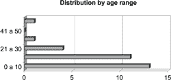
We analyzed 30 patients, being 26 (86.6%) male and 14 (13.4%) female subjects. The ages ranged from 3 to 60 years (Graph 1).
Twenty-eight patients (93.4%) had septal abscess as diagnosis, one patient (3.3%) had septal hematoma, and in one patient (3.3%) there was association between septal abscess and nasal bone osteomyelitis.
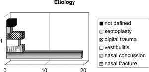
The most commonly reported cause to the genesis of the septal abscesses and hematomas was nasal fracture, found in 63.3% (n=19) of the patients. Next, we found nasal concussion (13.4%), vestibulitis (10%), digital trauma (3.3%) and septoplasty (3.3%). In two patients, the cause was not defined (Graph 2).
Only one patient presented immunodeficiency (myelodysplastic syndrome).
Cultures were made in 15 (20%) of the subjects, and the most prevalent microorganism was Staphylococcus aureus, detected in 42.9%. Other isolated agents were Streptococcus viridans (21.4%), Enterococcus faecalis (7.1%) and Streptococcus pyogens (7.1%).
All patients (30%) were submitted to abscess or hematoma drainage, being 13 in the emergency room and 17 in the operating room. All children were submitted to drainage in the operating room and under general anesthesia.
Anterior nasal packing was used in 96.7% (n=29) of the cases and Penrose drain was used in 83.3% (n=25), both for a period of 2 days.
Among the complications observed, recurrence of the clinical episode was detected in 7 patients (23.3%), followed by saddle nose (6.6%), abscess of palate secondary to septal abscess (6.6%), septal perforation (3.3%), sinusitis (3.3%), intranasal synechiae (3.3%) and facial cellulitis (3.3%). One patient developed facial cellulitis, pansinusitis and frontal empyema as a result of the septal abscess.
The most frequently used antibiotic was cefalotine (40%). The association between crystalline penicillin and chloramphenicol was used in 26.8% of the patients, followed by crystalline penicillin isolated (6.7%), clindamycin (6.7%), association of cefalotine and metronidazole (3.3%), oxacillin (3.3%), association of oxacillin and crystalline penicillin (3.3%), association of oxacillin and chloramphenicol (3.3%) and chloramphenicol only (3.3%). One patient, who developed frontal empyema as complication, was treated with oxacillin, metronidazole and ceftazidime for a prolonged period of time.
Graph 1. Distribution by patients age range.
Graph 2. Distribution by patients etiology.
Discussion
Nasal septum hematoma and abscess are defined as blood or pus collections between the bone and cartilaginous septum and the mucoperiosteum or mucoperichondrium 1.
They are not common entities and the real prevalence is not well established in the world literature, but it seems that it has changed throughout the years and in different centers. As to the pediatric population, we can see a relatively low prevalence, especially if considering the frequent facial traumas that are inevitable in children2, and if we take into account that the nose is one of the most damaged organ in the body. Blahova3 reported septal hematoma or abscess in 25 children from a series of 241 children that had suffered nasal trauma.
McGillieuddy4 stated that it is very important to examine the nasal septum of a child or adolescent who have suffered a trauma and to conduct a nasal bone x-ray, since septal hematomas are considered medical emergency. It is known, however, that the reality is different. Barrs and Kern reported that 50% of 100 children assessed in an emergency service after nasal trauma were submitted to nasal x-ray, whereas intranasal examination was conducted in only 20 children5.
As to gender, the literature suggested that there was great predominance of male patients with septal hematomas and abscesses in all age ranges1, 6. The incidence is the same only for car accidents. Twenty-six patients in our study were males. As to age range, most patients were below the age of 20 years.
Nasal surgery, nasal trauma caused by assault, sport activities or car accidents, minor facial lesions, dental abscesses, ethmoid and sphenoid sinusitis, and nasal furunculum, have been described as etiological factors7. In our study, nasal fracture was the main etiological factor. In children younger than 2 years we should suspect of child abuse5. Spontaneous onset has been rarely observed in clinical practice8, maybe owing to digital manipulations not detected by the physician and the patient. However, in the absence of preceding trauma, the source of infection should be defined7 before it is considered a spontaneous cause.
Even though the pathophysiology of a hematoma subsequent to a nasal trauma is unknown, it is believed that there are mechanical forces applied against the cartilage that result in rupture of small vessels of the mucoperichondrium. In cases in which the cartilage is fractured, the blood can dissect through the fracture line and cause bilateral hematoma. When the hematoma is not initially recognized and immediately drained, it can expand and mechanically obstruct the vessels that supply the nasal cartilage, leading to avascular necrosis induced by pressure within 3 to 4 days. Accumulated blood and necrotic tissue work as excellent media for bacterial culture that colonizes the nasal mucosa and leads to formation of abscess.
The diagnosis is made about 6 days after the initial event5-9. Nasal obstruction is the most common symptom associated with local pain, local fluctuation, and reddish edema of septal mucosa, which is not reduced after application of vasoconstrictors. There may also be unusual clinical presentations. Cuddihy and Srinivasan10 reported the case of nasal septum abscess in a 21 year-old girl that developed oroantral fistula.
The distinction between non-complicated hematoma and infected hematoma is difficult, especially if some days have passed since the initial trauma. However, the differential diagnosis between abscess and hematoma does not modify the treatment significantly. Ninety-three percent of the patients analyzed presented septal abscess.
Diagnostic errors are normally made by physicians who are not familiarized with otorhinolaryngology and they are not rare3.
Cultures were made for 50% of our patients, being that the most prevalent microorganism was S. aureus (42.9%), confirming the data from the world literature. It was followed by S. viridans (21.4%). Enterococcus faecalis and S. pyogens were also found. H. influenza and S. pneumoniae should be also considered, especially in children10, 11. Anaerobe and coliform microorganisms are less frequently found.
Thomson and Berkowitz reported the case of an extradural frontal abscess as a complication of the septal abscess in which there was growth of S. milleri, a case that had not been reported yet, despite the fact that this agent is frequently found in central nervous system infections2.
The etiological agents can vary in immunodepressed patients, as well as the evolution of the clinical presentation. Kean12 reported the case of a 71 year-old patient who had gastrointestinal cancer and after chemotherapy, she developed septal abscess. She presented clinical history of nasal obstruction for 3 weeks. The cultures made before and after surgical drainage revealed the presence of Pseudomonas. There was only one immunodepressed patient in our sample, who had myelodysplastic syndrome, but his cultures did not reveal bacterial growth, maybe owing to the wide antibiotic spectrum he received.
Complications can be severe. They range from cosmetic deformities to intracranial complications, such as subaracnoid empyema, cerebral abscess and meningitis.
The intracranial dissemination routes are varied1. The nasal septum has venous networks with numerous anastomosis. The anterior septum veins communicate with the upper lip vein and the palate vein that communicate with the facial angular vein and ophthalmic veins, leading the infection to the cavernous sinus. Ethmoid veins contribute to such dissemination by direct communicating with the superior sagittal sinus. Posterior drainage continues through the pterygoid plexus via deep facial veins, and the pterygoid plexus communicates with the cavernous sinus via the branches that go through the oval and lacerum foramens.
There are still other intracranial dissemination routes. Direct invasion can still occur as a result of surgery, traumatic fracture, congenital deformity or local erosion. The lymphatic vessels of the superior meatus drain the cribiform plaque and the perpendicular lamina of the ethmoid to the subaracnoid space.
Owing to the severity of intracranial complications, Thomson and Berkowitz proposed criteria to conduct computed tomography scans in the pediatric population2:
1. Extensive facial cellulitis;
2. Meningitis, consciousness abnormalities, focal neurological signals;
3. Severe headache;
4. Poor improvement after drainage of the abscess;
5. Long time between presentation and diagnosis;
6. Isolation of unusual or virulent microorganisms.
The recurrence of the clinical picture was not unusual in our study (23.3%). Only one patient presented focal cellulitis and frontal empyema. Saddle nose was observed in only one patient.
Cosmetic deformities can be late and severe, especially in younger children. Wilson and Mitward11 reported the case of a boy who some months after nasal trauma and septal hematoma presented saddle nose. Early diagnosis and treatment are the best way to prevent cosmetic deformities.
There are reports that Black noses are more resistant to cosmetic deformities than Caucasian noses7. There are no reasons serving as evidence for such observations.
Once defined the diagnosis, immediate treatment should be administered. First, it is recommended to have aspiration with local anesthesia and the material should be sent to gram staining and culture for aerobic and anaerobic agents. Intravenous antibiotics should be administered aiming at covering the main agents. In our revision, cefalotine (8g/d) was the most frequently used antibiotic.
After the first dose of antibiotic, incision and drainage of the collections can be performed with local anesthesia in adults and/or general anesthesia in patients that do not support local anesthesia, as well as in children. Bilateral incisions should be made when there are fluctuations on both sides. A small Penrose drain is inserted and sutured on the site. Anterior packing should be used and left in place for 48 to 72 hours. In non-complicated cases, intravenous antibiotic therapy is prescribed for 3 to 5 days after drainage and oral antibiotics are maintained for more 7 to 10 days.
Conclusion
Nasal septal abscess and hematoma are defined as a collection of blood or pus between the bone and the cartilaginous nasal septum and the mucoperiosteum or mucoperichondrium. They are not common entities and the real prevalence has not been defined in the international literature yet. There is predominance of male patients in all age ranges. Nasal surgery, facial trauma and other causes have been described as the etiological factors. Pathophysiology is unknown. Early diagnosis and immediate treatment are essential for prevention of complications.
REFERENCES
1. Ambrus PS, Eavey RD, Sullivan Baker a, Wilson WR Kelly JH. Management of nasal septal abscess. Laringoscope. 1981;91:575-582.
2. Thomson CJR; Berkowitz RG. Extradural frontal abscess complicating nasal abscess in a child. International Journal of Pediatric Otorhinolaryngology. 1998,45: 183-186.
3. Blahova, O. Late results of nasal septal injury in children. Int. J. Pedratr.Otol. 1985;10:137-141.
4. McGillicuddy OB. Hematoma of the nasal septo. Michigan Medicine.1972. Jan.
5. Barrs DM, Kern EB. Acute nasal trauma: emergency room care of 250 patients. J Far Pract. 1980;10:225-228.
6. Matsuba HM,Thawley. Nasal septal abscess: unusual causes, complications, treatment and sequelae. Annals of Plastic Surgery. 1986, February, vol 16 no.2 161-166.
7. Chukuezi AB. Nasal septal hematoma in Nigeria. J.Laryngol.Otol. 1992,106:396-398.
8. Koltai PJ, Rabkin P. Pediatric facial trauma. Pediatric Otolaryngology for General Otolaryngologist.Tokyo: Igaku-Shoin,1996.
9. Viscasillas J,Viscasillas S, Clarós P. Abscesos y hematomas del tabique nasal en la edad pediatrica. Anales ORLIber-Amer 1984 XI,5-6,419-429.
10. Cuddhy PJ, Srininivasan DLO.An unusual presentation of nasal septal abscess. The Journal of Laryngology. 1998, Aug 112:775-776.
11. Wilson SW, Milward TM. Delayed diagnosis of septal hematoma and consequent nasal deformity. Injury,1994 vol 24 no. 10 685-686.
12. Kean H. Pseudomonas nasal septal abscess. Pennsylvania Accademy Of Ophtalmology and Otolaryngology, May,25 Bedford, Pennsylvania.
13. Canty PA, Berkowitz. Hematoma and abscess of nasal septum in children. Arch Otolaryngol Head Neck Surg. 1996 122:1373-1376.
1 Resident Physician, Division of Clinical Otorhinolaryngology, Hospital das Clinicas, Medical School, University of São Paulo.
2 Physician, Post-graduation under course, Division of Clinical Otorhinolaryngology, Hospital das Clinicas, Medical School, University of São Paulo.
3 Ph.D., Assistant Physician, Division of Clinical Otorhinolaryngology, Hospital das Clinicas, Medical School, University of São Paulo.
4 Associate Professor, Division of Clinical Otorhinolaryngology, Hospital das Clinicas, Medical School, University of São Paulo.
Study conducted at the Department of Otorhinolaryngology, Hospital das Clinicas, Medical School, University of São Paulo.
Study presented at II Congresso de Otorrinolaringologia da USP, held on November 2001 and awarded with a Special Citation.
Address correspondence to: Raquel Tavares Departamento de Otorrinolaringologia do HC-FMUSP.
Av. Dr Enéas de Carvalho Aguiar, 255 6o andar sala 6021 05403-000 São Paulo SP
Fax: (55 11) 3088-0299
Article submitted on August 26, 2002. Article accepted on October 10, 2002
Print: ![]()