

Year: 1974 Vol. 40 Ed. 2 - (66º)
Artigos Originais
Pages: 374 to 376
Cysticercosis in the Labial Mucous Membrane
Author(s):
Lidio Granato* e
Antonio Luisi**
![]()
Histopathological examination will often lead to the diagnosis of lesions not suspected during clinical examination. We will describe such a case in a girl who had a small cystic lesion in the upper lip, clinically resembling a common salivary cyst, which proved to be a Cysticercus after the histopathological examination was made. Owing to the unusual localization of oral cysticercosis in man, since no similar case has been reported or published in our country, we thought it would be useful to present the case we have seen.
Case Report:
This is the case of a 7 year old girl who was feeling uneasy because of a small tumour in the inner part of the upper lip, with no pain but progressively increasing in size, she had noticed it for 6 months before our examination. During inspection we noticed a small elevation in the part of the upper lip, a pea-grain-like nodule with determined limits and no tenderness. The patient general condition was good and subsidiary examinations were all negative, except for the presence of Ascaris Lumbricoides in the faeces.
The patient was operated upon with the clinical suspecion of a salivary cyst, and the lesion was easily removed because it was superficially located and well defined. The anatomic-pathological examination showed it to be a cystic lesion, the size of a pea-grain, and showing by trans-illumination a small central opaque zone the size of a rice-grain. After immersing it into paraffin and cutting it, we noticed that it was a cysticercus. In the surrounding area the presence of a fibro-hyaline wall and inflamatory cells were noticed. No granulomatous reaction was noticed in the material and its relation to the parasite was altered by the superficiality of the lesion, which was excised with practically no labial tissue (figures 1 and 2).
Comments
Cysticercosis is the name by which the infestation in animal bodies by the Taenia Larva is known. The disease has been known since antiquity when it was first observed by the Greek people, whose cooks used to remove sub-lingual cysts in animals. According to Gaivão1 (1928), Hebrews and Egyptians also knew about this problem, and this is probably why Moses and Mahommet prohibited their people to eat pork. The Teniase disease is reported to affect persons who eat infected pork meat and with poor hygienic conditions. Cases of this discase are frequently reported in Europe, Asia and India, in some regions of African continent and in America.
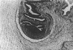
FIG. 1 H. E. 16 x - Cysticercus cellulosae. The parasite with the membrane (amnios) .
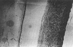
FIG. 2 - H. E. 160 x - Fibro-hyaline wall with inflammatory reaction.
It is a common disease in our country, particularly in São Paulo. There are several possible routes of infestation with the disease:
1. Internal auto-infestation
2. External auto-infestation
3. Hetero-infestation. This is probably the most usual route of infection, in which the larvae are swallowed with polluted water and food.
When the larval ovum is swallowed, it liberates the oncosphere which reaches the intestines and enters the blood circulation through the mesenteric veins, thus demaging various seccors of the body. Starting from the embryo a larval formation is developed which consists of an invaginated head and neck: within a vesicle. It is clear and semi-transparent, measuring from 10 to 15 mms, and there is a crystalline liquid inside it. From the point of view of morphological observation, the reaction of the tissues to the living cysticercus is not intense, bus with its death the body reacts as to a foreign body, thus resulting in inflammatory reactions of diverse degrees, and subsequently to fibrosis and certain degrees of calcareous impregnation. In relation to the localization and incidence of the disease, Tretiakoff and Pacheco e Silva2 (1924) found 3,6% of cysticercosis in autopsies performed at the Juqueri's Central Hospital in Franco da Rocha, São Paulo; Salles M. (1934) in material represented by 4.000 necrópsies made at the São Paulo Medical University, a rate of 0,12% was observed. In both cases the central nervous system was affected as the principal location. Burnier (1962) apud Pessoa S.3, studied a total of 350.000 patients in the Penido Burnier Clinic and reported a total of 218 cases of ocular disease. Subcutaneous localization comes in 3rd. place, after the Central Nervous System and eyeball, followed by less usual localization in the liver, kidney, myocardium and tongue. Pupkin and his collaborators4 (1967) presented a case occurring in Chile, in which cysticercus was located in the dorsal part of the tongue. In reviewing the literature on this subject, the same authors referred to another 9 cases occurring in the tongue, 8 being reported by Dixon and Lipscomb5 (1961) and the most recent one by Puppo, P. P.6 (1965). From 31 cases of Cysticercosis observed in the Pathological Anatomy Service of the Medical University of Pôrto, represented by material of 72.00(1 histological examinations, Costa7 (1965) reported only one case of localization in the mouth. Czernic7 (1972) reported one case of Cysticercosis in the mucous membrane of the upper lip, in a 24 year-old patient. We may see from the bibliographical review that these cases are really rare and the principal aim of this publication is to call attention to the possibility of finding this kind of pathology in the oral mucosa.
Bibliography
1. Galvão, S. T.: Incidence and Prophylaxis the Kisto Hydatico in São Paulo. Inaugural Thesis - Faculdade de Medicina de São Paulo (1928)
2. Trétiakoü, C.; Pacheco e Silva, A. C.: Contribution to the study of cerebral cysticercosis particularly those of the toxic cerebral lesions.
3. Pessoa Mem. Hosp. de Juqueri. 1: 1 37-66, 1924.
4. Pupkin, J. Apt., W., Riviera H.: Cisticercosis de la lengua Bol, Chileno Parasit. 22:66-68, 1967.
5. Dixon, H. B. F., y Lipscomb, F. M. 1961 - Cysticercosis An Analysis and Follow-up of 450 cases. Privy Council Medical Research Council. Special Report series n ° 299 - London 58 pp.
6. Puppo, P. P. 1965 - Cysticercosis of the tongue (Clinical Case Report) An. Esp. Odontorstomat, 24:35-40.
7. Costa, I. G.: Parasithologycal Cysts in the mouth. Rev. Port. de Estomat. 6:99 - 106, 1965.
8. Czernic, S.: Cysticercose de la lèvre et des extremités superieures. Canadian Journal of Oto-laryngology. 1: 67-72, 1972.
Dr. Lidio Granato
Santa Casa de São Paulo
Rua Cesário Motta Jr., 112
Departamento de Otorrinolaringologia
São Paulo - SP
Brasil
* Assistant Professor, Otorhinolaryngology Department, Medical Sciences University of Santa Casa de São Paulo.
** Associate Professor, Pathology Department, Escola Paulista de Medicina - São Paulo (Professor J. Michalany).
Print: ![]()
All rights reserved - 1933 /
2026
© - Associação Brasileira de Otorrinolaringologia e Cirurgia Cérvico Facial