

Year: 1974 Vol. 40 Ed. 2 - (25º)
Artigos Originais
Pages: 218 to 223
The Effect of Single Doses of Sodium Salicylate on Cochlear Structure and Function
Author(s):
José Antonio A. Oliveira* e
Ricardo F. Marseillan*
![]()
It has been known for a long time that certain derivatives of the salicylic acid, especially sodium salicylate, could induce deafness when administered in large doses. The hearing loss has the characteristics of an endorgan lesion, but is generally considered to be reversible. In this aspect the salicylates differ from most other ototoxic drugs, notably the aminoglycoside antibiotics. (For references see, for example, Pohlman e Kranz (1922), Falbe-Hensen (1941), Oliveira (1971), Mitchnell et al. (1973). In experimental animals, the cochlear ototoxicity of the salicylates has been investigated in several ways. The histological studies of Kirchner (1883) and Mosher (1938) showed middle and inner ear hemorrage attributed to the drug, results not confirmed by others (Wittmach, 1903, Blau, 1904, Hyke, 1904, Orembosky, 1922). Involvement of the neural auditory pathway, specifically the spiral ganglion, was detected by Wittmach (1903) and also by Beck (1913) and more recently by Taylor (1937), Lurie (1935) and Falbe-Hansen but denied by other investigators, that could not find any kind of neural lesions (Lindt, 1913, Schroder and Hinsberg, 1915, Covell, 1938). Also controversial are the morphological studies of the organ of Corti in experimental salicylate intoxication. Blau (1904) and Hyke (1904) reported rather extensive injury of the hair cells and of the support structures, whereas Covell (1938) suggested that the salicylates were direct protoplasmatic poisons, because he found the mitocondrial apparatus of the vascular stria cells and hair cells severely disrupted. Degeneration of the external hair cells was shown by Lurie (1935) and changes of the position of the basilar tectorial and round-window membranes by Falbe-Hansen (1941). The more recent histological study of the inner ear under salicylate has been done by Myers and Bernstein (1965) in the monkey, reporting no cellular alterations of any kind. Electrophysiological methods were first used by Gold and Wi1pizeski in 1966, finding that with a plasma level of salicylate of over 30 mg/mi in the cat the response threshold of the evoked responses in the cochlear nucleus was elevated. In the guinea pig, Silverstein, Bernstein and Davies (1967) measured the cochlear potentials after a single 350 mg dose and determined their depression. In the same species it has recently been reported that sodium salicylate does not modify the cochlear microphonics, but induces a temporary depression of the eighth nerve action potential (Mitchell, Brummett and Vernon, 1973). The purpose of the present investigation is to test the effect of single large doses of sodium salicylate on cochlear morphology and function, using modern experimental techniques.
Methods
The experiments were performed in 108 guinea pigs of both sexes, weighing from 300 to 500 g, selected for a good pinna reflex. Drug administration. Sodium salicylate was utilized as a 10°io solution in water with 5% 2 to 4 ml of the above solution. For the experiments requiring intratympanic administration, the animals were anesthetized with sodium pentobarbital, placed in a special head-holder and trephine hole made on the mastoid ridge. The drug was administered and the hole closed with dental cement, keeping the guinea-pigs for 30 minutes in an appropriate position so that the round window would be in an horizontal plane. Histological technique. The cochleas of the guinea pigs treated with sodium salicylate were examined for histopathological changes utilizing the surface preparation technique, modified from the basic technique of Engström Ades and Hawkins (1963). The principal steps of the procedure are the following (Oliveira and Marseillan, 1971, Oliveira, 1973).
a) Specimen removal. The animal is decapitated the skin and muscles removed and the cranium opened with a bone forceps. Immediately, both temporal bones are separated manually, the bulla opened and the middle ear bones removed. With a sharp stylet the round and oval window openings are enlarged in order to prepare a lower cochlear opening. Another opening is made at the cochlea apex into which the fixative is made to flow. The fixative used is a 10% formaline solution with pH 7. This fixative is more convenient for surface preparations not intended for electron microscopy, instead of the more usual osmium tetroxide: it is easier to saline perfusions are required.
The specimen may be left in the fixative for a long time without changes in the result.
b) Surface preparations. Using a stereomicroscope, the fixed cochleas are separated from the remainder of the temporal bone and the bony shell of the cochlea opened with curved stylets. Then, with a straight stylet, approximately two thirds of the basilar membrane of each turn are separated from the bony spiral lamina, and the segments mounted on glass slides with glucerin.
c) Study of the preparations. Using a phase-contrast microscope (Zeiss Photomicroscope) the mounted surface preparations are examined and photographs taken using magnifications from 560 to 1400 X. The photomicrographs are obtained focussing at three different planes: cilia, nuclei, and basilar membrane.
Electrophysiological technique. Electrodes were permanently implanted on one round window of the internal ear, and the electrical activity of the cochlea measured against a reference electrode in the neck muscles. The electrodes were of Formvar-insulated 34gauge stainless steel wire. One end was stripped bare, curved to form a loop and placed on the round window membrane. The other end was also stripped, tinned, passed under the skin and soldered to a connector fixed to the skull with screws and acrylic resin. The electrode was held in position and the opening in the bulla sealed with zinc oxiphosphate cement. Sound stimulation was produced with tone bursts in a closed acoustical system and with clicks in an opera one. The potentials were amplified, filtered and diplayed on the screen of a cathoderay oscilloscope for photography and measurement. The potentials recorded were 1, 2 and 4 kHz cochlear microphonics and the click-induced VIIIth nerve compound action potential. Further details of the technical procedures are given elsewhere (Marseillan, 1963, 1965).
Results
Histological investigation
In this series sodium salicylate was administered in a single dose, by either the intraperitoneal or intratimpanic routes, in 8 groups of guinea pigs. Two additional control groups received saline. Table 1 shows the dosage of the different groups.TABLE I
I.P.: intra peritoneal administration
I.T.: intratympanic administration
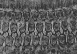
The animals were sacrificed 2, 4, 8, 16, 24 and 48 hours after receiving the drug, and their temporal bones excised for the surface preparations. No visible alterations of the external and internal hair cells were observed in the animals of groups 1, 2 and 3.
Their surface preparations did not differ from those obtained from control groups 9 and 10. Fig. 1 and 2 show photomicrographs of the organ of Corti of 2 animals of these groups.
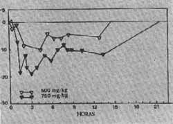
FIG. 1 - Changes in the amplitude of cochlear potentials after administration of a single dose (500 mg/kg) of salicylate.
FIG. 2 - Changes in the amplitude of cochlear potentials at 1 kHz after the administration of two doses (500 mg/kg or 750 mg/kg) of salicylate.
The guinea pigs treated with the higher doses - groups 4 to 7 - were observed for the development of signs of imminent death (convulsions intense tachychardia) and sacrificed. The longest survival time of these animals was 100 minutes. The surface preparations of the inner ears these guinea pigs showed normal cochleas. Similarly, no abnormalities were detected in the cochleas of the 5 guines pigs injected by the intratympanic route and sacrificed after 20 hours (Group 8).
Electrophysiological investigation
In these experiments 9 guinea pigs with permanent round-window electrodols received single intraperitoneal doses of sodium salicylate (Table II). Each animal was tested repeatedly, with the same or different drug dosage. The amplitude of the cochlear potentials was compared with pre-drug control measurements made during two weeks. The stability of the cochlear potentials during the control period was very good, as has been shown before (Marseillan, 1965). After drug administration, the animals were tested at different intervals up to 48 hours, recording cochlear microphonics and VIIIth verve action potentials.TABLE II
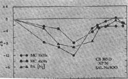
In groups 11 and 12 a fall in the amplitude of the cochlear microphonics was recorded, especially for high frequency tones. This fall was rather sudden, starting 30 minutes and attaining its maximum 2 to 3 hours after drug injection. For 1 kHz stimuli, the maximum decline was 4 dB, and 10 dB for 4 kHz. Figure 3 shows a typical curve. After 20 hours the amplitude of the potentials had returned to control values in all the guinea pigs. The amplitude of the N1 wave of the action potential was reduced 2 hours after administering the salicylate and showed a maximum reduction of 7 dB 6 hours after drug injection. Changes in the N2 wave were similar to N1, but reached a maximum reduction of 11 dB. The shape and amplitude of the action potential was back to normal after 20 hours. In the experiments of group 13, with higher dosage, the fall of the potentials was more marked Fig. 4.
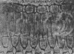
FIG. 3 - Photomicrographs of the Corti organ after the administration of a single dose (750 mg/kg) of salicylate.
DISCUSSION
Silverstein Bernstein and Davies (1967) have reported that in the cat salicylate intoxication produced a fall in cochlear electrical activity, suggesting that the hearing loss observed with high doses of the drug could be result of its effect on the cochlea.
FIG. 4 - Photomicrographs of the Corti organ after the administration of a single dose (750 mg/kg) of salicylate.
The pattern of the changes in cochlear electrical out-put of the guinea pigs of our experiments was similar to the results obtained in the cat, the action potential of the eighth nerve was more depressed than the cochlear microphonics; and the high frequency cochlear microphonics were more affected than the low frequency ones. The effect was more marked with the higher dosages, but the return to normal pre-drug levels of electrical activity was the same for all groups. Regarding the histological findings in salicylate intoxication, the results of previous investigations are contradictory. Lesions of the external hair cells and of the support cells have been reported (Blau and Hyke, 1904), but Myers and Bernstein (1965) could not find pathological changes in the cochlea after acute administration of the drug, in a study that used light-and electron-microscopy. Our observations, using surface preparations of the organ of Corti also failed to show any kind of alterations, after single-dose administration. These results indicate that, if the effect of sodium salicylate on hearing is duo to a disfunction of the hair cells of the organ of Corti, this disfunction does not have a morphological expression. This is also valid for the more direct intratympanic route of administration. Two hypothesis have been discussed in relation to the toxic mechanism of the salicylates on the inner ear: changes in hair cell metabolism and alterations of labyrinth fluids. As it has been recently shown (Smith, 1963), salicylate has an inhibitory effect over at least three groups of cellular enzimes. It depressos oxidative phosphorilation, and inhibits certain transaminases and several pyridine nucleotide de hidrogenases. The clinical finding of hearing loss has been explained as a consequence of these biochemical alterations, that would bring about changes in the bioelectrical properties of the cochlear partition (Myers and Bernstein, 1965; Myers, Bernstein and Fostiropoulos, 1965). This hypothesis is further corroborated by the finding of a diminished activity of malic dehidrogenase in the endolymph during salicylic intoxication (Silverstein, Bernstein and Davies, 1967). They also showed that the inhibition of one or more of these enzimes determined a decrease of the endocochlear potential, that returned to normal values with elimination of the salicylate. There is also the possibility that biochemical alterations of the labyrinthine fluids could affect the metabolism of the stria vascularis. Schuknecht (1964) has reported that this structure is frequently found atrophied - but with normal hair cells - in cases of bilateral audibility threshold increase for all tones. In these cases, the reduced metabolism of the stria would cause a fall in the endocochlear potential and consequently a diminished inner ear sensitivity.
Summary
The effect of acute administration of sodium salicylate by intraperitoneal and intratympanic routes on cochlear morphology and function was studied in 108 guinea pigs. Using round-window permanent electrodes, the effect of single intraperitoneal doses of the drug on cochlear microphonic and eighth nerve action potentials was investigated by recording their amplitudes at short intervals for periods of up to 48 hours after injection. Both cochlear microphonic and action potentials showed a fall, that was more marked in the case of the hiah-frequency cochlear microphonics and the N2 wave of the action potential. In all cases, cochlear electrical activity returned to normal in 20 hours. The effect was increased with an increase in dosage. No alterations in hair cell morphology or cochlear cytoarchitecture were seen in the salicylate intoxicated guinea pigs, after single-dose intraperitoneal and intratympanic administration.
Sumário
O efeito da administração de salicilato de sódio por via intraperitoneal e intratimpânica na morfologia e função coclear foi estudado em 108 cobaias. Usando eletrodos permanentes implantados na janela redonda o efeito de doses intraperitoneais únicas da droga foi injetado por registro das amplitudes dos potenciais microtônicos cocleares e de ação em intervalos curtos de tempo até 48 horas após a injeção. Ambos potencial microfônico coclear e de ação mostraram queda, que foi maior no caso de microfônicos cocleares de alta freqüência e onda N., do potencial de ação. Em todos os casos a atividade elétrica coclear retornou ao normal em 20 horas. O efeito aumentou com o aumento da dose. Nenhuma alteração foi verificada na morfologia das células ciliadas ou citoarquitetura coclear das cobaias intoxicadas após administração de uma única dose intraperitoneal ou intratimpânica.
Bibliografia
1. Beck, C.: Feinere Stoffwechselreaktlonen an den Sinneszellen dos Cortischen Organs. Arch. Ohren. u. s. w. Heilk u. z. Hals u. s. w. Heilk, 1959, 175: 374-378.
2. Blau, A.: Experimentelle studlen über die Veränderungen in Gehörorgan nach Vergiftung mit salizylsaürem Natrium. Arch. f. Ohremheilkunde, 7904, 61: 220-233.
3. Covell, W. P.: Histopathology of peritoneal auditory mechanlsm in drug lniected animais. Ann. Otol. Rhinol. Laryngol., 1938, 47:342346.
4. Engstrom, H., Ades, H. W., Hawkins, J. E. Jr. Cytoarquitecture of the organ of Corti. Acta Oto-laryng., 1963, Suppl. 188:92-99.
5. Falhe-Hansen, J.: Clinicai and experimental histological studies on effects of salicylate and quinìne on the ear. Acta Oto-laring. 7941, Suppl. 44 : 1-276.
6. Gold, A., Wilpfzeski, C. R.: Studies in auditory adaptation. Some effects of sodium salicylate on evoked auditory potentials in cats. Laryngoscope, 1966, 76:674-685.
7. Haike, H.: Experimentelle Untersuchunges zur Kenntnis der Wirkung der Natrlum Salicyllcum und dos Aspirins auf das Gehörorgan. Arch. für Ohrenheilkunde, 1904, 63:78-79.
8. Kirchner, W.: Extravasate in Labyrinth durch Chinin und Sallcylwirking. Monatschr. f. Ohrenheilk, 7883, 17:85 (Cif. por Falhe-Hansen, 7941).
9. Lindt, W.: Experimentelle Untersuchungen über den Einfluss dos chinins und natr. salic. auf das Gehõrorgan der Tiere. Verh, dtsch. Naturdorsch Arzte., 7913, 2:826.
10. Lurie, M. H.: Animal Experimentation on Hearing: its clues to the prevention of deafness. Trans. Amor. Acad. Ophtalm. Otolaryng., 1935, 40 :375-389.
11. Marsefllan, R, F.: Estudo da atividade elétrica coclear em cobaias não anestesiadas, 71 pp., Thesis, Faculdade de Medicina de Ribeirão Preto, 1963.
12. Marseillan, R. F.: Electrophysiologlcal study of kanamycin and amlnosldlne ototoxicity in the unanesthetized guines-pig. Acta Physiol. Lat. Amor., 15:300-307, 7965.
13. Mitchnell, C., Brummett, R., Hfmes, D., Vernon, l. : Electrophysiological study of the effed sodium salicylate upon the cochlea. Arch. Otolaryngol., 1973, 98:297-301.
14. Mosher, H. P.: Does animal experimentation show similar changes in the ear of mother and fetus after the ingestion of quinino by the mother Laryngoscope, 1938, 48 :361-395.
15. Myers E. N., Bernstein, J. M.: Salicylate ototoxicity. A clinical and experimental study. Arch. Otolaryng., 1965. 82:483-493.
16. Myers, E. N., Bernstein, J. M., Fosriporolous, G.: Salicylate ototoxicity. A clinical study. New England ). Med., 7965, 273:587-590.
17. Oliveira, J. A. - Efeitos do salicilato de sódio no labirinto da cobaia. Tese. Faculdade de Medicina de Ribeirão Preto da U. S. P., 1971.
18. Oliveira, J. A., Marseillan, R. F.: Estudo de lesões cocleares pela técnica de preparações de superfície na cobaia. Revista Brasileira de Otorrinolaringologia, 1971, 37:2-5.
19. Oliveira, J. A.: Citoarquitetura coclear. Revista Brasileira de Otorrinolaringologia, 1973, 39: 12-23.
20. Orembowski, N.: Die Veränderungen im Gehörorgan bei der Vergiftung mit Chinin und salizylsauren natron. (Cit. por Falhe-Hansen, 7941).
21. Pohlman, A. G., Kranz, F. W. : On effect of certain drugs, notably quinino on acuity of hearing. Proc. Soc. Exp. Biol. Med., 7922, 20 140 (Cit, por Kapur, 1965).
22. Schrtider und Hinsberg: Zur Frage der spexiellen Wirking von Chinin auf das GI. spirale. Zeistschr. F. Ohrenheilk, u. F. Krankh der Luftwege, 1915, 73:65 (Cit. por Falhe-Hansen, 1941).
23. Schuckbecht, H. F.: Further observations on the pathology of presbycusis. Arch. Otolaryng., 1964, 80: 369-382.
24. Silverstein, N., Bernstein, J. M., Davies, D. G.: Salicylate ototoxicity. A biochemical and electrophysiological study. Ann. Otol., 1967, 76: 118-128.
25. Smith, M. J. H.: Salicylates and intermediary metabolism. In: Dixon, A. St. )., Martin, B. K., Smith, M. J. H., Wood, P. H. N. (Ed.), Salicylates. An Internacional Symposium. Little, Brown, Boston, 7963: 47-54.
26. Wittmach, K. K. : Beitrage zur Kenntnis der wirkung der chinins auf das Gehörorgan. Pflüger Arch. Ges. Physiol., 1903, 95:237. (Cit. por De Moura e Hayden, 1968).
* Bioacoustics Laboratory
Department of Physiology - School of Medicine of Ribeirão Preto (USP) - 14.100 Ribeirão Preto, São Paulo.
This research was supported in part by the Fundação de Amparo à Pesquisa do Estado de São Paulo (FAPESPI).
Print: ![]()
All rights reserved - 1933 /
2026
© - Associação Brasileira de Otorrinolaringologia e Cirurgia Cérvico Facial