

Year: 2001 Vol. 67 Ed. 4 - (22º)
Relato de Casos
Pages: 586 to 590
Congenital Bilateral Choanal Atresia in a 13-Year Old Patient: Case Report.
Author(s):
Daniel Chung*,
Marcus M. Lessa**,
Fábio T. Lorenzetti*,
Elder Y. Goto*,
Richard L. Yoegels***,
Ossamu Butugan****.
Keywords: congenital bilateral choanal atresia, endoscopic nasal surgery, computed tomography
Abstract:
Congenital choanal atresia is a developmental failure of the posterior nasal cavity to communicate with the nasopharynx. Its incidence ranges from 1:5,000 to 1:8,000 births. It is more commonly found in females than in males (2:1). The surgical, repair is recommended in the first weeks of life in those patients whose disease is bilateral, because in newborns this is a life-threatening condition. This is a case report of a 13-year old patient, complaining of long term bilateral nasal obstruction and rhinorrhea, in whom we diagnosed bilateral congenital choanal atresia. The diagnosis was confirmed by endoscopic nasal exploration and CT scan, which showed bony atresia. The patient was treated by transnasal endoscopic surgical technique. The use of a stent was avoided. The patient is still being followed up, and the atresia is patent. Bilateral choanal atresia is a lifethreatening disease that, in most cases, is repaired in the first few weeks of life. However, it can be diagnosed during investigation of adults complaining of bilateral nasal obstruction and rhinorrhea.
![]()
INTRODUCTION
Congenital choanal atresia (CCA) can be defined as a failure in the communication development of the nasal cavity to the rhinopharynx6. It usually demands immediate caution, as it represents a life-threatening situation to the infant. It was described for the first time in 1755, by Johann Roderer, who noticed a complete nasal obstruction condition during a newborn evaluations. Emmert, forefather of the repair technique of this disease, reported in 1854 a successfully treated subject by transnasal introduction of a curved trocar 5.
Some theories have been proposed to explain the embryological origin of this clinical illness. Among the most accepted ones, four of them may be highlighted: 1) Persistence of mouth-pharyngeal membrane; 2) Physiological rupture failure of Hochstetter's mouth-nasal membrane; 3) Abnormal mesodermic tissue adherence placed on the nasal choana; 4) Medial growth of vertical and horizontal processes of palatine bone7,8.
The incidence of CCA is approximately 1 in each 5,000 to 8,000 live births and unilateral affections are the most common. It is also more frequent in female patients2,5,8, at a 2:1 ratio. Atresias are divided into bone type (90%) and membranous type (10%), though the two types may be present in combination, a quite common presentation5,8. It ran be presented as an isolated malformation or as part of a malformation group (20 to 50% of the cases), such as in syndrome known by the acronym CHARGE (coloboma, heart malformation, atresia of choanae, retardation, genital abnormalities and ear deficit or deafness)14.
The diagnosis of bilateral choanal atresia in newborns demands high clinical suspicion, as the symptoms can vary from mild respiratory distress during breastfeeding to severe respiratory obstruction4. Unilateral atresia may be left undetected for years and manifest later by rhinorrhea and nasal unilateral congestion8. Although some patients are less symptomatic, the classical clinical presentation of bilateral choanal atresia in newborns is cyclic cyanosis, in which respiratory distress is relieved only when the child cries; this fact is due to the exclusive nasal breathing pattern newborns have during the first three weeks of life7.
Nasal endoscopy and paranasal sinuses CT Scan provide precise diagnosis; however, some easy maneuvers may be performed7,8: 1) passing nasal tubes through the nasal cavity, 2) observe the movement of a tiny amount of cotton placed in front of the nostrils, during breathing with the mouth closed, 3) observe air condensation in a mirror placed under the nose and 4) methylene blue instillation in the nasal fossa. The X-ray performed after the contrast injection in the nasal cavity may show anatomical alterations, but CT Scan, by evidencing the type and precise localization of atresia, in addition to discarding other abnormalities, became the preferred test2,4,5,6,8. Endoscopy also provides diagnostic assurance, but in very young patients, which represent the majority of bilateral CCA cases, its performance is difficult due to size of nasal fossa and the distress it causes.
Initial treatment is extremely important and aims at establishing an oral airway by McGovern's probe or orotracheal intubation. The definitive resolution is mandatory and often perform7ed as urgency, but it is only reached with repair surgery which, according to Pirsig9, should respect the following guidelines: to reestablish nasal air flow, to prevent potentially hazardous lesions to facial harmonic growth, to be technically safe and require short surgical time, length of hospital stay and postoperative period. The presently available surgical techniques are: endoscopic and microscopic transnasal, transpalatal, transeptal and transantral surgeries8,13.
The purpose of the present paper was to report the case of a 13-year-old patient, with diagnosis of bilateral choanal atresia. We discussed the diagnostic investigation and the possible surgical treatment techniques. The importance of the present case lies on the fact that the disease is extremely rare in this age range, since when the newborn survives, he or she shows intense respiratory distress that is easily noticed by the medical neonatal team, who normally perform the surgery in the first weeks of life.
CASE REPORT
DVS, female 13-year-old student, born in Rondônia, came to our ambulatory with complaint of bilateral rhinorrhea and nasal obstruction since birth. She also manifested mouth breathing and respiratory difficulty, especially at night. Right after birth, she needed intensive care and mechanic ventilation for about 15 days. One year before, she had undergone adenoidectomy. Owing to these problems, her school performance was poor, despite all series of clinical treatment she had been submitted in other services.
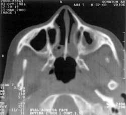
The patient did not have any personal or family background, or relevant complaints besides the ones previously mentioned. At physical examination, the only noticeable alteration was yellow and purulent mucous in both nasal cavities. Paranasal sinuses CT Scan (Figure 1) showed bilateral choanal bone atresia, thickness of vomer posterior region, and velamentum of posterior nasal cavity, suggesting mucous retention. Nasal endoscopy confirmed CT Scan findings.
After diagnostic confirmation, surgical repair was undertaken by endoscopic technique. The atresia region was inferiorly and laterally perforated with cutting forceps, and the initial perforation was enlarged with a mushroom-type sphenoid clamp. Next, the posterior septum area was removed with a backbiter forceps. The surgery was uneventful and we did not apply supporting stents in the region.
Figure 1: Paranasal sinuses CT scan at axial section, showing bilateral bone choanal atresia, posterior thickness of vomer and velamentum of nasal cavity, suggesting mucous retention.
The patient progressed well and uneventfully in the early postoperative period, treated with antibiotics and nasal lavage with saline sterile solution. In her last medical appointment (8 months after the surgery), she referred significant nasal breathing and rhinorrhea improvement. Endoscopy was repeated and showed good aspect of nasal mucosa, free choanae and no secretion.
DISCUSSION
This case report is outstanding because of the patient's age at diagnosis. Bilateral CCA is a rare condition, and even more rare if diagnosed after childhood7. Josephson5, analyzing 15 patients in surgical repair of choanal atresia, found five bilateral cases, all of them operated on between the second and the 30th day of life. Lazar6, in a similar study with 10 patients, observed two of them with bilateral disease; the first one was repaired at the 4th week of life and the other at 7 years of age, after three previous similar surgeries. All the eight treated patients with bilateral atresia described by Sadek11, were in the newborn period.
Although severe respiratory distress and breastfeeding difficulties are caused by nasal obstruction, crying can relief dyspnea and compromise diagnosis13. However, since there is routine nasal tubes used in the newborns, the diagnosis has been more frequently performed. According to Llorens7, those patients may fit three distinct clinical categories: in the first one, the patient shows classic cyclic cyanosis; in the second one, the child remains eupneic but may manifest symptoms when being fed or at specific positions (usually in cases of unilateral choanal atresia); and in the third and most rare group, the malformation remains unnoticed through childhood and is manifested in the adult age, with nasal obstruction and rhinorrhea. Our patient does not fit any of these groups, as she presented severe respiratory insufficiency at the first days of life, needing intensive care. During childhood, she underwent adenoidectomy, similarly to a 17-year-old patient with the same diagnosis reported by Llorens7, but the latter was submitted to the procedure twice. This fact emphasized the importance of clinical suspicion and complementary tests, such as nasal endoscopy and CT Scan, in the investigation of chronic nasal obstruction 4,5.
Paranasal sinuses CT Scan showed posterior vomer enlargement and absence of air choanal space, as previously reported by Slovis12. The test also contributed to the exclusion of other possible nasal obstruction differential diagnoses: septum deviation, nasal concha hypertrophy, pyriform sinus stenosis, meningoceles, meningoencephalocele, nasal tumors and local inflammation3,8. Aeration pattern and paranasal sinuses size were normal, endorsing a study carried out by Behar1, who concluded that maxillary sinuses development was independent of posterior nasal aeration.
The surgical techniques applicable in the anatomical deformity reported here are innumerous and vary from the ancient blinded transnasal perforation procedure, helped by a curved trocar, to the recently acclaimed endoscopic techniques. A survey carried out with ASPO members (American Society of Pediatric Otolaryngology) in March 1999 showed that the most frequently used techniques, in decreasing order, were: endoscopic transnasal, transpalatal, perforation with Fearon's dilator, microscopic transnasal and laser-based transnasal surgery. Blinded perforation has been abandoned due to high recurrence rates and risk of CSF fistulae and meningitis caused by lesion of cribiform lamina8. Transpalatal access revolutionized the surgery of CCA repair because it was the first procedure to allow enough exposure to safely remove the atresic plate, the posterior part of vomer and lateral portion of nasal walls. Moreover, mucosal flaps could be lifted from the atresia site and recover the bone surface, decreasing the need for stent use and increasing success rates. This method offers good postoperative results; however, its effects on palatal growth are encouraging the use of the endoscopic technique. Transpalatal access has some disadvantages, such as the risk of developing palate deformities (52%), cross-bite, maxilla narrowing, muscle palatal dysfunction and fistula, greater surgical time and increased risk of bleeding5,6,8.
Our patient underwent transnasal endoscopic surgery, presenting favorable results until the present (8month follow-up); nevertheless, this is a still a short period, according to Richardson10, who noticed that restenosis tend to occur within the first 12 postoperative months. This method was described by Stankiewiecz13, and it begins with application of vasoconstrictor on the nasal fossae, helped by 2.5-4mm 0° telescopes; next, we proceed to the incision of the mucosa over the atresic plate, followed by removal of bone obstruction from the medial region, due to its thinner thickness. The posterior vomer portion and nasal lateral walls are also resected in order to enlarge the new choana and prevent restenosis. Repair can be done using endoscopic nasal and sinusal surgical material or electric instruments, such as the "Shaver" (cutting razors or drills coupled to an aspirator), without significant influence on the surgery success8. We performed the surgery with traditional nasosinusal material, with no further difficulties. Telescopic transnasal surgery has good results and low risk of recurrence, especially if performed in children over three years of age6.
Stent placement (generally endotracheal tubes) at the new choana is commonly performed; however, the scientific literature still lacks studies concerning surgical time, ideal types of materials and efficacy of this method8. According to the survey carried out by ASPO, the majority of the physicians used stents (97%) for a mean of 7.9 weeks. We decided not to use any kind of stent in our patient, because our service rule is to avoid stent utilization in patients older than one year, since the stent itself can be a constant source of trauma and restenosis stimuli. Nasal washing with isotonic saline solution and antibiotic therapy were prescribed for the immediate postoperative period.
FINAL COMMENTS
Bilateral choanal-atresia is a life threatening childhood disease, usually repaired in the first weeks of life. Nonetheless, in rare cases, it can be found during investigation in adults with nasal obstruction and bilateral rhinorrhea.
REFERENCES
1. BEHAR, P M.; TODD, W - Paranasal Sinuses Development and Choanal Atresia. Arch. Otolaryngol Head Neck Surg., 126. 155-57, 2000.
2. BROWN, O. E.; POWNELL, E; MANNING, S. C.-Choanal atresia: a new anatomic classification and clinical management applications. Laryngoscope, 106 (1 Pt 1): 97101, 1996.
3. BUTUGAN, O.; CAHALI, M. B.; MURAKAMI, M. S.; IKINO, C. M. Y; WANTONIO, W E. E A.; MEDEIROS, I. R. T.; SANTORO, E. E; VOEGELS, R. L.; SENNES, L. U. - Tumores Nasais na Infância. Rev. Bras. Otorrinolaringol., 65 (1): 20-4, 1999.
4. CROCKET, D. M.; HEALY, G. B.; MCGILL, T. J. Computed tomography in the evaluation of choanal atresia in infants and children. Laryngoscope, 97: 174-83, 1987.
5. JOSEPHSON, G. D.; VICKERY, C. L.; GILES, W C.; GROSS, C. W - Transnasal endoscopic repair of congenital choanal atresia. Arch. Otolaryngol. Head Neck Surg., 124 (5): 537-40, 1998.
6. LAZAR, R. H.; YOUNES, R. T. - Transnasal repair of choapal atresia using telescopes. Arch. Otolaryngol. Head Neck Surg., 121 (5): 517-20, 1995.
7. LORENS, D. C.; CASASUS, J. C. - Atresia bilateral 6sea, de coanas en adulto. An. Otorrinolaringol. Ibero Am., 21 (5): 487-96, 1994.
8. PARK, A. H.; BROCKENBROUGH, J.; STANKIEWICZ, J. - Endoscopic versus traditional approaches to choanal atresia. Otolaryngol. Clin. North Am., 33 (1): 77-90, 2000.
9. PIRSIG, W - Surgery of choanal atresia in infants and children: Historical notes and undated review. Int. J. Pediatr Otorhinolaryngol, 11: 153-70, 1986.
10. RICHARDSON, M. A.; OSGUTHORPE, J. D. - Surgical management of choanal atresia. Laryngoscope, 98: 91518, 1988.
11. SADEK, S. A. - Congenital bilateral choanal atresia. Int. J. Pediatr Otorhinolaryngol, 42 (3): 247-56, 1998.
12. SLOVIS, T L.; RENFRON, B.; WATTS, F. B. - Choanal atresia: Precise CT evaluation. Radiology, 155: 345-48, 1985.
13. STANKIEWICZ, J. A - The endoscopic repair of choanal atresia. Otolaryngol. Head Neck Surg., 103: 931-37, 1990.
14. TELLIER, A. L.; DAIRE, V C.; ABADIE, V; AMIEL, J.; SIGAUDY, S.; BONNET, D.; DEBENEY, E L.; DURAND, M. E M.; HUBERT, E; MICHEL, J. L.; JAN, D.; DOLLFUS, H.; BAUMANN, C.; LABRUNE, E; LACOMBE, D.; PHILIP, N.; LEMERRER, M.; BRIARD, M. L.; MUNNICH, A.; LYONNET, S. - CHARGE Syndrome: Report of 47 Cases and Review. Am. J. Med. Genet., 76 (5): 402-9, 1998.
Article submitted on February 2, 2001. Article accepted on February 26. 2001.
Print: ![]()