

Year: 2001 Vol. 67 Ed. 4 - (18º)
Relato de Casos
Pages: 563 to 566
Pemphigus Vulgaris and ENT Repercussions. Case Report.
Author(s):
Paula M. Fraiha*,
Patrícia T. Fraiha**,
Luciana R. V. Rodrigues***,
Patricia G. Bittencourt***.
Keywords: pemphigus vulgaris, bullous lesions, ENT, manifestations
Abstract:
Pemphigus vulgaris is a rare disease that is less aggressive in our environment than in Mediterranean people. It has characteristics of an immune-mediated disease. The patient presents skin and mucous membranes bullous lesions, especially on the oral mucosa. Corticoid therapy is fundamental for symptom relief and prognosis of the disease. Nevertheless, in many occasions immunosuppressive drugs, tetracycline, dapsone and others drug combinations are necessary to control the disease. The advocated therapy is aggressive and lead to significant side effects.
![]()
INTRODUCTION
Pemphigus vulgaris (PV) is a vesicle-bullous dermatological disease. It is the most common type of pemphigus12. It is more prevalent in adult age, after the 51h decade of life, an age range in which both genders are equally affected. Before the age of 20 years, it is more frequent in women. The estimated incidence is of one to five cases in 1 million people diagnosed every year in the general population6. It affects people from all ethnic backgrounds, especially Caucasians2. It presents immune-mediated characteristics, chronic progression and severe prognosis2.
Lever, in 1965, presented a study showing that 30 of the 33 patients studied by him and treated between 1937 and 1949 did not survive longer than 5 years. The advent of corticoid therapy in 1950 improved the prognosis of the disease11. Before the existence of this treatment, the disease was generally fatal7.
Mucosas are affected in about 70% of the cases and normally the disease starts from oral lesions4. Since in severe cases the disease may spread quickly, early and precise diagnosis is imperative4.
The purposes of the present study were to review the literature about the topic, show the importance of diagnosis and report a case of the disease.
REVIEW OF LITERATURE
Pemphigus vulgaris is a vesicle-bullous disease. Oral lesions present well-defined margins, sometimes with epithelial laceration around them. They may be present before the onset of cutaneous lesions12. Its onset is more marked in traumatic sites of mastication or teeth: posterior third of jugal mucosa, posterior portion of the tongue, palate, gums and lips6. Bullous lesions of oral mucosa suffer rupture and present ulcerated areas, resulting in patient's discomfort and difficulty to intake food. It may be followed by sialorrhea, halitosis, tongue dorsum fissures and general malaise. Ulcer aspect varies from small lesions similar to aphthas to large map-like lesions8. Sometimes there are no bullous formation where the oral epithelium is detached, as a consequence of acanthosis in the dermalepidermal region7,10.
On the skin, there are varied size and shape blisters, which are ruptured and show damaged areas'. Blisters present a thin liquid inside them, which may become purulent or sanguineous10. Cutaneous blisters may be present in any point of the tegumentum, normally affecting the armpit, scalp and navel. The lesions may suffer coalescence and present extensive burn-like areas.
Delicate pull of clinically non-affected mucosa or skin may cause detachment of epithelium, characterizing the Nikolski's sign8.
In general, when the otorhinolaryngologist sees the patient, the blisters have ruptured and there are mucosa erosions or blisters with no liquid content, hindering diagnosis.
The skin has adhesion molecules that anchor the keratinic cells one to the other and to the adjacent basal membrane. These molecules may be the target of autoantibodies, leading to loss of intercellular adhesion and formation of blisters9.
Samples of skin biopsy of cutaneous lesions in patients with PV showed intra-epidermal fissures somewhat above the basal layer of keratinocytes, which is called suprabasilar acanthosis12. Thus, basal membrane is intact in PV, whereas it is differently affected in other pathologies2. As a result of loss of adhesion of desmossomes and retraction of tonofilament, acantholytic epithelial cells become spherical and are named Tzank8 cells
Immunoglobulin (Ig)G autoantibodies act upon the transmembrane desmossomic proteins named desmoglein (Dsg)12. Studies of molecular cloning with autoantibodies of PV showed that Dsg is the target antigen of pemphigus1. The affection may be reproduced in mice by passive transfer of IgG of subjects with PV showing that humoral immunity plays an important role in the pathogenesis of the diseases.
Levels of antibodies in the blood vary according to the activity of the disease. The most frequently found antibody is IgG, but there may be IgA and IgM as well. Complement 3 (C3) is usually present, even in early cases2.
A susceptible patient may develop the disease even with low levels of antibodies, induced by genetic or environmental factors such as drugs, burns, virosis and ultraviolet radiation4. The onset of the disease results from the interaction between genetic and external factors. Human lymphocyte antigens (HLA) are involved in the pathogenesis of PV similarly to other immune-mediated diseases12. Genetic factors responsible for susceptibility of PV are classified as HLA system class II3. Immunogenetic studies have been carried out in order to define future therapeutic alternatives3.
Lymphocytes T play an important role in specific immune responses against foreign and host antigens. If T cells do not manage to regulate the reaction of T lymphocytes to its own antigens, there may be immunemediated diseases, such as pemphigus vulgaris5.
Diagnosis is based on clinical characteristics, cytology of the blisters' liquid with acantholytic cells; histolosical evidence of intra-epithelial acantholysis, direct immunofluorescence, with the presence of IgG antibodies on the surface of the affected epithelial cells, indirect immunofluorescence, with evidence of circulating anti-epithelial antibodies or immunochemical techniques with specific anti-Dsg 3 antibodies2,9.
Differential diagnosis includes aphthous stomatitis, pemphigoid, bullous dermatitis, liquens planus, some clinical form of polymorph erythema, dystrophic bullous epidermolysis, and other skin and mucosa affections that produce bullous lesions, which may be clinically similar to PV in different stages of the disease8,10.
If we carefully follow the clinical characteristics of the pathology and perform complementary exams, we will be able to come up with a definite diagnosis.
The advocated treatment is divided into local and systemic measures. Local treatment does not intend to control the lesions, but to prevent secondary infection. Bath with potassium permanganate at 1:30,000 may be employed; avoiding skin detachment. Solid Vaseline or antibiotic creams on the ulcerations may generate some relief. Mouthwashes with nistatine may prevent the development of oral candidiasis9.
Systemic treatment involves corticoids as the main therapeutic tool.
Immunosuppressors may be used in association with corticoids. The newest immunosuppressor drug is mofetyl micophenolate, which reduces antibodies titers in short time. Gold compounds have been used, either in single or associated therapy; however, they generate high rates of side effects and their results are not better than the use of dapsone and immunosuppressors. Plasmapheresis may also be indicated as adjuvant treatment. The use of tetracycline may produce good results9.
CASE REPORT
M. R. S., female 42-year-old patient, researcher living in Nova Iguaçu/RJ, hypertension controlled with drugs, came to the service reporting condition of hemorrhagic blisters on the oral mucosa for three months, which ruptured three days later in average and became covered with a white layer. She also reported dysphagia and sore mouth.
Oropharyngeal examination showed presence of blisters with reddish jugal mucosa, roof of the mouth and hard palate. Biopsy of the jugal mucosa was performed for histopathological analysis and direct immunofluorescence. We prescribed mouthwash with oral nistatine and topical corticoid on the biopsy lesions.
Fifteen days after the biopsy, the patient returned and presented aphthous lesions in the internal face of the lower lip and mastication region of the jugal mucosa, besides cutaneous blisters with pruritus characteristics, especially on the scalp and dorsal region.
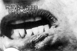
Figure 1. Pemphigoid lesion of jugal mucosa.
She also reported general malaise, without fever. Histopathological exam showed areas of suprabasal blisters with some acantholytic cells. Corium showed edema, ectactic vessels and inflammation infiltrate of mono and polymorphonuclear cells, compatible with PV Direct immunofluorescence was negative for anti-serum IgG, IgM, IgA, C3 and fibrinogen. Indirect immunofluorescence showed intra-epithelial antibodies higher than 1/10.
We prescribed oral corticoid therapy and one week after treatment the patient presented improvement of oral lesion, sore mouth and general malaise, feeling well enough to intake foods. We introduced tetracycline in order to maintain treatment without increasing drastically the dose of corticoids, minimizing side effects. After one month of treatment, we concluded that the introduction of tetracycline had not improved the condition and we decided to maintain the corticoid as single therapy at higher doses. We finally managed to have regression of the mucosa and cutaneous lesions and the patient has been followed up for six months now (Figures 1, 2 and 3).
DISCUSSION
The case reported here showed that oral mucosa lesions may be the first manifestations of pemphigus vulgaris. Mucosa and cutaneous lesions guided our suspicion of PV. Although direct immunofluorescence was not characteristic, clinical signs, indirect immunofluorescence and histopathology confirmed the diagnosis. The introduction of corticoids was essential to control the disease and the use of tetracycline did not add benefits to the treatment in our case.
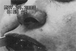
Figure 2. Pemphigoid lesion of labial mucosa.
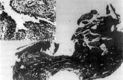
Figure 3. Acantholytic fissure.
FINAL COMMENTS
Pemphigus vulgaris is a disease of autoimmune characteristics with immune-genetic compromise. In Brazil, the clinical progression does not seem to be as severe as in Mediterranean populations. The otorhinolaryngologist should be attentive to manage the case satisfactorily, since he or she may be the first professional to see the patient. The fact that the treatment relies on corticoids and immunosuppressors for a prolonged period of time requires caution because of severe side effects. The patient should be periodically followed up through clinical examinations and complementary tests. Similarly to other immune-mediated diseases, we still expect future specific less toxic therapies that can regulate the production of autoantibodies.
REFERENCES
1. AMAGAI, M; KARPATI, S; PRUSSICK, R; KLAUS-KOVTUN, V; STANLEY, J. - Autoantibodies against the amino-terminal cadherin-like domain of pemphigus vulgaris are pathogenic. J Clin Invest., 90: 919-26, 1992.
2. AZULAY & AZULAY - Dermatologia, Rio de Janeiro, Guanabara Koogan, 1997 p. 95-105.
3. GLORIO, R. R.; RODRIGUEZ, C. G.; HAAS, R.; LARRIBA, J.; FAINBOIM, L.; WOSCOFF, A. - PCR determination of association between class II HLA and pemphigus vulgaris. Medicina, 59 (1): 28-32. 1999.
4. GRISPAN, D. - Enfermedades de La Boca, 1ª edición, Buenos Aires, Editorial Mundi, 1990 p. 1301-30.
5. LIN, M. S.; SWARTZ, S. J.; LOPEZ, A.; DING, X.; FERNANDEZ VINA, M. A.; STASTNY E; FAIRLEY J. A.; DIAZ, L. A, - Development and Characterization of Desmoglein-3 Specific T Cells From Patients With Pemphigus Vulgaris, JCI, 99 (1): 31-40, 1999.
6. NEVILLE, B. W [et al.] - Oral and Maxillofacial Pathology, 1st ed., Philadelphia, W B. Sauders Company, 1995 p. 559-61.
7. PENDBORG, J. J. - Atlas de Doen~as da Mucosa Oral, la ed., Sao Paulo, Ed. Medica Panamericana, 1981, 21618. '
8. REGEZE, I. A.; SCRUBBA, J. J. - Patologia Bucal Correlações Clinico Patológicas, 1ª ed., Rio de Janeiro, Ed. Guanabara Koogan, 1991 p. 10-17.
9. SAMPAIO & RIVITTI - Dermatologia, São Paulo, Artes Medicas, 1998 p. 229-48.
10. SHAFER, W G. [et al.] - Tratado de Patologia Bucal, 4a ed., Rio de Janeiro, Ed. Guanabara Koogan, 1987 p. 765-71.
11. STANLEY, J. R. - Therapy of Pemphigus Vulgaris, Arch Dermatol, 135: 76-8, 1999.
12. UDEY, M. C. & STANLEY, J. R. - Pênfigo: Doenças da Autoimunidade Antidesmossomas, JAMA BRASIL, 4 (3): 2927-32, 2000.
* Coordinator of Specialization Course in Otorhinolaryngology, Clínica Professor José Kós.
** Resident Physician (3rd year), Clínica Professor José Kós.
*** Resident Physician (1st year), Clínica Professor José Kós.
Study conducted at Clínica Professor José Kós.
Address correspondence to: Dra. Paula Moreno Fraiha - Rua Padre Elias Gorayeb, 15 -Sala 606 - Tijuca - 22520-140 Rio de Janeiro/RJ.
Article submitted on December 4, 2000. Article accepted on February 8, 2001.
Print: ![]()