

Year: 2001 Vol. 67 Ed. 4 - (16º)
Artigos Originais
Pages: 551 to 555
Classification of Rhinosinusitis Orbital Complications.
Author(s):
Ossamu Butugan*,
Aracy P. S. Balbani**,
Richard L. Voegels***.
Keywords: rhinosinusitis, abscess, complications, orbit
Abstract:
Ethmoidal and sphenoidal rhinosinusitis are the most likely to result in orbital complications. Orbital complications are divided into pre and post-septal, according to their localization in relation to the orbital septum, which is an important barrier to the spread of infection towards the orbit. The most common orbital complication is palpebral cellulite. About 15% of post-septal complications lead to irreversible amaurosis, despite the appropriate treatment. Thanks to the development of high-resolution computerized tomography, Mortimore's classification for orbital complications of rhinosinusitis seems to be the most accurate and complete.
![]()
INTRODUCTION
Despite the development of different effective antimicrobial drugs against etiological agents of nasosinusal infections, rhinosinusitis is still the main cause of orbital infections7,13, highly frequently in Brazil10,16. In the past 15 years, we noticed that 128 patients needed to be hospitalized to treat orbital complications at the Division of Otorhinolaryngology, Hospital das Clínicas, Faculdade de Medicina da Universidade de São Paulo2.
Different studies pointed out that before the discovery of penicillin 1/5 of the orbital complications resulted in irreversible atriaurosis, now dropped to half of that figure11. However, this advance did not reduce the severity of the problem. If not diagnosed early or appropriately treated, orbital complications may irreversibly compromise the visual acuity of the patients11. Therefore, the knowledge of different orbital complications of rhinosinusitis and their classification is extremely important for otorhinolaryngologists.
REVIEW OF LITERATURE
In ethmoidal rhinosinusitis, we may find a) direct extension of nasosinusal infection to the orbit through the natural dehiscence of papyraceous lamina, and b) retrograde spread of septic emboli to the orbital venous network (superior and inferior ophthalmic arteries), whose main anatomical characteristic is the non-existence of valves5,15.
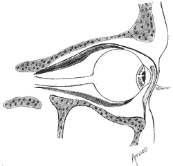
Another important anatomical consideration concerns the orbital septum - a periorbital deflection, a thin layer of connective tissue that recovers the orbital cavity (Figure 1).
Laterally, the septum forms the lateral palpebral ligament, and medially, the medial palpebral ligament, posteriorly to the lachrymal sac. Therefore, the septum is an anatomical barrier to the dissemination of infection to the back of the orbits. It is based on these anatomical parameters that orbital complications are classified.
Orbital complications of rhinosinusitis are generically divided into pre and post-septal, according to its location in relation to the septum. The classification based on clinical and anatomical criteria was introduced in 1937 by Hubert6 and modified in 1948 by Smith and Spencer14. However, the best known and most widely used classification was introduced by Chandler et al., published in 1970, based on the adaptation of the previous ones1. It divides orbital complications in:
Group 1: Periorbital cellulitis
Group 2: Orbital cellulitis
Group 3: Subperiosteal abscess
Group 4: Orbital abscess
Group 5: Cavernous sinus thrombosis.
Figure 1. Schematic representation of the orbital septum.
Later, Welsh and Welsh17, Schramm et al.12, Moloney9 and Singh13 proposed new classifications for the complications.
High resolution computed tomography (CT scan) contributed enormously to show anatomical details and extensions of infectious processes of paranasal sinuses. Based on this radiological test, in 1997 Mortimore9 proposed a new classification of the orbital complications of rhinosinusitis. He concluded that CT scan enabled clear differentiation of pre and post-septal complications and also the subgroups of cellulitis and abscesses, which is not always possible through clinical assessment (test of ocular motility and visual acuity). He also subdivided postseptal complications in subperiosteal and infra-choanal, excluding the cavernous sinus thrombosis as orbital complication - he believed that it was an intracranial complication. The author also classified the syndrome of orbital apex (affection of III, IV and VI nerves and ophthalmic branch of the V cranial nerve at the superior orbital fissure and optical canal)3, independently from intra-choanal cellulitis. Thus, he provided a new perspective for the assessment of orbital complications, as described below:
Group 1: Pre-septal infection:
a) cellulitis
b) palpebral abscess
Group 2: Subperiosteal post-septal infection:
a) cellulitis
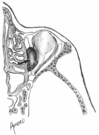
Figure 2. Pre-septal orbital complications of rhinosinusitis: palpebral cellulitis.
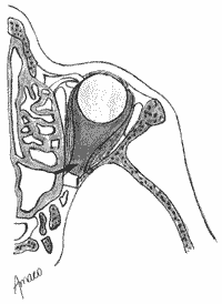
Figure 3. Pre-septal orbital complications of rhinosinusitis: subperiosteal abscess.
b) subperiosteal abscess
Group 3: Intra-choanal post-septal infection:
a) cellulitis 1. diffuse
2. localized
b) abscess
Group 1: Pre-septal infection: It accounts for 97% of orbital complications. It affects primarily children in whom the main etiological agent is type B Haemophilus influenzae, especially in children below four years of age 8. It is divided into:
a) cellulitis, the most frequent orbital complication 2, caused by the obstruction of venous palpebral drainage. In such cases, there is palpebral edema with no further limitation of ocular movements, nor compromise of visual acuity (Figure 2). The main symptoms are local pain and difficulty to open the eyes, which may be followed by fever, headache and purulent rhinorrhea. At the physical examination, palpebral edema with inflammatory signs (hyperemia and local heat) are the main findings, and in many occasions it is possible to observe conjunctival secretion and drainage of purulent secretion in the meatus through nasal endoscopy
b) palpebral abscess. In some patients there may be formation of purulent collection on the upper eyelid, leading to marked local edema and higher likelihood of fever and pain, but it still does not affect ocular motility. In some rare cases, there may be fistulation with spontaneous drainage of purulent material.
Group 2: Subperiosteal post-septal infection. It is subdivided into:
a) cellulitis. It comprises the diffuse affection of the orbital contents by the inflammatory process, causing edema on the orbital fat with no formation of abscess. It is clinically characterized by the symptoms previously described, in addition to signs of conjunctival hyperemia, chemosis (conjunctival edema resulting from venous and local lymphatic stasis) and proptosis, which may lead to mild restriction of ocular movement.
b) subperiosteal abscess. It is characterized by purulent collection on the medial wall of the orbit, between the bone lamina and the periorbit (Figure 3). It causes lateralinferior displacement of the ocular globe, restricting extrinsic ocular movements, and it may be followed by reduction in visual acuity. The patients frequently complain of severe ocular pain, especially during the attempts to move the ocular globe.
Group 3: Intra-choanal post-septal infection. The inflammatory process of fatty and adjacent optic nerve connective tissues may compromise visual acuity.
Figure 4. Post-septal orbital complications of rhinosinusitis: diffuse intra-choanal cellulitis.
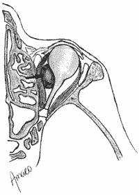
Figure 5. Post-septal orbital complications of chinosinusitis: intra-choanal abscess.
a) cellulitis.
A1.) Diffuse. Edema of intra-choanal fat (Figure 4). It results in marked proptosis and ophthalmoplegia, with reduction of visual acuity, which may progress to irreversible amaurosis if not submitted to immediate treatment.
a2.) Localized (syndrome of orbital apex). It corresponds to the affection of vascular-nervous structures that pass from the superior orbital fissure to the optic foramen (including the III, IV and V cranial nerves). The exact mechanism of the syndrome is unknown (direct bacterial invasion of the posterior ethmoid and sphenoid sinuses, or compression of the optic nerve with thrombosis of the central artery of the retina)4. The complete syndrome is manifested by amaurosis, ophthalmoplegia, marked ocular pain and sensorial disorders in the area of the ophthalmic nerve, which may vary from anesthesia to nerve affection3. At physical examination, intrinsic ocular motility is usually affected (absence of pupil reflex). The funduscopy usually reveals papillary edema and retina vascular congestion.
b) abscess
It is the most severe orbital complications of rhinosinusitis, quickly progressing to amaurosis, if not immediately submitted to decompression (Figure 5). Irreversible degeneration of the optic nerve and retina occurs when arterial occlusion lasts longer than 90 minutes. It is characterized by ophthalmoplegia, absence of pupil reflex (non-photoreagent pupils), amaurosis, ocular pain and marked chemosis and proptosis.
DISCUSSION
The interest in classifying orbital complications of rhinosinusitis has existed for a long time. The first classifications followed clinical criteria, according to extrinsic ocular motility and visual acuity. After the advent of high resolution CT scan of paranasal sinuses, the arena has changed dramatically. Currently, the CT scan is mandatory in cases of suspicion of orbital and intracranial complications of rhinosinusitis, enabling identification of orbital purulent collections of any given volume. Therefore, the decision concerning therapeutic approach (clinical treatment with antibiotics or surgical intervention and antibiotic therapy) may find a valuable support in the imaging diagnosis.
Even though the classification by Chandler is still widely used in clinical practice, it seems that the schematic representation proposed by Mortimore is the one that translates the best the rich details shown through CT scan. Therefore, we would like to suggest that otorhinolaryngologists use the classification proposed by Mortimore in their daily practice and when carrying out scientific studies.
FINAL COMMENTS
High resolution computed tomography of paranasal sinuses allowed great development in the anatomical study of the complications of rhinosinusitis. Therefore, the classification by Mortimore for orbital complications of rhinosinusitis seems to be more adequate and complete because it takes into account the anatomical parameters visualized in the CT scan.
REFERENCES
1. CHANDLER, J. R.; LANGENBRUNNER, D. J.; STEVENS, E. R. - The pathogenesis of orbital complications in acute sinusitis. Laryngoscope, 80: 1414-1428, 1970.
2. D'ANTONIO, W E. E A.; IKINO, C. M. Y; CORTINA, R. A. C.; VOEGELS, R. L.; BUTUGAN, O. - Complicações orbitárias em pacientes com sinusite aguda. Anais do I Congresso Triológico de Otorrinolaringologia, São Paulo, 1999.
3. EL-SAVED, Y; AL-MUHAIMED, H. -Acute visual loss in association with sinusitis. J. Laryngol. Otol., 107: 840-842, 1993.
4. GALATI, L. T.; BAREDES, S.; MAURIELLO, J.; FROHMAN, L. - Visual loss reversed after treatment of acute bacterial sinusitis. Laryngoscope, 106. 148-151, 1996.
5. GIANNONI, C. M.; STEWART, M. G.; ALFORD, E. L. Intracranial complications of sinusitis. Laryngoscope, 107: 863-867, 1997.
6. HUBERT, L. - Orbital infections due to nasal sinusitis. N. Y. State J. Med., 37: 1559-1564, 1937.
7. MANN, W; AMEDEE, R. G.; MAURER, J. - Orbital complications of pediatric sinusitis: treatment of neriorbital abscess. Am. J. Rhinol, 11: 149-153, 1997.
8. MOLONEY, J. R.; BADHAM, N. J.; McRAE, A. - The acute orbit: preseptal (periorbital) cellulitis, subperiosteal abscess and orbital cellulitis due to sinusitis. J. Laryngol. Otol., 101 (supl. 12): 1-18, 1987.
9. MORTIMORE, S.; WORMALD, E J. - The Groote Schuur hospital classification of the orbital complications of sinusitis. J. Laryngol. Otol., 111: 719-723, 1997.
10. OGNIBENE, R. Z.; VOEGELS, R. L.; BENSADON, R. L.; BUTUGAN, O. - Complications of sinusitis. Rhinology, 8: 175-179, 1994.
11. PATT, B. S.; MANNING, S. C. - Blindness resulting from orbital complications of sinusitis. Otolaryngol. Head Neck Surg., 104: 789-795, 1991.
12. SCHRAMM JR., V L.; CURTIN, H. D.; KENNERDELL, J. S. - Evaluation of orbital cellulitis and results of treatment. Laryngoscope, 92: 732-738, 1982.
13. SINGH, B. - The management of sinogenic orbital complications. J. Laryngol. Otol, 109: 300-303, 1995.
14. SMITH, A. T.; SPENCER, J. T. - Orbital complication resulting from lesions of the sinuses. Ann. Otol. Rhinol. Laryngol., 57: 5, 1948. .
15. STANKIEWICZ, J. A.; NEWELL, D. J.; PARK, A. H. Complications of inflammatory diseases of the sinuses. Otolaryngol. Clin. North Am., 26(4): 639-655, 1993.
16. VOEGELS, R. L.; BENSADON, R. L.; OGNIBENE, R. Z.; BUTUGAN, O. - Complicações periorbitárias das sinusites. Rev. Bras. Otorrinolaringol., 60: 149-152, 1994.
17. WELSH, L. W; WELSH, J. J. - Orbital complications of sinus disease. Laryngoscope, 84: 848-856, 1974.
* Associate Professor, Discipline of Otorhinolaryngology, Faculdade de Medicina, Universidade de São Paulo.
** Doctorate studies under course, Discipline of Otorhinolaryngology, Faculdade de Medicina, Universidade de São Paulo.
*** Ph.D., Professor, Discipline of Otorhinolaryngology, Faculdade de Medicina, Universidade de São Paulo.
Discipline of Otorhinolaryngology, Faculdade de Medicina, Universidade de São Paulo.
Address correspondence to: Prof Dr. Ossamu Butugan - Hospital das Clínicas da FMUSP - Divisão de Clinica Otorrinolaringológica.
Avenida Dr. Enéas de Carvalho Aguiar, 255-Sala 6167, 6° Andar-05403-010 São Paulo/ SP-Fax: (55 11) 3088-0299.
Article submitted on February 4, 2001. Article accepted on March 19, 2001.

Print: ![]()