

Year: 2001 Vol. 67 Ed. 3 - (13º)
Artigos Originais
Pages: 381 to 385
Sphenochoanal Polyp: Dose and Treatment.
Author(s):
Marcus M. Lessa*,
Francini Pádua**,
Christian Wiikmann**,
Farízio Romano**,
Richard L. Voegels***,
Ossamu Butugan****.
Keywords: choanal polyps; sphenochoanal polyps; endoscopic sinus surgery
Abstract:
Introduction: Choanal polyps (CP) are benign mucous tumors that usually originate in the mucosa of the maxillary sinus, and hence are termed antrochoanal polyps. CP rarely originate in the mucosa of other nasal structures and paranasal sinuses. Purpose: The purpose of this report is to present a retrospective study of six cases of CP originating in the sphenoid sinus and termed sphenochoanal polyps (SCP). The seven patients underwent transnasal endoscopic surgery on the Ear, Nose and Throat Division of the Hospital das Clinicas (HC), University of São Paulo Medical Center (FMUSP), during the period may 1996 to august 2000. Study design: Clinical retrospective. Material and method: Four patients were female and two male, with ages ranging from eight to 43 years. In all cases diagnosis was made based on clinical feature, nasal endoscopy and computed tomography of paranasal sinuses. One patient referred a previous surgery in other service. All six patients were submitted to endoscopic sinus surgery and have been followed-up with no signs of recurrence until the present moment. Conclusion: In our experience, SCP should be treated with an endoscopic approach, which allows a total removal of the polyp and its origin from the sphenoid sinus with a minimal disruption of the anatomy and physiology of this.
![]()
INTRODUCTION
Choanal polyps (CP) are mucous benign tumors that by definition invade the choana. They are relatively rare and account for 3-6% of nasal polyps2. The polyps are classified according to origin of pedicle as: antrochoanal (from the maxillary sinus), ethmoidochoanal and sphenochoanal. Sphenochoanal polyp (SCP) is the rarest type among the choanal polyps4.
Choanal polyps were described for the first time in 1763 by Palfyn2; however, it was in 1906 that Killian5 made a more detailed description of problem. Kubo6 (1909) and Van Alyea13 (1956) confirmed the intrasinusal origin of CP, but the first report of a SCP was made by Zuckerlandl1,4,14.
The authors of the present study report six cases of sphenochoanal polyps treated between May 1996 and August 2000, discussing origin, clinical presentation, diagnostic assessment and surgical approach.
CASE REPORT
The description of the six cases is presented in Table 1.
DISCUSSION
Sphenochoanal polyp (SCP) surges from the edematous and hyperplastic line of submucous layer from the sinus floor, goes through the ostium and protrudes into the choana and sometimes into the nasopharynx14. Its pathogenesis is still uncertain and many theories have been suggested. Among them, the most widely accepted one is that it is originated from a submucous cyst secondary to a lymphatic vessel thrombosis caused by a sinus postinfection inflammation1,2,10. The association with immune deficiency and allergy is controversial. Elopy et al.2 considered variable the relation with allergy. Ileri et al.4 believed that polyps would grow under the influence of inflammatory and allergic conditions; however, Crampetel declared that there was no correlation. In our cases, none of the patients were submitted to allergic tests.
SCP affect especially teenagers (older than 10 years) and young adults2, of both gender2. In our sample, age varied from 8 to 43 years (mean age of 30.5 years), and in the six presented cases, four were female and two were male patients (Table 1).
Symptoms of SCP consist mainly of unilateral nasal obstruction because of progression of the polyp inside the nasal cavity and choana. It may be followed by unilateral purulent rhinorrhea and an unpleasant sensation of cough2. If the polyp is large, obstructing completely the nasopharynx, there may be bilateral nasal obstruction. Some patients may experience conductive hearing loss because of the obstruction of the auditory tube'. In children below 10 years of age, the symptoms may be similar to adenoid hyperplasia or chronic rhinosinusitis3. Headache is caused by the obstruction of the sphenoid sinus, which presents enervation derived from the first branch of the trigeminal nerve (VI) and afferent fibers, from the sphenopalatine ganglion1,8. According to Sethi et al.11, isolated lesions of the sphenoid, both inflammatory and expansive, are manifested with headache as the main symptom, frequently retro-orbital, varying in incidence from 33% to 81%. In our sample, unilateral nasal obstruction was the main symptom, followed by headache and unilateral purulent rhinorrhea (Table 1).
The diagnosis of SCP is based on the clinical picture, nasal endoscopy and imaging techniques. The evolution of endoscopy and CT scan has contributed to increase the number of new diagnosed cases11. Ileri4 prefers to use endoscopy, similarly to our service, complemented by paranasal sinuses CT scan. In some cases, however, it is not possible to precisely define the origin of the polyp and endoscopic sinusal surgery becomes at the same time diagnostic and therapeutic procedures, since it enables perfect visualization of the insertion of the pedicle of the polyp7.
In the endoscopy, SCP is normally placed between the nasal septum and the middle concha, differently from the antrochoanal polyp (ACP) that is detected inside the middle meatus, going through the middle turbinate and the lateral wall of the nasal cavity1,14. We can see the polyp in the choana and, sometimes, we can also see the attachment in the sphenoethmoid recess1. In our sample, only in Cases 1 and 5 we did not identify the origin of the attachment of the polyp, since it was difficult to visualize them because of their large size.
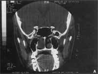
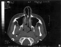
In Paranasal sinuses CT scan, polyps presented as solitary masses and since they are relatively hypocellular, they are shown as hypoattenuating images, leading to opacity of the affected sinus14. They occupy the posterior region of the nasal cavity, invade the choana through the sphenoethmoidal recess as from the sphenoid ostium and reach the sinus (Figures 1 and 2). Some difficulties may be experienced in differentiating SCP from ACP by CT scan in the presence of maxillary sinusitis associated to SCP, because in that case the maxillary sinus will be opaque and an incorrect diagnosis of ACP might be made1,2,14. CT scan, in addition to enabling observation of the implementation site of the polyp, allows identification of occasional anatomical variations, which are essential for the surgical approach of paranasal sinuses2.TABLE 1 - Clinical picture, nasal endoscopy, previous treatment, result of pathology of surgical piece and postoperative evolution of patients with diagnosis of sphenochoanal polyps (REE = sphenoethmoidal recess; post-op = postoperative; R = right; L = left).
Figure 1. Case 4: a) CT scan at coronal section showing attachment of polyp on the anterior wall of right sphenoid sinus; b) CT Scan at axial section showing extension of polyp into the choana and rhinopharynx.
MRI is not part of the routine battery of tests for investigation of choanal polyps; however, it is the preferred exam in case of suspicion of malignant alteration2. As to differential diagnosis, it is important to bear in mind that inverted papilloma should be always considered in cases of CP of uncommon origin7,9. In young patients, unilateral masses should raise the hypothesis of nasoangiofibroma. In older subjects, neoplasias such as squamous cell carcinoma, adenocarcinoma, rhabdomyosarcoma, or lymphoma should be considered2. Solitary polyps medial to the middle concha suggest meningocele 2. When in doubt, biopsy should be conducted in order to define the diagnosis, provided that the imaging tests have already excluded the possibility of meningocele, meningoencephaloce or vascularized tumors, such as nasoangiofibroma. In our sample, there was no need to conduct biopsy in the patients. The treatment of all choanal polyps is surgical and consists of the complete resection of the polyp, its pedicle and sinus implantation basis1,2,14. In order to achieve that, it is necessary to know preoperatively the origin of the polyp to schedule the appropriate surgical procedure. Incorrect diagnosis results in inappropriate surgical procedure, such as non-exploration of sphenoid sinus or maxillary sinus antrostomy, exposing the patient to higher risks of recurrence1. In our report, Case 4 had been submitted to a previous surgery in another service and we believe that due to incorrect diagnosis of origin, not having resected the attachment resulted in recurrence of the pathology within a 2-year period.
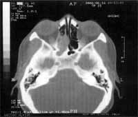
Figure 2. CT scan at axial section showing enlargement of ostium of sphenoid sinus on the right and affection by the same polyp.
Endonasal endoscopic surgery provides an excellent visualization of all affected sinuses; therefore, it is an effective procedure for the treatment of SCP1,4,7. In 1978 the technique of endoscopic functional sinus surgery was popularized by Messerklinger and started to be used for the management of recurrent chronic sinusitis. In the beginning of the 90's, it started to be used for the exeresis of ACP7. In children, it should be the preferred choice because transseptal approach is not recommended during growth. However, since the sphenoid sinus is the last one to develop, a hypotrophic sinus may be found in children, leading to total removal of SCP and opening of the sinus1. Eloy et al.2 (1996) recommended wide intranasal sphenoidectomy with endoscopic control and treatment of other existing lesions at the same surgical time. During exeresis of SCP, the posterior-inferior portion of the middle concha may or may not be removed. According to Ileri et al.4 the resection of this region improves the exposed area; however, in our service, this procedure was not conducted, in an attempt to preserve as much as possible nasal physiology and anatomy. We should expand the ostium of the sinus and remove any sick component that is still left, in order to avoid recurrence1,4. Simple polypectomies are associated with higher recurrence rates2. The approach of sphenoid sinus should be careful, since the internal carotid artery and the optic nerve are present on the lateral wall, leading occasionally to dehiscence in 25% and 6% of the cases, respectively12.
Histopathologic study of the surgical piece should be performed. Macroscopy shows a drop-shaped polyp, with intranasal and intrasinusal portions. The intranasal portion is firm, translucent, yellowish, sometime reddish, with smooth and regular aspect. The intrasinusal portion may be cystic or polypoid and it contains a light-colored fluid that may solidify within few minutes when in contact with room temperature. Microscopically, the polyp has a cystic formation recovered with respiratory epithelium and occasional epidermoid metaplasia 2. Stroma is edematous and contains inflammatory infiltrate, with neutrophils, macrophages and monocytes, with predominance of plasmocytes, rarely presenting eosinophils and lymphocytes2,7,4.
All reported patients were submitted to endoscopic functional sinus surgery with complete resection of the polyp and its pedicle from the sphenoid sinus. Pathology revealed inflammatory polyp in all cases. All patients, except for Case 1 who abandoned follow-up after 1 year, have been under ambulatory follow-up and so far no recurrences have been observed.
CONCLUSION
Based on our experience, sphenochoanal polyps may be treated through endoscopic functional surgery because it enables complete resection of the polyp and its origin from the sphenoid sinus, producing minimal anatomical and physiological modifications in the region.
REFERENCES
1. CRAMPETE, L.; MONDAIN, M.; ROMBAUX, Ph.. Sphenochoanal polyp in children. Diagnosis and treatment. Rhinology, 33: 43-45, 1995.
2. ELOY, PH; EVRARD, I.; BERTRAND, B.; DELOS, M. Choanal polyp of sphenoidal origin. Report of two cases. Acta otorbinolaryngologica belg., 50: 183-189, 1996.
3. GORDTS, F.; CLEMENT, RA.R. - Unusual choanal polyps. Acta otorhinolaringologica belg., 51: 177-180, 1997.
4. ILERI, F.; KOYBASIOGLU, A.; USLU, S. - Clinical presentation of a sphenochoanal polyp. Eur Arch Otorhinolaryngol, 255: 138-139, 1998.
5. KILLIAN, G. - The origin of choanal polyp. Lancet, 2: 81-82, 1906.
6. KUBO, J. - Uber die eigentliche ursprungsstelle and die radikaloperation der solitaren choanalpolypen. Arch Laryngol RhinoL, 21: 81, 1909.
7. LOPATIN, A.; BYKOVA, V; PISKUNOV, G. - Choanal polyps: One entity, one surgical approach?. Rhinology, 35: 79-83, 1997.
8. PEARLMAN, S. J. Et al. - Isolated Sphenoid Sinus Disease. Laryngoscope, 99: 716-720, 1989.
9. PETER K. M. et al. - Case report of a mass that mimicked an antrochoanal polyp. ENT-Ear, Nose and Throat Journal, 78: 556-557, august, 1999.
10. PIQUET, J. et al. - Microchirurgie endonasale du polype antrochoanal. Acta Otorhinolaringol Belg., 46: 267-271, 1992.
11. SETHI, R. S. - Isolated sphenoid lesions: Diagnosis and management. Otolaryngol Head Neck Surg., 120: 730736, 1999.
12. STAMMBERGER, H. et al - Essentials of functional endoscopic sinus surgery, 1993.
13. VAN ALYEA, O. E. - Management of non-malignant growths in the maxillary sinus. Ann Otolaryngol, 65: 714722, 1956.
14. WEISSMAN, J. L.; TABOR, E. K.; CURTIN, H. R. Sphenochoanal Polyps: Evaluation with CT and MR Imagine. Radiology. 178: 145-148, 1991.
* Postgraduate physician of Division of Clinical Otorhinolaryngology, Hospital das Clínicas, Faculdade de Medicina da Universidide de São Paulo (FMUSP).
** Resident Physician, Division of Clinical Otorhinolaryngology, Hospital das Clínicas da FMUSP
*** Ph.D. Professor, Discipline of Clinical Otorhinolaryngology, Hospital das Clínicas da FMUSP
**** Associated Professor, Discipline of Otorhinolaryngology, FMUSP
Study conducted at the Division of Clinical Otorhinolaryngology, Hospital das Clínicas da FMUSP
Study presented as free paper at 35° Congresso Brasileiro de Otorrinolaringologia, held on October 17 - 20, 2000, in Natal /RN.
Address correspondence to: Marcus Miranda Lessa - Divisão de Clínica Otorrinolaringoldgica do Hospital das Clínicas - FMUSP - AY. Dr. Enéas de Carvalho Aguiar, 255, 6° Andar - Sala 6021 - 05403-000 São Paulo /SP - Tel: (55 11) 3069-6288 - Fax: (55 11) 270-0299 - E-mail: marcusmlessa@hotmailcom
Article submitted on January 26, 2001. Article accepted on February 27, 2001.
Print: ![]()