

Year: 2001 Vol. 67 Ed. 3 - (11º)
Artigos Originais
Pages: 367 to 369
Closure Technique of the Myocutaneous Pectoralis Major Flap Donor Defect.
Author(s):
Mauro B. M. Vieira*,
Amélio F. Maia**,
Jaime C. Ribeiro*,
Jomar R. Carvalho***.
Keywords: myocutaneous flap, reconstruction, donor defect
Abstract:
Introduction: The pectoralis major myocutaneous flap is the most frequently used reconstruction procedure in head and neck surgery. Usually, the donor defect can be closed primarily. In some cases, this is not possible due to the extensive amount of skin needed for reconstruction. This happen mainly when the lateral limit of the flap goes beyond the nipple. Study design: Clinical prospective. Material and method: For these cases, we recommend the closure of die donator area with local flaps. The technique associate a advancement flap in the superior region with a transposition flap in the inferior region. In the 10 cases that it was done, the procedure allowed the defect closure in every case without the need of skin graft. Other advantage is less tension in the suture line leading to less respiratory restriction when compared with a primary closure attempt. A occasional complication is tip neorosis of the transposition flap but the area is usually small and heals satisfactory by secondary intention. Conclusion: The techinique has been very useful and now is our the procedure of choice for closure of the pector4 major myocutaneous donor defect.
![]()
INTRODUCTION
The use of pectoralis major myocutaneous flap for head and neck reconstruction was initially described by Ariyan, in 19791. Owing to its reliability, resistance, wide rotation arch and possibility to reconstruct at one single surgical time, the technique soon became popular. It started to progressively replace deltopectoral flaps described by Bakamjian2, which up to that moment, were the man options for head and neck reconstructions. Even with the advent of microsurgical flaps, pectoralis major myocutaneous flap persisted as the most widely used reconstruction technique by head and neck surgeons5.
The accumulated experience in the use of the flap and better knowledge of anatomy provide transplantation of larger skin areas. The pedicle, which included skin in the initial description, started to be formed by muscles and only thoracic-acromial vessels in the proximal part. It enabled the use of island flaps6.
The resultant pectoral defect is submitted to primary closure in most cases. In cases in which a large island of skin has been removed, the primary suture of the donor area becomes more problematic. There is excessive tension and risk of dehiscence of suture and blockage of full pulmonary expansion capability. One option is to use skin grafts, considering its disadvantages: the need to expose a third surgical site, sequelae in the graft donor area, risk of losing the graft because of mobility of the receptor area and atrophic scar. Such factors encouraged us to find a more satisfactory method. The purpose of the present study was to present the technique we have created to close the pectoral region using local grafts.
MATERIAL AND METHOD
We selected ten patients whose pectoral region defect was too large to be closed by primary suture. We used the pectoralis major myocutaneous flap to reconstruct the head and neck. In such cases, the lateral limit of the defect normally reached or exceeded the level of the nipple and the medial limit reached the midline. Due to the difficulties of performing local flaps in female patients, only male patients were submitted to the procedure.
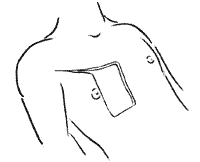
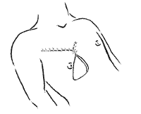
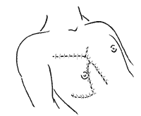
The planning of procedure started from the preparation of the pectoralis major myocutaneous flap. After the design of the skin island, the upper margin was laterally prolonged towards the deltoid region. The incision was at the level of the inferior margin of the previous deltopectoral fasciocutaneous flap (Figure 1). After the transfer of the myocutaneous flap, the pectoral region was closed by using a combination of advancement flap on the superior region with transposition flap on the inferior region. The superior-medial edge of a lateral flap was advanced and sutured 5 cm below the superior extremity of the medial limit of the defect. The 5-cm superior to the margin of the defect was sutured on the medial 5cm of the superior margin. The remaining superior margin of the defect was sutured on the superior margin of the lateral advancement flap (Figure 2). The inferior incision of the defect was laterally and slightly superiorly prolonged by an extension correspondent to the length of the medial margin, not sutured yet. From the lateral extremity of this incision,-anew inferior medial incision was made, creating a triangular flap (Figure 3). Ideally, the edge of this flap should be rounded to reduce the risk of necrosis. The inferior medial base flap was rotated medial-superiorly and applied on the remaining defect (Figure 4). Stitches were removed after 15 days.
Figure 1. Donor defect from pectoralis major myocutaneous flap.
Figure 2. Closure of superior part of the defect with advancement flap from the lateral region.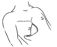
Figure 3. Triangular flap of inferior-medial pedicle.
Figure 4. Closure of residual defect with triangular transposition flap.
RESULTS
In all patients, defects were closed without requiring other complementary techniques. There were no cases of pulmonary complications and patients presented effective cough without significant pulmonary restrictions. There were no cases of suture dehiscence. In three patients, there was. necrosis of distal 2cm of transposition flap, with satisfactory healing after debridment.
DISCUSSION
Pectoralis major myocutaneous flap is the main reconstructive technique for large head and neck defects. Any improvement on the technique provides obvious practical implications due to its frequency of use. One of the difficulties is related to the management of the donor area, especially of large medial-lateral defects. Depending on the extension, primary closure is difficult or impossible. The attempt to determine the possibility of primary closure by evaluating the dimensions of the defect does not suffice, because a 10cm defect in a 1.50m tall patient is different from a 10cm defect in a 1.90m tall patient. Our experience suggests that it is more practical to evaluate based on anatomical references. If the transversal dimension of the defect is longer than the distance from the midline to the nipple, the likelihood of not having primarily closure is high.
Closure under excessive tension increases the risk of suture dehiscence, in addition to resulting in pulmonary disorders because of restriction of pulmonary expansion capability. The use of skin graft is far from being considered ideal.
The procedure reported here is a simple and quick solution for the problem, providing a number of advantages. Deltopectoral fasciocutaneous flap is preserved because it might become necessary in the future. Less tension in the suture decreases the risk of suture dehiscence and pulmonary complications. Closure is conducted with local flaps that have the same characteristics of the tissue in the region. There is little medial displacement of the nipple, resulting in a less "exotic" position than in primary closure.
The main hurdles are difficulty to perform it in the pectoral female anatomy and the risk of necrosis on the edgy of the transposition flap, especially if it is very sharp.
CONCLUSION
The use of adjacent flaps to close large defects after the use of pectoralis major myocutaneous flap is an easy to employ technique, which produces good results and reduces the risk of complications associated with primary closure made under excessive tension.
REFERENCES
1. ARIYAN, S. - The pectoralis major myocutaneous flap: a versatile flap for reconstruction in the head and neck. Plast. & Reconstr Surg., 63(1): 73-81, 1979.
2. BAKAMJIAN, V Y. - A two stage method for pharyngoesophageal reconstruction with a primary pectoral skin flap. Plast. Reconstr Surg., 36: 173-84, 1965.
3. FREEMAN, E. E and cols. The Vascular anatomy of the pectorals major myocutaneous flap. Brit. J Plast. Surg., 34: 3-10, 1981.
4. MEHRHOLF, A. I. and cols. The pectoralis major myocutaneous flap in head an neck reconstruction: analysis of complication. Am. J Surg., 143: 478-82, 1983.
5. PANJE, W R. - Free flaps versus myocutaneous flaps in reconstruction of the head and neck. Otolaryngol. Clin. N. Am., 15 (1): 111-121, 1982.
6. WEI, W I. and cols. The true pectoralis major myocutaneous island flap: an anatomical study. Brit. J. Plast. Surg.. 37: 568-73. 1984.
* Assistant Physician. ** Clinical Head. *** Resident Physician.
Clinic of Otorhinolaryngology and Head and Neck Surgery, Hospital Felício Rocho, Belo Horizonte /MG.
The present study was sponsored by Centro de Apoio à Ciência - Felicoop / Banco Bandeirantes.
Address correspondence to: Mauro Becker Martins Vieira - Avenida do Contorno, 9215 / Sala 802 - 30110-130 Belo Horizonte /MG.
Tel: (55 31) 291-8288 - Fax: (55 31) 337-5398
Article submitted on December 14 2000 Article accepted on January 18, 2001.
Print: ![]()