

Year: 2001 Vol. 67 Ed. 2 - (20º)
Relato de Casos
Pages: 271 to 273
Sialocele of Wharton Duct - Case Report and Review of Literature.
Author(s):
Manoel A. Marcon*,
Lídio Granato**,
José C. Burlamarqui***.
Keywords: sialocele, sialoadenitis, surgery
Abstract:
The authors report a case of left Wharton duct sialocele with increase of volume of the oral cavity floor and with worsening during feeding. History of trauma in the area was absent. The radioimaging studies confirm the dilatation of the Wharton duct and so the inflamatory process in the left submandibulary gland. The treatment consisted in resecting the submandibulary gland and the duct sialocele. The patient developmented satisfactory without complications.
![]()
INTRODUCTION
The increase in volume of sublingual or cheek region, related with intake of food, is normally correlated with disorders of the drainage ducts of salivary glands. Distal dilation of Wharton's duct may be related to calculus or post-inflammatory fibrosis.
In addition to acquired causes, which are the most frequent, there are some rare congenital causes, the so-called sialocele of Wharton's and Stensen's ducts. Sialography or CT scan normally shows the duct dilation, plus disorders of glandular parenchyma because of alteration of drainage.
Sialocele of Wharton's duct that causes a significant bulging is rare. Since we did not find any similar case published in the literature, we believed that it was worth describing it here.
CASE REPORT
O. O. S., 21-year-old male patient, from Maceio /AL, doorman.
Complaint: A ball under the tongue for the past 5 years.
History: The patient reported bulging under the tongue that sometimes became even bigger, especially when he ate acid food. It also released a light secretion in the oral cavity, called "thick saliva" by the patient, which normally happened during the meals.
The patient also reported episodes of enlarged volume, pain and fever for some days; treated with antibiotics.
He did not refer trauma or calculus in the region. He did not report any other complaints, or anything of significance concerning personal and family history.
ENT exam:
* Absence of cervical lymph nodes;
* Normal floor of the oral cavity;
* Enlargement of left submandibular gland; under bimanual palpation, it released a thick and light colored secretion.
Based on the diagnostic hypothesis of sialolithyasis of left submandibular gland, we ordered the following exams:
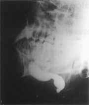
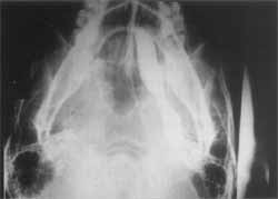
1. Sialography, which revealed dilation of Wharton's duct (Figures 1 and 2);
2. CT scan of cervical region, which showed presence of hypoattenuating, elongated and homogenous formation, with clearly-defined limits, no enhancement in contrast and localized in the topography of the left submandibular gland and duct. We did not detect image of calculus.
Conclusion: Sialocele of left Wharton's duct.
Figure 1. Sialocele of Wharton's duct (lateral view)
Figure 2. Sialocele of Wharton's duct (sagittal section).
We indicated exeresis of left submandibular gland under general anesthesia by external approach. We used Sebileau's incision (1 cm below and parallel to'the horizontal branch of mandible), and after resection of the gland, we proceeded from posterior to anterior, on the floor of the mouth, making a ligation of the duct close to its drainage orifice.
The result of anatomic pathological analysis was nonspecific chronic sialoadenitis with interstitial fibrosis and cystic dilation of duct.
DISCUSSION
Sialocele of drainage ducts of salivary glands is very rare. Parotid gland, which is the largest salivary gland in men, is subject to trauma and may present intraglandular sialocele. The dilations have been described as saccular or lobular and depend on the complete or partial interruption of the duct system1. Similarly to acquired causes, Stensen's duct of parotid gland has been a site of cystic dilations in a number of cases described in the literature, normally found among glass blowers and wind instrument players4. Accidental bites at the duct of the parotid during mastication may also dilate it.
Wharton's duct is equally predisposed to traumas, including the ones caused by ill-adapted dental prosthesis, and to the presence of calculus, which are more common in the submandibular glands than, in the parotid gland2.
The present case described a patient that did not correlate the onset of the, condition with trauma or history of calculus, which is normally quite common. The hypothesis is that it could have been a duct malformation that was triggered by small traumas or inflammatory processes5.
Sialoadenitis confirmed in the analysis may be explained by the difficulty in drainage resultant from the ectasia of the duct.
CONCLUSION
Sialocele of Wharton's duct is a rare affection that presented here with repetitive acute episodes of a chronic process. The difficulty of drainage resulted in chronic sialoadenitis. The cause of sialocele is supposedly congenital and surgical treatment, which consisted of the removal of the submandibular gland, satisfactorily resolved the case.
REFERENCES
1. CHOLANKERIL, J. V; RAVIPATI, M.; KHEDERKAR, S.; JANEIRA, L. L; VILLACIN, A. - Unusually Large Sialocele: CT Characteristics. J Comput. Assist. Tomogr, 13 (2): 3678, 1989.
2. FARB, S. N. - Disorders of salivary glands -Ot6laryngologyMedical Outline Series - Medical Examination publishing Company, INC: 245-56, 1970.
3. IQBAL, S.M.; R. R.; DEWANGAN, G. L. - Sialocele of Stensen's duct. J Laryngol. Otol, 100: 363-5, 1986.
4. PRATT, L.W, (1965), apud HOOPER, R.; SAXON, R.; TROPP, A. - Cysts of the parotid gland. J. Laryngol. Otol., 89: 427-33, 1975.
5. RIBEIRO, M. Q.; GRANATO, L.; CAMPANA, D.; PADULA, F.; MARIGO, C. - Sialocele of Stensen' ducts (A case report) -E. R. S & I. S. I. A. N. Meeting; 98 - Monduzzi Editore Sp.2 Bologna-Itália 17A-7 1998.
* Resident of the Department of Otorhinolaryngology at Santa Casa de São Paulo.
** Joint Professor of the Department of Otorhinolaryngology at Santa Casa de São Paulo.
*** Instructing Professor of the Department of Otorhinolaryngology at Santa Casa de São Paulo.
Study conducted at the Department of Otorhinolaryngology at Santa Casa de São Paulo.
Address for correspondence: Departamento de Otorrinolaringologia da Santa Casa de São Paulo - Rua Dr. Cesário Mota Jr., 112 - 01277-900 São Paulo /SP.
Article submited on July, 27, 2000. Article accepted on September 28, 2000.
Print: ![]()