

Year: 2001 Vol. 67 Ed. 2 - (10º)
Artigos Originais
Pages: 204 to 211
Otolaryngologic Manifestations in Children with AIDS.
Author(s):
Regina H. G. Martins*,
Lígia Batista**,
Ana Cláudia V. de Souza**,
Graziela A. Costa**,
Antonio Zuliani***,
Jaime Olbrich Neto****.
Keywords: child, AIDS, otolaryngology, HIV+
Abstract:
Introduction: The study of otolaryngologic manifestations in children HIV+ can lead to early diagnosis of AIDS, allowing specific treatment, responsible by reduced morbidity and mortality. Objectives: Detect the otolaryngologic manifestations in children with AIDS and alert to the importance of the early diagnosis. Study design: Clinical prospective. Material and method: We evaluated 22 children with AIDS assisted at Faculdade de Medicina de Botucatu (São Paulo, Brasil). The medical records were reviewed and the children were submitted to otolaryngologic and hearing acuity exams. Results: We evaluated 12 boys and 10 girls, whose ages ranged from 8 months to 12 years. In B and C clinical classification were included 18 children who were using anti-retroviral medicaments. Physical examination mainly indicated cervical lymphadenopathy (18 cases), paleness of the nasal mucous membrane with abundant mucous secretion over the nasal epithelium (15 cases) and retraction of tympanic membranes (seven cases). The main otolaryngologic diagnoses were: rhinosinusitis (16 cases), oral candidiasis (13 cases), inadequate eustachian tube function (seven cases) and recurrent tonsillar infections (six cases). Conductive hearing loss were detected in 4 children. No child presented sensorineural hearing loss. Conclusions: The main otolaryngologic manifestations presented by the children with HIV virus were rhinosinusitis, oral candidiasis, inadequate eustachian tube function and recurrent tonsillar infections. The allergic aspect of the nasal mucous membrane and the cervical lymphadenopathy were frequent signs and could alert the otolaryngologyst to AIDS during the exam.
![]()
INTRODUCTION
In 1980, occasional cases of a new disease started to be reported, usually manifested by infectious lung condition caused by Pnemocystis carinii and Kaposi sarcoma, affecting mainly young subjects who were either homosexuals or used injecting drugs. The pathology, of unknown etiology at the time, was quickly characterized as a world epidemics, affecting risk populations as well as children2,12,25,26.
Thanks to the common pattern of pneumonia, lack of appetite and death caused by opportunist infections, evidence of viral etiology was suspected and confirmed in 1983, when Luc Montagnier isolated the AIDS virus (immunodeficiency human virus, HIV)2. It is a lymphotropic retrovirus responsible for immune disarrangement, specially affecting cell immunity and attacking the helper lymphocytes T (CD4+), destroying and reducing the number of cells in the peripheral blood. Cytotoxic lymphocytes T (CD8+) present a relative elevation in its population, inverting the ratio of the two cell types at the primary infection and throughout the whole asymptomatic period of the infection; finally both cells are largely reduced at the final stages of the pathology. There is activation of humoral immunity by increase of serum immunoglobulins, especially IgG and IgM, but the immune responses are ineffective. In addition, neutralizing antibodies that are significantly important are present approximately 120 days after the infection, but they do not maintain themselves, leading to impairment of humoral immunity during the progression of the disease22,24.
Retrovirus was initially isolated from peripheral blood and later it was identified also in the semen, lymph nodes, spleen, bone marrow, brain, saliva, tear, CSF, lung arid otological secretions, maternal milk, and cervical-vaginal secretions, among other sites. Despite all these different sites, the main transmission routes in adults are blood transfusions and sexual intercourse, in addition to contact with contaminated blood. In children, the main transmission route is vertical transmission. It is difficult to say when exactly the contamination takes place, but it may be during pregnancy via placenta, at delivery because the child gets in contact with blood and cervical-vaginal secretions of the mother, or after birth, through breastfeeding, because milk has high viral load. Parenteral contamination in children is rare7,21.
As a result of progressive immunological impairment, adult patients and especially children, become an easy target for a large number of opportunist infectious agents, such as Pneumocystis carinii, criptosporidium, candida and cryptococos, among others. Clinical progression of AIDS depends on a number of factors, such as: time of infection, viral genotype and phenotype, immune response, individual genetic constitution, appropriate treatment of opportunist infections, systemic viral load and use of anti-retrovirus medication, which have provided lower morbidity and mortality rates5,11.
In a summarized version, the clinical progression of the disease in adults may be divided into 3 phases: primary infection (first weeks after the contamination), characterized by non-specific symptoms, present in 50% to 75% of the patients, such as fever, lethargy, sore throat, myalgia, headache, photophobia, lymphadenopathy, cutaneous rash. The second phase, clinical latency period, that may be prolonged (from 8 to 10 years) in adults but much shorter in children (between 8 months and 2 years), and the period of the clinical apparent disease, characterized by the persistence of clinical symptoms22,30. In the phase of primary infection, the virus is located in lymphatic tissues, from where it replicates and causes disorders and destruction. During the other phases, we observed plasma replication of the virus and involvement of tissues and organs30.
A large number of adult patients with AIDS have otorhinolaryngological manifestations, present in 40% to 90% of the cases, especially recurrent episodes of bacterial rhinosinusitis, effusion otitis media, oral candidiasis, cervical abscess, dental abscess, herpes lesions, enlargement of salivary glands, enlargement of lymphatic tissue of rhinoipharynx and cervical ganglionar systems, necrotic stomatitis and Kaposi sarcoma of oral cavity4,8,14.
In children affected with HIV it is normally known that initial symptoms may be similar to the pathologies that affect healthy children in the first years of life, such as tonsillar, otological and nasosinusal infectious episodes, due to lack of immune maturity presented by many small children; this fact, however, delays and impairs early diagnosis of AIDS. Therefore, the conduction of a otorhinolaryngological clinical study directed to children infected with HIV becomes of importance. The purpose of the study was to learn more about the involvement of our specialty in the progression of AIDS in children because in a number of cases otorhinolaryngologists are the first health professionals to provide medical care to these children.
MATERIAL AND METHOD
Material
We evaluated 22 children, both genders, who had diagnosis of AIDS and ages between 8 months and 12 years (mean), and were followed at Hospital das Clínicas da Faculdade de Medicina de Botucatu /SP (UNESP), between June 1999 and July 2000, seen at the Ambulatory of Pediatric Immunology and Otorhinolaryngology. Diagnosis of HIV infections was confirmed by epidemiological, clinical and lab characteristics.
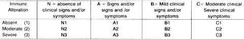
The present study was approved by the Ethics Committee for Research in Human Beings at Faculdade de Medicina de Botucatu.CHART 1 - Clinical classification of children with HIV infection, according to criteria proposed by CDC (Center for Disease Control)5.
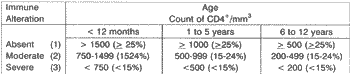
CHART 2 - Immune category of children with AIDS.
Method
In order to diagnose infections caused by HIV/AIDS, we followed the parameters listed below:
Epidemiological characteristics: we valued mechanisms of vertical and parenteral transmission.
Clinical characteristics: we considered physical signs and symptoms if present, by analyzing medical files of current and previous complaints and interviews with parents. Children were submitted to otorhinolaryngological physical exam and auditory assessment, exams conducted always by the same professional (otorhinolaryngologist and audiologist, respectively), both authors of the present study.
Lab tests: a) audiological assessment for audiological assessment in younger children, we used behavioral tests with musical instruments; for older children, we conducted conventional or conditioned pure tone audiometry, with electronic audiometer (Clinical Audiometer Madsen Electronics, Denmark). All children were submitted to immitanciometry using the device Amplaid 75; b) diagnosis of the disease: in order to confirm diagnosis of AIDS, we employed anti-HIV serologic tests (Elisa - two positive samples) and Western blot, if necessary.
Clinical classification followed the criteria determined by CDC (Center for Disease Control)5, shown in Charts 1 and 2.
RESULTS
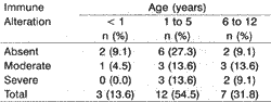
Among the evaluated children, the main exposure route was vertical contamination, and only one child had been contaminated via parenteral route (post-blood transfusion). Table 1 shows distribution of 22 children infected with HIV according to age range and immune alteration, following CDC criteria. Only three children were younger than 1 year of age, 12 were aged between 1 and 5 years and seven of them were aged between 6 and 12 years. Children younger than 1 year did not present severe immune compromise, which was more marked among older children.
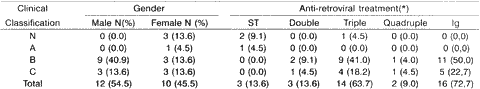
Table 2 presents the distribution of children according to clinical classification, gender and anti-retrovirus medication. Only three children did not take medications, and among them two belonged to category N and one, to category A. The remaining children used an association of drugs (double, triple, quadruple or immunoglobulin schemes). There was a slight predominance of male subjects (12 children) over female subjects (10 children). We observed more concentration of severe affections (categories B and C) among boys. Among girls, clinical classification was evenly distributed.
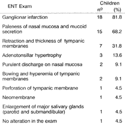
The main findings of otorhinolaryngological physical exam are presented in Table 3 and auditory assessment results are shown in Table 4. The most significant alteration detected in the physical exam was high incidence of ganglionar infarction in several cervical systems, especially jugular-carotid, submandibular and posterior cervical systems, found in 82% of the children (18 cases). Another alteration that was highlighted was the pale and edematous aspect of nasal fossa mucosa, in addition to abundant mucoid secretion on the turbinates, presented in 15 children (68% of the cases). Bacterial contamination of paranasal sinus mucosa was observed in only 2 cases.
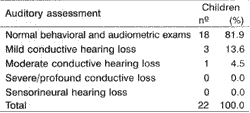
Otoscopy was abnormal in 11 children. Retraction of tympanic membranes was observed in 7 children, but only 3 of them presented mild conductive hearing loss (pure tone thresholds below 30dB) and type C curve at immitanciometry in the audiological assessment (Table 4). In two children, we detected acute episodes of otitis media during the exam; they were treated with amoxicillin. Audiometric and impedanciometric exams of children, carried out one month later, were normal. In one child we detected bilateral perforation of tympanic membrane, but there was no secretion. Audiometry showed moderate conductive hearing loss (pure tone thresholds at 50dB bilaterally). The presence of a small neomembrane in one ear was observed in one case, whose audiological assessment was normal. No children presented sensorineural hearing loss, even those that used associated medication regimens (Table 4).TABLE 1 - Distribution of children infected by HIV virus, according to age and severity of immune alteration (dosage of CD4/mm³).
Moderate tonsil hypertrophy (3 cases) and enlargement of salivary glands (one case) were less frequent findings at the ENT exam. Only one child had normal ENT exam.
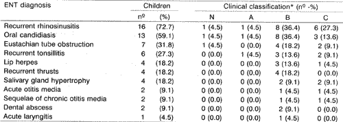
In Table 5, we present the main otorhinolaryngological diagnoses and their relation with clinical classification of the evaluated children. The most frequent diagnoses were recurrent episodes of rhinosinusitis (13 cases), eustachian tube obstructions (7 cases), and repetitive tonsillitis (6 cases). The least common affections were lip herpes (4 cases), recurrent thrusts (4 cases), salivary gland hypertrophy (4 cases), acute otitis media (2 cases), sequelae of chronic otitis media (2 cases), dental abscess (2 cases) and acute laryngitis (one case). We could notice that the main diagnoses were correlated with children that had more severe cases - B and C (Table 5).
DISCUSSION
The predominance of vertical transmission of HIV virus in the pediatric population was confirmed in our study and is compatible with the findings by other authors7,21. Right after the contamination by HIV virus, regional lymph nodes become the main sites for viral replication, leading to marked ganglionar hyperplasia. The persistence of the virus in the lymphoid tissue leads to stimulation of the immune system. Viral colonization extends itself to other ganglionar systems, characterizing the spreading phase22. Ganglionar infarction is the physical exam sign most frequently observed, confirmed in the present study by 82% of the children and in accordance with Carvalho and Tonelli3, that observed the same alteration in 98% of the 60 children studied by them. Adenopathies may surge in any stage of the disease and compromise different systems, especially the cervical ones. They are normally bilateral, non-painful and movable on the superficial plans. The exaggerated enlargement of ganglin or any other modification in its characteristics may require biopsy, in order to define differential diagnoses, such as lymphoma, hystoplasmosis, toxoplasmosis, tuberculosis, sarcoma, and carcinoma4,21,24.TABLE 2 - Distribution of children with AIDS, according to clinical classification, gender and anti-retroviral treatment.
Key: (*) ST - No treatment; Double - Treatment with two nucleoside analog inhibitors of reverse transcriptase; Triple - Treatment with two nucleoside analog inhibitors of reverse transcriptase + one inhibitor of protease; Quadruple -Treatment with two nucleoside amlog inhibitors of reverse transcriptase + one inhibitor of protease + one nucleoside non-analog inhibitor of reverse transcriptase; Ig - Immumglobulin
TABLE 3 - Results of ENT exams of the 22 children infected by HIV virus.
Some authors9,14 described the enlargement of tonsillar tissue, together with enlargement of systemic ganglionar systems resulting in pulmonary obstruction in some cases, manifested by night apnea. In our study, the enlargement of tonsils was not a frequent finding of the physical exam, even in children that presented several impaired ganglionar systems. An explanation for this finding could be the one given by Chow et al. 4, who highlighted that children with HIV present failures of antigen recognition by the immune system, leading to reduction of lymphocytic proliferation.
Among the most frequent ENT diagnoses, we found recurrent rhinosinusitis. The main symptoms presented by these children were constant nasal obstruction, followed by light-colored nasal discharge, no symptoms of sneezing, nasal pruritus or previous history of bronchial asthma. At physical exam, they presented pale aspect of all recovering epithelium of the nasal fossa and abundant mucoid secretion over the surface of the turbinate, similarly to what happens in patients with allergic rhinitis. The high incidence of nasal-sinusal impairment has also been confirmed by other authors8,15,20,28,30. Vicenti28, using cultures of secretion of maxillary sinus of patients with AIDS, found the following pathogens Streptococcus pneumoniae, Streptococcus viridans, Staphylococcus aureus, Staphylococcus epidermidis, Haemophylus influenzae and Pseudomonas aeruginosa.TABLE 4 - Results of auditory assessment conducted in 22 children infected by HIV
Otological complaints were frequent and observed in 50% of the evaluated subjects. The main predisposing factor, in addition to AIDS, was constant nasal obstruction and lymphoid hyperplasia on the rhinopharynx region, responsible for edema and eustachian tube obstruction. In our study, the alterations were confirmed by otoscopy with predominance of retraction and thickness of tympanic membrane. The detection of these alterations, both in children and adults with AIDS, requires a very careful exam of rhinopharynx, because it may host not only lymphoid hypertrophy but also tumors, such as lymphoma, sarcoma and carcinoma9,17,23,27 . No children presented serous otitis media with effusion, justifying the small number of abnormal audiometric findings.
Different authors have reported the involvement of the inner ear in patients with AIDS8,13,16,17,19. Although HIV is neurotrophic, the main factors responsible for sensorineural hearing loss seem to be secondary to infections of the central nervous system, such as meningocriptococcosis, neural Lues, tuberculous meningitis, central toxoplasmosis, herpes or cytomegalovirus encephalitis. Kohan et al'6 evaluated 26 adult patients infected with HIV with otological complaints and detected sensorineural hearing loss in seven cases, in addition to external otitis (6 patients) and otitis media (6 patients). The authors emphasized that among the possible etiological factors of the hearing loss there could be the use of ototoxic drugs (3 patients), criptococcical meningitis (one patient), central toxoplasmosis (one patient), neural Lues (one patient) and tuberculous meningitis (one patient).TABLE 5 - Main otorhinolaryngological diagnoses and clinical classification of children infected with HIV virus.)
Key: N - absence of sings and/or symptoms; A - mild signs and/or symptoms; B - moderate signs and/or symptoms; C - severe signs and/or symptoms.
Another important factor of cochlear affection is the action of ototoxic drugs used by these patients, such as sulfa, streptomycin, amycacin, amphotericin B, acyclovir and cetoconazol, in addition to anti-retrovirus drugs16,29. In our sample, many children took complex medication regimens but we did not detect any case of sensorineural hearing loss, which seems to be more frequent in adults. The hypothesis that in patients with AIDS there would be early compromise of auditory retrocochlear pathways was not confirmed by Lima and Fukuda18, in their study with auditory evoked brainstem responses in 30 patients with positive serology for HIV without hearing complaints.
The use of CT scan may help in the diagnosis of otological affections, as shown by Kohan et al.17. The authors conducted CT scans of 18 patients infected by HIV who had hearing complaints. The authors noticed that seven of the patients has signs of otitis media with effusion and otomastoiditis, and in one of these patients, they detected the presence of a tumor in the rhinopharyngeal region; in 3 patients they found central nervous system pathologies, such as toxoplasmosis, and in 8 patients the results of the exams were within normal ranges. They also found aural polyps and osteomyelitis of temporal bone. The authors reinforced the importance of CT scan of the ear and rhinopharyngeal region in such patients, because these regions may host tumor masses.
AIDS should also be part of the list of differential diagnosis of patients with sudden deafness, as shown by Grimaldi et al.13 who had a case of AIDS patient who progressed throughout the disease with high frequencies hearing loss; his BERA showed increased latency of interval I-V In the pathogenesis of the alteration, the authors suggested the existence of perivascular inflammatory processes that would result in ischemic episodes involving the 8th cranial nerve, based on histological findings of biopsies of nervous endings in patients with HIV and peripheral neuropathy.
The oral cavity may host different lesions of positive HIV patients. Among children in our study, the most frequent ones, in decreasing order, were oral candidiasis, lip herpes, recurrent thrusts and dental abscesses. According to Ferreira8, oral candidiasis is reported by approximately 90% of the patients at same stage of the disease, which may be sign of early immunodeficiency. The pseudomembranous form is more common and it shows a whitish intertwined area that may be easily detached and presents hyperemia under it. Other clinical forms may be evidenced in adult patients, such as erythematous or atrophic forms, characterized by hyperemia plaques or spots on the oral mucosa, angular queilitis (erosion or fistulae of labial commissure) and hyperplasic forms (whitish non-detachable areas)6.
Herpes lesions may compromise lips, oral and nasal mucosa. They start as vesicles that rapidly break and form an ulcerated and painful area. Lesions are recurrent and may be extensive. Similarly to herpes lesions, thrusts are related to immunodeficiency and are also frequent in children. It is important to point out that ulcerated lesions of the oral mucosa may represent the first signs of tumors, such as lymphoma and sarcoma, requiring local biopsy.
AIDS should be suspected in children who have necrotic gingivitis and dental abscess, which should be closely observed by professionals of odontology Necrotizing ulcerative gingivitis is a severe affection, with inflammatory process that extends to dental alveoli and may cause bone sequestration6,10.
Children who presented enlargement of salivary glands reported recurrent episodes of glandular hypertrophy. In one of these children, during physical ENT exam, we confirmed glandular compromise and involvement of parotid and submandibular glands, which were enlarged, hardened and had multinodular surfaces, adhered to the coalescent masses of regional ganglionar infarction. Aspiration punch revealed ganglionar impairment caused by hystoplasm. It was a fungus (Histoplasma capsulatum) that may colonize in the mucosa of oral cavity, pharynx, larynx, salivary glands, lungs and lymph nodes, which become sites of multiplication and dissemination. Macroscopic lesions are nodular, formed by granulomatous tissue, firm and brownish. The affected tissue may experience necrosis and ulceration. The diagnosis of hystoplasmosis is made by local biopsy1,10. In addition to the infectious etiology, the enlargement of salivary glands may occur as a result of hypertrophy and lymphocytic infiltration of glandular parenchyma, such as in Sjögren's syndrome. The generalized enlargement of parotid glands was observed in 45% of the 60 children infected with HIV studied by Carvalho3 and described as multicystic glandular hypertrophy, uni or bilateral, non-painful and involving primarily the parotid glands, with no increase of serum amylase.
Only one child had an episode of laryngitis, together with hoarseness and mild breathing difficulty, which improved right after the' use of amoxicillin. According to Ferreira8, the main pathogen responsible for laryngitis, tracheitis, and laryngotracheitis in patients with AIDS is Haemophylus influenzae and pneumoccocus, pathogens that also affect the non-infected population.
The most frequent diagnoses in the present study, as well as the alterations observed in the physical exam, had higher incidence among children who belonged to clinical categories B and C, including more severe signs and symptoms of the progression of the disease.
CONCLUSIONS
In the present study, we observed that a number of otorhinolaryngological diagnoses were frequent in children with AIDS, especially episodes of recurrent rhinosinusitis, oral candidiasis and eustachian tube obstruction. Most of these affections may be the first symptoms of the disease at this age range. The allergic aspect of nasal mucosa inchildren who do not have complaints of allergic rhinitis and the presence of ganglionar infarction of cervical systems were frequent findings and should be seen as warning signs during the physical exam. Therefore, otorhinolaryngologists should be attentive to the main signs and symptoms presented by these children and include AIDS among the differential diagnoses of other diseases of our specialty.
REFERENCES
1. BALLENGER, J. J. - Enfermidades granulomatosas crônicas de la laringe. In- Ballenger, J.J. Enfermidades de la nariz, garganta y oido. Barcelona, Editorial Jims, 1986, 447-459.
2. BARRE-SIMOUSSI, F.; CHERMANN J. C.; REY F., et al. Isolation of T lymphotropic retrovirus from a patient at risk for acquired immune deficiency syndrome (AIDS). Science, 220: 868, 1983.
3. CARVALHO, A. E; TONELLI, E. - Avaliação imunológica em 60 crianças com AIDS. J Pediatr, 75: 172-180, 1999.
4. CHOW J. H.; KAUL, A.; GROMISCH, D. S. - Head and neck manifestations of the acquired immunodeficiency syndrome in children. Ear Nose and Throat, 69: 416-423, 1999.
5. Consenso sobre terapia anti-retroviral para crianças infeccdas pelo HIV Ministério da Saúde. Secretaria de Políticas de Saúde. Coordenação Nacional de DST a AIDS. Brasília, 1999. 23.
6. COTIN, G.; KUFFER, R. - Problèmes de pathologie buccale. In- Encyclopedie Medico Chrurgicale. Paris, Editions Techniques, 1980, 20624A10.
7. DOUGLAS, G. C.; KING, B. F. - Maternal-fetal transmission of human immunodeficiency virus: A review of possible routes and cellular mechanisms of infection Clin. Infect. Dis.,15: 678-691, 1992.
8. Ferreira, N. G. M.; Ferreira, A. G. - Manifestações otorrinolaringológicas em pacientes infectados com o vírus da AIDS. F. Med., 105: 147-56, 1992.
9. FRANCE, A. J.; KEAN, D. M.; DOUGLAS, R. H. B. Adenoidal hypertrophy in HIV infected patients. Lancet, 5: 1076, 1988.
10. GEHANNO, E; MATHERONS, S. - Les manifestations ORL observées lors de I' infection HIV notamment lors du SIDA. Encyclopedie Medico Chirurgicale. Paris, Editions Techniques, 1988, 20956 A10.
11. GRAHAN, N. M. H.; ZEGER, S.T.; PARK, L. P P; VERMUND, S. H.; DETELS, R.; RINALDO, C.; PHAIR, J. P The effects on survival of early treatment of human immunodeficiency virus infection. N. Engl. J Med., 326: 1037-1042, 1992.
12. GOTTLIEB, M. S.; SCHROFF, R.; SCHAMBER, H. M., et al. - Pneumocystis carinii pneumonia and mucosal candidiasis in previously healthy homosexual men: evidence of a new acquired cellular immunodeficiency. N. Engl. LMed.. 305: 1425, 1981.
13. GRIMALDI, L. M. E.; LUZI, L.; MARTINO, G. V; FURIAN, R.; NEMNI, R.; ANTONELLI, A. CANAL, N.; POZZA, G. -Bilateral eighth cranial neuropathy in human immunodeficiency virus infection. J. Neurology, 240: 363-366, 1993.
14. HELSPER, J.; FORMENTI, S.; LEVINE, A. - Initial manifestation of acquired immunodeficiency syndrome in the head and neck region. Am. J. Surg., 152: 403-406, 1986. 15. HIRATA, C. H. W; TEIXEIRA, M. S.; WECKX, L. L. Manifestações otorrinolaringológicas na Aids. Rev. Bras. Med., 53: 143-157, 1996.
16. KORAN, D.; ROTHSTEIN, S. G.; COHEN, N. L. - Otologic disease in patients with acquired immunodeficiency syndrome. Ann. Otol. Rhinol. Laryngol, 97: 636-640, 1988.
17. KORAN, D.; HAMMERSCHLAG, E E.; HOLLIDAY, R. A. Otologic disease in AIDS patients: CT correlation. Laryngoscope, 100: 1326-1330, 1990.
18. LIMA, M. A. M. T; FUKUDA, Y - Audiometria de tronco encefálico em pacientes HIV positivos assintomáticos: análise nas freqüências de 11 a 61 cliques por segundo. Rev. Bras. de Otorrinolaringol., 65: 404-411, 1999.
19. MORRIS, M. S.; PRASAD, S. - Otologic disease in the acquired immunodeficiency syndrome. Ear Nose and Throat J, 69: 451-453, 1990.
20. MUNHOZ, M. S. L.; UIP, D.E.; MUNHOZ, A. N. L.; MUNHOZ, M. L. G. S. -Manifestações otorrinolaringológicas em pacientes com síndrome de imunodeficiência adquirida (SIDA). Rev. Bras. Med. ORL., 1: 54-56, 1994.
21. PALASITHRAN, E; ZIEGLER, J. B.; STEWART, G. J. Breastfeeding during primary human immunodeficiency virus infection and risk of transmission from mother to infant. J. Infect. Dis., 167: 441, 1993.
22. PANTALEO, G.; FAUCI, A. S. - New concepts in the immunopathogenesis of HIV infection. Annu. Rev. Immunol., 13: 487-512, 1995.
23. ROLAND. J. T.; ROTHSTEIN, S.G.; MITTAL, K. R.; PERKSY, M. S. - Squamous cell carcinoma in HIV positive patients under age 45. Laryngoscope, 103: 509-511,1993.
24. RUBINI, N. - Alterações imunológicas em criança com infecção pelo HIV J. Pediatr., 75: 147-148, 1999.
25. SATCHER, D. -A epidemia global de HIV/AIDS JAMA, 3: 2286, 1999.
26. SEIGAL, F. E; LOPES, C.; HAMMER, G. S., et al. - Severe acquired immunodeficiency in male homosexuals, manifested by chronic perianal ulcerative herpes simplex lesions. N. Engl. J. Med., 305: 1439, 1981.
27. STERN, J. C.; LIN, P J.; LUCENTE, F. E. - Benign nasopharyngal masses and human immunodeficiency virus infection. Arch. Otolaryngol. Head Neck Surg., 116: 206-208, 1990.
28. VICENTI, A. B. - Bacteriologia do conteúdo do seio maxilar na imunodeficiência humana. Rev.Bras. Med. - ORL., 5: 55-65, 1998.
29. WERSALL, J. - Ototoxic Antibiotics: a review. Acta Otolaryngol. (Stockh)., 519: 26-29, 1995.
30. ZULIANI, A. - Síndrome da Imunodeficiência adquirida (SIDA). In: Condutas em Pediatria. São Paulo, EPUB (Editora de Publicaq6es Biomédicas), 1999, 765-780.
* Assistant Professor of the Discipline of Otorhinolaryngology and Head and Neck Surgery of the Department of Ophthalmology, Otorhinolaryngology and Head and Neck Surgery at Faculdade de Medicina de Botucatu - UNESP
** Undergraduate, School of Medicine, Faculdade de Medicina de Botucatu - UNESP
*** Assistant Professor of the Discipline of Pediatric Immunology at Faculdade de Medicina de Botucatu - UNESP
**** Pediatrician, Master Degree in Tropical Medicine, Intern of the Discipline of Special Immunology at Faculdade de Medicina de Botucatu - UNESP
Study conducted at the Disciplines of Otorhinolaryngology and Head and Neck Surgery and Pediatrics at Faculdade de Medicina de Botucatu, financially supported by FAPESP (Fundação de Amparo à Pesquisa do Estado de São Paulo - Foundation for the Support of Research in the State of São Paulo) - Department of Otorhinolaryngology, Ophthalmoloy and Head and Neck Surgery UNESP - São Paulo.
Address for correspondence: Regina Helena Garcia Martins - Faculdade de Medicina de Botucatu (UNESP) - Departamento de Oftalmologia, Otorrinolaringologia a Cirurgia de Cabeça a Pescoço - 18618-000 - Botucatu /SP - Tel: (55 14) 6802-6256 - E-mail: rmartins@fmb.unesp.br
Article submitted on October 3, 2000. Article accepted on January 15, 2001.
Print: ![]()