

Year: 2004 Vol. 70 Ed. 1 - (19º)
Relato de Caso
Pages: 120 to 123
 PDF PT
 Foreign body in nasopharynx: regarding a case
Author(s):
Fernando P. G. Sobrinho 1,
Alena M. B. Jardim 2,
Iara C. de Sant'Ana 3,
Hélio A. Lessa 4.
Keywords: foreign body, nasopharynx, larynx, neck radiography
Abstract:
The accidental ingestion of foreign body establish a comum problem in the emergency room and prompt attending services, especially in childhood. Althought rare, a foreign body reputed swallowed or inhaled can be projected and lodge in nasopharynx. The physical examination and standard radiologic screening incidences may don't evidence any abnormality. Therefore, the authors of this case report recommend that a cavum roentgenogram and/or cautions rhinopharynx visualization with Garcia's mirror, Hopkins type telescopes or flexible naso-fibroscope should be performed in these cases where cervical radiographic study don't show properly the nasopharynx above all important in radiolucents foreign bodies avaliation. The early diagnostic is necessary to avoid major complications.
![]()
INTRODUCTION
Foreign bodies in the aerodigestive tract are a frequent problem in services of emergency, especially in pediatric age ranges. At the same time, they can also determine severe complications and, therefore, require appropriate assessment and management.
In general, a patient with history of accidental intake of foreign body is investigated through physical examination and submitted to complementary radiology imaging, which normally includes neck, thorax and abdomen imaging tests.
Even though it is rare, a foreign body that had been swallowed or inhaled can be projected, impact and fixate on the nasopharynx, maintaining itself as clinically silent and not clearly evidenced in routine radiological incidences, hindering diagnosis and increasing the risk of morbidity.
We report a case of a child with history of accident intake of foreign body assessed by paranasal sinuses x-ray that evidenced presence of nasopharynx foreign body.
LITERATURE REVIEW
In the studied literature, there are few reports about foreign bodies in the nasopharynx, being that most of them are in Russian and with no English abstract. However, it is known of its rare occurrence.
Gomez 4 reports a case of intake of a plastic object, detected by normal exploration esophagoscopy after five months of unilateral purulent rhinorrhea and halitosis, treated as chronic maxillary sinusitis and radiologically confirmed. At the end of the period, a foreign body in the child's paranasal sinus was evidenced by posterior rhinoscopy.
In 1988, Parker et al. 7, in Bristol, United Kingdom, reported two cases of history of intake of five-cent coins in patients aged 2 and 6 years, respectively, being that the first had an interval of 7 weeks between the visit and the final diagnosis of abnormal nasopharynx finding, even though it was a radiopaque object. None of these two patients presented signs or symptoms and initial physical and radiological exams did not reveal any abnormalities. The definite diagnosis was obtained with paranasal sinuses radiological analysis.
Before that, Dayal and Singh 1 had reported in 1970 a series of three cases, in which two patients aged 3 years and 18 years, respectively, had clinical presentation of epistaxis, being that foreign bodies they had ingested were a toy and a fresh water leech. The third patient, victim of a firearm accident that led to significant facial damage, had radiological evidence of a metallic object in the rhinopharynx.
Gendeh and Gibb 3 described a Malaysian patient aged 24 years with unilateral purulent rhinorrhea that had started 6 months before after a severe car accident. With the diagnosis of refractory maxillary sinusitis, radiologically confirmed, and traumatic sequels, he was submitted to a surgical procedure during which the posterior rhinoscopy revealed the presence of wood fragments impacted on the rhinopharynx and measuring up to 6.7cm long by 1.5cm wide, surrounded by abundant phlogosis and purulent secretion. The evident lacerations of the nasal pyramid, later healed, had been considered by the authors as the probable entrance for the objects.
Foreign bodies in this location can also have iatrogenic origin 3, such as consequences of ENT procedures that involve fragments of cotton, gauze or buffers that can act as foreign bodies. However, it is possible that this issue is also reported in the forensic literature and the brown press.
CASE REPORT
A female 2-year-old patient was referred to the emergency room with suspicion of intake of a toy, an opaque glass ball (or marble) for approximately 2 hours. The mother did not report dyspepsia or sialorrhea in the child. The physical examination showed a calm and playful child, opposite to the mother's affliction. She presented noisy breathing, similarly to what is observed in some mouth breathers, but she had eupnoea. Acyanotic extremities. Anterior rhinoscopy and neck mobility did not show signs of any abnormality. There was slight bulging of the transition area between the soft palate and the hard palate towards the oral cavity, evidenced in the oropharyngoscopy. The remaining physical examination did not reveal any affections.
The patient had already been submitted to abdomen, chest and neck x-rays at anterior-posterior and profile incidences, and upper aerodigestive endoscopy, which was normal. The neck x-ray revealed a strange image in the posterior limit of the paranasal sinuses: a silhouette, of small dimensions, which was extremely radiopaque.
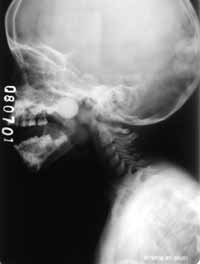
Based on the initial clinical examination and previous radiological battery, we ordered nasopharynx radiograph, which evidenced the presence of a round, radiopaque image occupying the rhinopharynx lumen (Figure 1), documenting the existing of a foreign body.
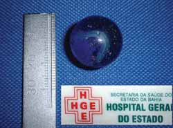
The foreign body was removed via oral access when the patient was under general anesthesia and orotracheal intubation, after previous packing of hypopharynx (Figure 2). A very useful device was a delicate intubation probe introduced endonasally, projecting the foreign body towards the oropharynx, associated with oral digit maneuver. The follow-up did not show any sequel from the procedure.
DISCUSSION
The diagnosis of accidental foreign body in the nasopharynx is not common. Conversely, it is more frequent in other aerodigestive sites such as nasal fossa, tonsils, upper esophageal sphincter, tracheobronchial tree, etc.
In general, foreign bodies are a frequent problem in childhood 2. The severity of the clinical case, in turn, is normally related with location, nature and dimensions of the foreign body, as well as the presence of infectious, obstructions and inflammatory complications, such as close necrosis by pressure, which can be associated with late diagnosis. The absence of early treatment can be fatal in some cases, especially when involving organs such as the larynx.
Harris et al. 5 reported that 66 to 77 children aged less than 10 years die every year in the USA owing to asphyxia associated with foods impacted in the larynx. In 1989, Lima6 published a series of 91 cases of foreign body aspiration diagnosed in a period of 7 years, in which 11 (12.1%) of them impacted the larynx, impairing glottic space. Out of them, 5 (45%) patients died and three (27%) progressed with transient hypoxic encephalopathy demonstrating high morbidity/mortality rate. The author generically classified the nature of foreign bodies as thin and laminar or globous and rounded. It seems that the latter were involved with most of the fatal and complicated cases.
Conversely, some foreign bodies can impact the gastrointestinal tract, especially in naturally narrow spaces such as upper esophageal sphincter or illium-ceccum valve.
As previously described, even though there were reports of long permanence of nasopharynx objects, leading to less important infectious complications such as nasopharyngitis and rhinosinusitis, aspiration of foreign bodies in this location, including during sleep, offers an imminent and potential risk of death. In our study, the displacement of spherical and solid objects pushed to the lowest segment of the digestive and respiratory tracts could have led to disastrous consequences. For example, Parker et al. referred that some degree of transient pharyngeal hypertonic muscles can temporarily support some foreign bodies in the nasopharynx, but palate muscle relaxation follows this state.
As to diagnosis, it is observed that clinical history of foreign bodies located in the rhinopharynx can vary from absence of signs and symptoms to epistaxis and evident purulent rhinorrhea, uni or bilateral. In these patients we can observe in the physical examination objects with dimensions enough to bulge the soft palate and have its presence suggested by oropharyngoscopy, as observed in our study. In turn, impact of foreign bodies in the esophagus can lead to sialorrhea, neck lordosis affections, dysphonia, dysphagia and local pain that can limit passive or active neck mobilization. Finally, unilateral purulent and fetid rhinorrhea or epistaxis can be the only finding of the presence of foreign body in the nasal fossa 8.
In emergency rooms, usually neck x-rays at anterior-posterior and profile incidences, chest and abdomen x-rays are ordered by the pediatrician or clinician to investigate the suspicions of foreign body intake, after clinical examination 7. Therefore, foreign bodies, especially radiopaque ones, can be easily evidenced in the esophagus. However, since neck x-ray cannot satisfactorily delimit a part of the nasopharynx, the nasopharyngeal abnormality can be lost and the diagnosis of foreign body in this location is impaired, as observed in some reports of the reviewed literature. The rarity of foreign bodies in this topography favors the low level of suspicion in these cases.
Posterior transoral rhinoscopy with Garcia mirror or Hopkins angulated telescope of 30o or 70o degrees, rigid nasal endoscopy with Hopkins angulated telescope 0o or 30o, or even flexible nasofibroscopy would be extremely useful in the diagnosis of rhinopharynx foreign bodies, but they are not readily available in many services of primary care and even in emergency units, especially in those in which there is no ENT on call. In addition, we should mention the expected difficulties of conducting classical posterior rhinoscopy with mirror in young children.
However, these endoscopic procedures should be conducted with maximum caution and under the appropriate conditions so as not to precipitate movement of the foreign body impacted in the rhinopharynx to the lower aerodigestive tract. In the reviewed reports we did not find references concerning the use of endoscopy in diagnosing nasopharynx foreign bodies, maybe because most of them are publications that date back from before those endoscopic devices became popular and because they have reached a satisfactory result with rhinopharynx conventional radiology techniques in most cases.
CONCLUSIONS
Patients with past history of foreign body intake and initial irrelevant radiological assessment, such as neck x-ray that inappropriately limits the nasopharynx, should be submitted to paranasal sinuses radiological study and/or careful conventional posterior rhinoscopy or endonasal/transoral endoscopy, especially in view of suspicion of radiotransparent objects. The absence of correct diagnosis in these cases can lead to significant complications.
Figure 1: Radiological image shows round and radioapaque image placed in the nasopharynx.
Figure 2: Removed foreign body, spherical and glassy, measuring approximately 18mm millimeters.
REFERENCES
1. DAYAL, D.; SINGH, A. P. Foreign body nasopharynx. J Laryngol Otol 84: 1157-1160, 1970.
2. FRANÇOIS, M.; HAMRIOUI, R.; NARCY, P. - Nasal foreign bodies in children. Eur Arch Otorhinolaryngol 255: 132-134, 1998.
3. GENDEH, B.S.; GIBB, A. G. - An unusual foreign body presenting in the nasopharynx. J Laryngol Otol 102: 641-642, 1988.
4. GÓMEZ, D.M. - Cuerpo extraño en cavum. Anales ORL Iber - Amer, XIV, 1: 105-107, 1987.
5. HARRIS, C.S.; BAKER, S.P.; SMITH, G.A. et al. Childhood asphyxiation by food. JAMA, 255: 2231-2235, 1984.
6. LIMA, J.A. - Laryngeal foreign bodies in children: a persistent, life-threatening problem. Laryngoscope 99: 415-420, 1989.
7. PARKER, A.J.; BINGHAM, B.J.; OSBORNE, J.E. - The swallowed foreign body: is it in the nasopharynx? Postgrad Med J 64: 201-203, 1988.
8. TONG, M.C.F.; YING, S.Y.; HASSELT, C.A. - Nasal foreign bodies in children. Int J Pediatr Otorhinolaryngol 35: 207-211, 1996.
(1) Collaborating physician, Service of Otorhinolaryngology, Hospital das Clínicas, Federal University of Bahia. Emergency Room Otorhinolaryngologist, Hospital Geral do Estado - HGE.
(2) Former intern, Service of Pediatric Emergency, Hospital Geral do Estado - HGE.
(3) Physician, Service of Pediatric Emergency, Hospital Geral do Estado.
(4) Ph.D., Professor, Discipline of Otorhinolaryngology, Medical School, Federal University of Bahia; Head of the Service of Otorhinolaryngology, Hospital das Clínicas, Federal University of Bahia.
Study conducted by the Service of Otorhinolaryngology, Hospital das Clínicas, Federal University of Bahia and Hospital Geral do Estado (Salvador - Bahia).
Address correspondence to: Fernando P. G. Sobrinho, CD. Rec. dos Pássaros, R3, B29A, 301, Cabula CEP: 41.150-050 - Salvador-Ba. E-mail: fpgsobrinho@bol.com.br
Print: ![]()