
Year: 2004 Vol. 70 Ed. 1 - (14º)
Artigo Original
Pages: 90 to 93
Evaluation of auditory brainstem response potentials in subjects with West syndrome
Author(s):
Alfredo Lopes Pereira Filho 1,
Diego Augusto de Brito Malucelli 1,
Líscia Lamenha Apolinario Ferreira 2,
Fabiana Gonçalez-D'Ottaviano 3,
José Alexandre Médicis da Silveira 4
Keywords: West syndrome, evoked auditory potentials, spasms, dysmyelination, pediatric group
Abstract:
The West syndrome is a pediatric disease that involves muscular spasm, mental deficiency and epileptic encephalopathy. This disease tends to be noticed in the first year of life and has no etiology known. It is believed to be caused by different etiology factors as uterine infection, tuberous sclerosis, perinatal asphyxia, or post-born diseases. Study Design: Observacional cohort with transversal cut. Material and Method: In this study, ten West Syndrome patients were submitted to Auditory Brainstem Response (ABR) to evaluate the involvement of the hearing system. Results: The abnormal results consisted in morphological alterations (case 7), increase of the interval I-V (cases 1, 2, 9), increase of the amplitude proportion I/V (case 8) and alteration of the thresholds. These findings suggest that the nervous system dysfunction results of the hypogenesis or degeneration of the nervous cells, as a result of the dysmyelination process. Conclusion: The authors recommend the use of ABR to evaluate the function of the nervous and hearing systems in patients with West Syndrome.
![]()
INTRODUCTION
West syndrome received its name in 1841 when the author described for the first time "this peculiar form of infantile seizures", which is a flexion spasm associated with mental retardation 1. The complete description of the syndrome, however, was made by Vasquez and Turner2 who, in 1951, correlated clinical findings to abnormal electroencephalic activity (hypsiarrhythmia), and as such, included this disease under the epilepsy classification.
West syndrome is a rare clinical entity that amounts to 2.4% of all cases of epilepsy 3. There is male predominance of 2:1 and the incidence of family history of epilepsy does not differentiate it from other epilepticform syndromes4.
The pathophysiological mechanisms are still source of discussion. To Vasquez and Turner2, as well as to Gibbs and Gibbs5, clinical-electroencephalographic abnormalities could be explained by release or excitation of the brainstem. Jeavons and Bowder6 believe that there should be release of subcortical mechanisms based on cortical lesions, or uncoordinated excitation of damaged subcortical structures. The authors, though, came to a consensus in the fact that both the clinical nature of the seizures and hypsiarrhythmic affections observed in the electroencephalogram (EEC) are strongly correlated with age and degree of central nervous system maturation.
Etiology of childhood spasms is associated with many factors, among which we can include genetic, teratogenic, perinatal, post-natal and acquired factors 7. Many theories about development of childhood spasms have been investigated, including autoimmune cerebral dysfunction and cortical microdysplasia 7.
Clinical manifestations are present during the first year of life, especially between the 3rd and 8th month of life 7. The essential clinical element of West syndrome is spasms, being that 70% of the cases are of flexion spasms. The spasms comprise sudden, brief contractions, most of them symmetrical, massive and in which there is predominance of head and trunk, the upper limps elevate and are flexed over the trunk, and the trunk is flexed over the abdomen. It is repeated in series of 3 or 4 spasms or episodes of 30 to 50 consecutive spasms. Together with spasms, normally they present motor affections, being that the most frequent one is hypotonia. The cognitive function impairment comprises mental retardation in about 80 to 90% of the patients and epilepsy in more than 50% of the cases. Once the clinical hypothesis is made, the diagnosis can be defined after conduction of EEC that revels a specific abnormality named hypsiarrhythmia 8.
The recommended therapy for managing childhood spasms comprises ACTH (adrenocorticotropic hormone) and is based on a prospective randomized controlled study by Baram et al. in 19969. The success of treatment requires elimination of both spasms and hypsiarrhythmia, determined by the EEC 5.
In the present study ten patients with West syndrome were referred to our service to undergo auditory brainstem response test (ABR) whose main objective was to assess electrophysiological thresholds and level of central auditory brainstem pathways impairment.
MATERIAL AND METHOD
Ten patients with West syndrome were referred to our service and examined in the period between February 1988 and March 2001.
The studied patients were all males, ages ranging from 9 to 120 months (median of 41 months) and they were submitted to ABR under general anesthesia with halogenated compound, since they were children that did not cooperate during the test. To record auditory brainstem responses we used surface electrodes, active electrode on the forehead, reference electrode on the mastoid of the tested side and ground electrode on the opposite mastoid. The stimuli used were clicks at 50 to 120 dB SPL.
ABR was analyzed concerning wave morphology, interpeak intervals I-V, proportion of wave V/I amplitude, and electrophysiological thresholds.
RESULTS
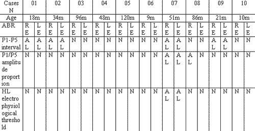
Table 1 illustrates the clinical characteristics and findings of auditory brainstem potentials obtained for the ten studied patients.
ABR was normal in five out of 10 patients (50%). Abnormal ABR findings included changes in morphology of curves in both ears in case 7, with reproducibility only in waves I and II, increase in interpeak interval of wave I-V in both ears of three patients represented by cases 1, 2 and 9, and increase in proportion of wave I/V amplitude on the right ear in case 8 and electrophysiological threshold affection in both ears of case 7.
DISCUSSION
West syndrome is a type of infantile spasms characterized by epileptic encephalopathy associated with flexion spasms and mental disability whose onset is in the first year of life and of unknown etiology. It is believed that some specific etiological factors such as intrauterine infection, tuberosis scleroris, perinatal asphyxia, or post-natal affections could be implicated.
All our cases were male patients, which is not in agreement with the findings observed by many authors such as Diament et al.1 in 1996 and Miyazaki et al.10 in 1992 (in a presentation of a series of 12 cases of childhood spasm), who observed predominance of 2:1 and 3:2 in male patients, respectively.
Many studies have been developed to clarify encephalic damage of childhood spasms and to determine the pathophysiological and electrophysiological characteristics of the disease. However, no consensus has been defined to present.
In the present study, auditory brainstem potentials demonstrated that dysfunction of the nervous system can be involved in the occurrence of childhood spasms. Previous studies tried to define the nervous system as the site of lesion in childhood spasms, but they are fairly scarce. Satoh et al.11, in 1986 used autopsy fragments and observed impairment of the central nervous system in subjects with childhood spasms, including hypogenesis of tegument, damage of the spongioform aspect and scars located on periaqueductal glia.
ABR abnormal findings that included morphological affections of curves with absence of waves IV and V, increase in interpeak interval of waves I-V, and increase in proportion of wave I/V amplitude were observed in the studies by Ogino et al.12. (presenting a case of West syndrome) and Miyazaki et al.10 The authors related increase in interpeak interval of waves I-V with dysmyelination of the central nervous stem and increase in the proportion of waves I-V amplitude with nervous cell damage. Such ABR findings suggest that dysfunction of the nervous system results mainly from hypogenesis of generation of nervous cells, part as a result of dysmyelination 10. The results obtained in this study that included affection of curve morphology, increase in interpeak interval of waves I-V and increase in proportion of wave I/V amplitude are in agreement with the data reported by the literature.
Hara et al.13 assessed ABR findings in patients with childhood spasms and concluded that the test did not define the abnormalities specific for patients with childhood spasms. However, it is importance to assess cerebral dysfunction in the subclinical stage of the disease. Moreover, the existence of a defect of neural conduction present in most patients with childhood spasms, confirmed ABR as the method of choice in the assessment of central nervous system 13, 14.
In the present study, the electrophysiological thresholds were within normal range in 90% of the patients. Case 7 had affected electrophysiological thresholds in both ears in the absence of waves III, IV and V. The studies analyzed in the literature did not inform about electrophysiological thresholds. Probably, this fact is due to the limited number of patients in the studies.
Ogino et al.12 observed early neurophysiological affections in patients with childhood spasms when compared to central nervous system structural defects in imaging exams (MRI - magnetic resonance imaging, or CT - computed tomography scan), data that agree with the study by Curatolo et al.15 which described ABR as more sensitive than MRI in detecting early damage to the nervous system in childhood spasms.
CONCLUSIONS
The study of electrophysiological potentials is a sensitive tool in detecting central nervous system disease, even though the observed affections are non-specific. However, it is important to define early diagnosis of cerebral dysfunction and to investigate hearing loss.
In view of these observations, the authors recommend that auditory brainstem responses be conducted to assess central nervous system dysfunction and to detect hearing loss in children with suspicion of childhood spasms.Table 1. ABR findings and correlation with age.
m: months; N: normal; AL: abnormal; RE: right ear; LE: left ear.
REFERENCES
1. Diament A, Cypel S. Síndrome de West. Neurologia Infantil. 3ª edição. São Paulo: Editora Atheneu; 1996. Capítulo 67: p. 972-6.
2. Vasquez HJ, Turner M. Epilepsia en flexion generalizada. Arch Argent Pediat 1951; 35:111.
3. Gastaut H. Relative frequency of different types of epilepsy; a study employing the classification of international. League Against Epilepsy Epilepsia 1975; 16: 457-66.
4. Gibbs FA, Gibbs EL. Atlas of electroencephalography. Adisson-Wesley. Vol 2. Editora Cambridge-Massesguster; 1952.
5. Cowan LD, Hudson LS. The epidemiology and natural history of infantile spasms. J Child Neurol 1991; 6: 355-61.
6. Jeavons PM, Bowder BD. Infantile spasms: a review of the literature and a study of 112 cases. London: Heinemann Medica; 1964.
7. Hrachov RA, Glaze DG, Frost JD. A retrospective study of spontaneous remission and long-term outcome in patients with infantile spasms. Epilepsia 1991; 32:212-8.
8. Dias MJM. Neurologia Infantil - Semiologia clínica e tratamento. 3a edição. São Paulo: Editora Sarvier; 1982. p. 655-9.
9. Baram TZ, Mitchell WG, Tournay A. High-dose corticotropin (ACTH) versus prednisone for infantile spasms: a prospective randomized blinded study. Pediatrics 1996; 97:37.
10. Miyazaki T, Hashimoto T, Tayama N, Kuroda Y. Brainstem involvement in infantile spasms: a study employing brainstem evoked potentials and magnetic resonance imaging. Neuropediatrics 1993; 24: 126-30.
11. Satoh J, Mizutani Y. Neuropathology of the brainstem in age-dependent epileptic encephalopathy - especially of cases with infantile spasm. Brain Dev 1986; 8:443-449.
12. Ogino T, Hata H, Minakuchi E, Iyoda K, Narahara K, Ohtahara S. Neurophysiologic dysfunction in hypomelanosis of Ito: EEG and evoked potential studies. Brain Dev 1994; 16: 407-12.
13. Hara M, Mitsuishi Y, Yashima K, Kozasa M, Saito K, Fukuyama Y Ito. Syndrome (Hypomelanosis of Ito) as a cause of intractable epilepsy. Jpn J Psychiatr Neurol 1989; 43: 487-9- resumo Medline.
14. Takematsu H, Sato S, Igarashi M, Seiji M. Incontinentia pigmenti achromiance (Ito) Arch Dermatol 1983; 119: 391-5.
15. Curatolo P, Cardona F, Cusmai R. BAEPs in infantile spasms. Brain Dev 1989; 11: 347-8.
16. Griebel V, Kregeloh-Mann I, Michaelis R. Hypomelanosis of Ito: report of four cases and survey of the literature. Neuropediatrics 1989; 20: 234-7.
17. Kaga K, March RR, Fukuyama Y. Auditory brainstem responses in infantile spasms Int J Pediatr Otorhinolaryngol 1982; 4: 57-67.
1 Resident Physician, Clínica OTORHINUS.
2 Ph.D. studies under course, Medical School, University of Sao Paulo - Former resident physician, Clínica OTORHINUS.
3 Preceptor of resident physicians, OTORHINUS, and Service of Otorhinolaryngology, Hospital Santa Marcelina.
4 Ph.D. in Medicine, Medical School, University of Sao Paulo, - Director of Clínica OTORHINUS.
Study conducted at Clínica Otorhinus - Centro de Estudos Alexandre Médicis da Silveira - Rua Cubatão 1140 Sao Paulo SP 04013-004
Tel (55 11)5572-0025 - Fax (55 11)5572-7373
