

Year: 2004 Vol. 70 Ed. 1 - (10º)
Artigo Original
Pages: 62 to 65
 PDF PT
 Evaluation of computerized tomography and computed tomographic Lopamidol cisternography on the topographic diagnosis of cerebrospinal fluid rhinorrhea and comparison with surgical findings
Author(s):
Roberto E. S. Guimarães 1,
Helena M.G. Becker 2,
Celso G. Becker 3,
Paulo Fernando Tormin Borges Crosara 4,
Gustavo C. Anjos 5,
Letícia P. Franco 6
Keywords: cerebrospinal fluid fistula, rhinorrhea, computed tomographic cisternography, Lopamidol, computed tomography
Abstract:
Since the microendoscopic nasal approach to treat the cerebrospinal fluid (CSF) rhinorrhea turned a routine, the need of correct topographic diagnosis was greatly increased. Aim: The aim of this study is to evaluate the efficiency of the computed tomographic cisternography (CTC) and computerized tomography (CT) to locate the CSF fistula according with surgical finds. Study Design: Observacional cohort with transversal cut. Material and Method: Forty-six patients with clinical diagnosis of CSF rhinorrhea were accessed by CT. Results: Thirty-eight of them were also accessed by CTC with Lopamidol. CTC pointed the exact site of fluid escape in 71% of the cases and CT in 52%. CTC helped to find the site of CSF rhinorrhea in 84,15% of the evaluated cases. Conclusion: There is not a gold standard method to access the site of CSF rhinorrhea yet. Lopamidol CTC is a worthy method in this purpose.
![]()
INTRODUCTION
Cerebrospinal fluid leak (CSF leak) was detected as an abnormal condition for the first time in 1782 by Willis.1 It may be divided into traumatic and non-traumatic. Traumatic leaks are subdivided into accidental, iatrogenic, acute or late. Non-traumatic leaks can be divided into with or without associated CSF hypertension 2.
The presence of a CSF leak implies increased risk of meningitis, which ranges from 9 to 25%, even in cases in which the leak has ceased spontaneously 3, 4. Owing to these complications and their severity and because they determine high risk of morbidity-mortality even nowadays, the treatment of CSF leak raises concern and interest of ENT and neurosurgeons.
As a result of the development of nasal microendoscopic surgery, the treatment of CSF leaks by the Otorhinolaryngologist became less aggressive than by extracranial access, being at the same time more effective 5-7. Endonasal approach increased the need for imaging methods capable of demonstrating reliability of local CSF leak, since the precise location makes the surgery more directed and, consequently more effective, quick and safe.
A series of imaging methods has been used in the diagnosis of CSF leaks, from simple x-rays of paranasal sinuses that presented 25% of sensitivity, to cisternography with radioisotopes with 50% sensitivity, polytomography with 53% sensitivity 8, CT scan, cisternography with metrizamide, with 87% sensitivity in patients with active CSF leak 8 and Magnetic resonance imaging 9-12. Out of those methods, the last three had collaborated for topodiagnosis of CSF leak 5, 9, 10.
Thus, we present an experience with 46 cases of CSF leak in which we compared the results of simple CT scan, cisternography and surgical findings.
MATERIAL AND METHOD
During the period between January 1993 and April 1999, 46 patients, being 23 men and 23 women suspected of CSF rhinorrhea were submitted to paranasal sinuses computed tomography at axial and coronal incidences without contrast. Out of the total, 38 patients were also submitted to cisternography with intrathecal injection of 10cm3 of non-ionic water-soluble contrast with Lopamidol after lumbar punch at the level L3-L4 or at the level of magna cistern. In order for the contrast to reach the intracranial cisterns, after the application, patients were placed in a head-down position for some minutes. Next, we conducted CT scan at coronal plan, conducting screening of the cribosa region and roof of the ethmoid and sphenoid sinuses. In one of the patients we conducted temporal bone CT scans owing to suspicion of ear CSF leak. The minimum age was 4 years, maximum of 66 years and median age of 39 years. Out of the total, 14 cases presented meningitis before treatment (30.4%). The period from onset of symptoms to treatment ranged from 10 days to 17 years.
In patients operated after 1997, during the surgical procedure, we used intrathecal contrast with fluorescein. Except for 7 cases in which we used hyperdense solution, all the others used hypodense fluorescein solution which was proposed initially by the author and that supports location and confirmation of efficacy of transoperative closing of the leak, and allows the patient to be placed in surgical position immediately after the intrathecal injection of contrast, which reduces the surgical act time 5.
RESULTS
Out of the total of 46 patients submitted to CT scan without contrast, 24 exams suggested site of the CSF leak compatible to the surgical findings (52.17%); in 15 cases, CT was non-conclusive (32.6%) and in 5 patients the location of the leak suggested by the CT scan was not in accordance with the surgical finding (10.8%). In 2 patients with CT scan that suggested the drainage site the leak was not identified in the surgery (4.3%).
Out of 38 patients submitted to CT cisternography 27 evidenced leak in agreement with the surgery (71.05%), 4 cases were different from the surgical findings (10.5%), 6 exams failed to detect the outflow of contrast into the nasosinual cavity (15.7%) and one exam evidenced contrast in left anterior ethmoid, but the fistula site was not found during the surgery (2.6%). In cases in which there was divergence of leak location by the tests, by CT or CT cisternography scan, the surgical finding of leak showed it was in the adjacent tissues to the one demonstrated by the imaging methods in all cases.
In 38 patients submitted to both diagnostic methods, there were 24 diagnoses in agreement, 9 cases of non-conclusive CT scan in which CT cisternography was conclusive, 4 cases in which both were non-conclusive, 2 cases in which CT scan was conclusive and CTC was not conclusive and one case in which the location suggested by the exams disagreed, being that the surgical finding confirmed that CTC demonstrated correct location of the CSF leak (Table 1).
DISCUSSION
We believe that the approach should follow a sequence. Initially, we used exams to confirm the CSF leak: measuring nasal secretion glucose weigh is the easiest and non-invasive method that we use as routine, whenever possible, in which values were greater than 30mg/dl and confirmed the diagnosis of CSF leak in patients with normal glucose levels 13, 14. Even though it is not available in our country, we can also make use of 2 transferrin in rhinorrhea, which is a specific CSF and perilymph protein, which is the reference pattern for the diagnosis of CSF leak and its study is facilitated since it is necessary a minimum amount of secretion for the diagnosis 15, 16.
The ideal imaging technique for accurate location of CSF preoperative leak is still controversial. Most diagnostic difficulties are found in the subgroup of patients in which they presented intermittent or inactive CSF leak 10.
The role of CT cisternography scan with water-soluble non-ionic contrast (metrizamide) in CSF leak was first defined by Drayer.17 Later the method proved to be efficient in many other publications 18-20. Currently, it is considered the method of choice for topodiagnosis of CSF leak. In our experience, the method was useful to identify the leak in 84.15% of the exams, being that in 71.05% of the cases the precise area of the CSF leak was identified and in 10.5% the CSF leak was next to the location suggested by the CTC, facilitating the determination of the leak site during the surgical act. The findings obtained in the study were in agreement with the studies by Mc Cormack21, Lantz22 and Colquhoun.23
The detection of contrast in the radiological image of paranasal and nasal sinuses is extremely important information for CSF leak. Schaefer et al.24 suggested that we should have non-contrasted CT scan conducted immediately before the conduction of CTC in order to collect a reference image and prevent any artifact from interfering in the decision of the test.
According to Manefelde19, we can identify bone defect in the CT scan in 82% of the CSF leak patients that were operated on, but the figures are not in agreement with those in many other studies.
Glucose present in CSF leak causes edema of the paranasal sinuses mucosa that can be radiologically documented 5. However, the data lack specificity. According to Loyd et al.25, the finding of bone defect and increase in soft parts around it is highly sensitive data for the diagnosis of CSF leak.
In 1991, Wakhloo et al.12 suggested the use of magnetic resonance associated with CT cisternography. It is necessary to evidence that MRI allows the individualization of the neighboring tissues of the brain, allowing demonstration of herniation of brain parenchyma in the extradural space 9, 10, 26. More recently, the use of MR cisternography weighted in T2, which does not require the use of contrast, has been proposed. In our sample, we used MRI only in cases in which we suspected of menigoencephaloceles or intracranial tumor.
CONCLUSION
CT cisternography with non-ionic water-soluble contrast Lopamidol was a useful method for topodiagnosis with detection indexes and similar location to those described previously in the literature for CTC with metrizamide. Non-contrasted CT scan is useful as a supporting method of CTC, and it should not be used as the only method for the imaging diagnosis of CSF leak.
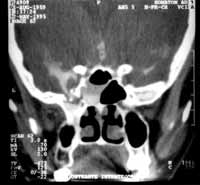
Figure 1. CT cisternography presenting nasal trauma with CSF leak in the left posterior ethmoid (Onodi cells) draining it to the sphenoid sinus.
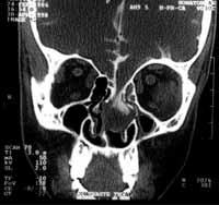
Figure 2. CT cisternography presenting meningoencephalocele on the left cribosa lamina.
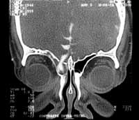
Figure 3. CT cisternography presenting right frontal meningoceles.
Table 1. Comparison of radiological findings and surgical findings in 38 patients that were submitted to both methods.
REFERENCES
1. Willis T. Opera omnia: cerebri anatomia nervorumque descriptio et usus. Henri Westen, Amsterdam, 1782 apud Thompson St C. The cerebro-spinal fluid: its spontaneous escape from the nose. London: Cassell & Co.; 1899. 140p.
2. Ommaya AK, Di Chiro G, Baldwin M, Pennybacker JB. Non-traumatic cerebrospinal fluid rhinorrhea. J Neurol Neurosurg Psychiatry 1968; 31: 214-225.
3. Leech PJ, Paterson A. Conservative and operative management for cerebrospinal fluid leakage after closer head injury. Lancet 1973; 1(811):1013-6.
4. Lewin W. Cerebrospinal fluid rhinorrhea in closed head injuries. Br J Surg 1954; 42:1-18.
5. Guimarães RES. Localização de fístula liquórica da base anterior do crânio com o uso transoperatório de fluoresceína intratecal em solução hipodensa. Belo Horizonte, 2000, p. 16 (Tese de Doutorado - UFMG).
6. Gjuric M, Goede U, Keimer H, Wigand ME. Endonasal endoscopic closure of cerebrospinal fluid fistulas at the anterior cranial base. Ann Otol Rhinol Laryngol 1996; 105(8): 620-3.
7. Stammberger H, Greistofer K, Wolf G, Luxenberger W. Surgical occlusion of cerebrospinal fistulas of the anterior skull base using intrathecal sodium fluorescein. Laryngorhinootologie 1997; 76(10):595-607.
8. Chow JM, Goodman D, Mafee MF. Evaluation of CSF rhinorrhea by computerized tomography with metrizamide. Otolaryngol Head Neck Surg 1989; 100: 99-105.
9. Creamer MJ, Blendonohy P, Katz R, Russel E. Coronal computerized tomography and cerebrospinal fluid rhinorrhea. Arch Phys Med Rehabil 1992; 73(6): 599-602.
10. Jonhson DB, Brennan Toland J, O'dwyer AJ. Magnetic resonance imaging in the evaluation of cerebrospinal fluid fistulae. Clin Radiol 1996; 51(12) 837-41.
11. Toquet J, Bordure P, Herman P, Gayet-Delacroix M, Delaroche O, Legent F. Intérêt de la recherche de Beta2 transferrine et de la MR cisternographie pour le diagnostic des fistules de liquide céphalorachidien. Ann Otolaryngol Chir Cervicofac 1998; 115(5): 293-8.
12. Wakhloo AK, Van-Velthoven V, Schumacher M, Krauss JK. Evaluation of MR imaging, digital subtraction cisternography, and CT cisternography in diagnosing CSF fistula. Acta Neurochir 1991; 111: 119-27.
13. Calcaterra TC. Extracranial surgical repair of cerebrospinal rhinorrhea. Ann Otol Rhinol Laryngol 1980; 89:108-16.
14. Hubbard JL, Mc Donald TJ, Pearson BW, Laws Jr ER. Spontaneous cerebrospinal fluid rhinorrhea: evolving concepts in diagnosis and surgical management based on the Mayo Clinic experience from 1970 through 1981. Neurosurgery 1985; 16(3): 314-21.
15. Meurman OH, Irjala K, Suonpaa J, Laurent, B. A new method for the identification of cerebrospinal fluid leakage. Acta Otolaryngol Stockh 1979; 87(3/4): 366-9.
16. Porter MJ, Brookes GB, Zeman AZ, Keir G. Use of protein electrophoresis in the diagnosis of cerebrospinal fluid rhinorrhea. J Laryngol Otol 1992; 106: 504-6.
17. Drayer BP, Wilkins RH, Boehnke M, Horton JA, Rosenbaum AE. Cerebrospinal fluid rhinorrhea demonstrated by metrizamide CT cisternography. AJR - Am J Roentgenol 1977; 129: 149-51.
18. Ghoshhaijra K. Metrizamide CT cisternography in the diagnosis and localization of cerebrospinal fluid rhinorrhea. J Comput Assist Tomogr 1980; 4: 306-10.
19. Manelfe C, Cellerier P, Sobel D, Prevost C, Bonafe A. Cerebrospinal fluid rhinorrhea: evolution with metrizamide cisternography. AJR - Am J Roentgenol 1982; 138: 471-6.
20. Naidich TP, Moran CJ. Precise anatomic localization of atraumatic sphenoethmoidal cerebrospinal fluid rhinorrhea by metrizamide CT cisternography. J Neurosurg 1980; 53: 222-8.
21. Mccormack B, Cooper PR, Persky M, Rothstein S. Extracranial repair of cerebrospinal fluid fistulas: technique and results in 37 patients. J Neurosurg 1990; 27: 412-7.
22. Lantz EJ, Forbes GS, Brown ML, Laws Jr ER. Radiology of cerebrospinal fluid rhinorrhea. AJR - Am J Roentgenol 1980; 135(5) 1023-30.
23. Colquhoun IR. CT cisternography in the investigation of cerebrospinal fluid rhinorrhea. Clin Radiol 1993; 47: 403-8.
24. Schaefer SD, Diehl J, Brigss WH. The diagnosis of CSF rhinorrhea by metrizamide CT scanning. Laryngoscope 1980; 90: 871-5.
25. Lloyd MNH, Kimber PM, Burrows EH. Post-Traumatic Cerebrospinal Fluid Rhinorrhea: Modern High-Definition Computed Tomography is All That is Required for the Effective Demonstration of the Site of Leakage. Clin Radiology 1994; 49:100-3.
26. Murata Y, Yamada I, Isotani E, Suzuki S. MRI in spontaneous cerebrospinal fluid rhinorrhea. Neuroradiology 1995; 37(6): 453-5.
27. Di Chiro G, Ommaya AK, Ashburm WL, Briner WH. Isotope cisternography in the diagnosis and follow-up of cerebrospinal fluid rhinorrhea. J Neurosurg 1968; 28(6): 522-9.
1 Joint Professor, Department of Otorhinolaryngology, Medical School, U.F.M.G.
2 Joint Professor, Department of Otorhinolaryngology, Medical School, U.F.M.G.
3 Assistant Professor, Department of Otorhinolaryngology, Medical School, U.F.M.G.
4 Otorhinolaryngologist, Ph.D. studies under course, U.F.M.G.
5 Resident physician, Service of Otorhinolaryngology, Hospital das Clínicas, UFMG.
6 Resident physician, Service of Otorhinolaryngology, Hospital das Clínicas, UFMG.
Affiliation: Department of Ophthalmology, Otorhinolaryngology and Speech Therapy, UFMG.
Address correspondence to: Av. Pasteur, 88 Sta. Efigênia Belo Horizonte MG 30150-290.
Tel (55 31) 3222-2891
Print: ![]()