

Year: 2000 Vol. 66 Ed. 6 - (19º)
Relato de Casos
Pages: 697 to 700
Nasal Complication Implants of Dental Implants. Case Report.
Author(s):
Ricardo F. Bento*,
Luiz U. Sennes**,
Saramira C. Bohadana***,
Natasha A. Braga***,
Sueli de Lima ****.
Keywords: dental implants, complications, nose and paranasal sinus
Abstract:
Introduction: Dental implants are an auxiliary method in oral rehabilitation, which allows permanent fixation of prosthesis of one or more teeth. When applied in the superior dental arcade, there is an intimate relation-with the pose and paranasal sinus. Aim: The goal of this work is to present a case submitted to neddled implants and screwed dental implants in the superior dental arcade, with transfixation of the nasal floor in thee differents points. The exteriorized portions of the implants were sectioned by endonasal approach with diamond burs. Results: The final result showed reintegration of the nasal mucosa covering the implants, without damage of the prosthesis fixation. Conclusion: In cases of lesser complications, the authors warn out the possibility of conservative treatment preserving-the implants.
![]()
INTRODUCTION
Dental implants represented a great breakthrough in the field of functional and esthetical restoration of teeth. When performed in the upper arch they may result in sinonasal complications, normally requiring their removal. Implants of 1st and 2nd molars may project into the maxillary sinus, resulting in infectious sinusitis7, 10, whereas in the anterior region (incisives and canines) they may trigger nasal complications. Comprehensive knowledge of sinonasal anatomy and radiological control during placement are essential to prevent complications1.
The objective of this study was to present a case in which there was exteriorization of dental implant into the nasal cavities, highlighting the performance of a conservative management that preserved the implant.
REVIEW OF LITERATURE
The first references to dental implants date back to III century BC, described in ancient Chinese medical texts. Sushruta (600 BC) recommended the replacement of avulsed teeth in young people2. Meanwhile, the Mayans were probably the first ones to know alloplastic implants, showing this advantage in VIII century.
The first alloplastic endosseous implants surged in the XIX century, in the shape of screws, needles, laminae and other derivations. Metals used were gold, silver and platinum, considered improper because of the resultant ionization caused by the contact with mouth fluids, leading to exaggerated proliferation of soft tissues and inhibition of osteogenesis.
More inert metals, such as metal alloys of chromium, cobalt, nickel and tantalum, are less subject to ionization and more tolerated by tissues. Among these, titanium is one of the most tolerated metals by mouth tissues, although others, such as stainless steel, vitreous carbon, ceramic materials and porous metallic alloys also have the same characteristics.
There are different ways to fix the various types of dental osseointegrated implants. Today, osseointegrated implants have high efficiency and are normally shorter and made of titanium, using the support of only one tooth. Osseointegration is defined as a direct and functional structural connection between the actual bone and the surface of the implant, which normally takes 4 to 6 months. This interval should be respected before any loads are applied to the tooth2, 3.
Owing to the anatomical proximity of upper teeth and nasal and paranasal cavities, otorhinolaryngologic complications because of manipulation, dental extraction and especially intra-osseous implants are frequent, such as:
o infectious rhinitis
o infectious sinusitis
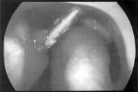
Figure 1. Metallic implant is perforating the floor of the nasal fossa up to the, septum.
o osteomyelitis
o proantral fistulas
o oronasal fistulas,
o reaction to foreign body.
Other complications of dental implants are rare, but there may be neural lesion, bone loss, infection, lesion to the adjacent tooth and the supporting structures, in addition to early loss of implants and bone fracture6. Iatrogenic perforations of maxillary sinus are now considered as one of the most frequent causes of fungal sinusitis, especially by Aspergillus4.
CASE REPORT
J. R. S., Caucasian female patients aged 43 years. She reported that she had been to the dentist 6 months before to place a tooth implant. During the surgery, she experienced pain and bleeding in the nasal fossa and, since then, she had had bilateral purulent nasal discharge, associated with mild and recurrent epistaxis.
At ENT exam, we detected the presence of metallic implants crossing both nasal cavities in the following sites:
o from the floor of the fossa to the septum on the right (needled implant) (Figure 1).
o from the floor of the fossa to the left inferior turbinate (screwed implant).
o from the inferior portion of the nasal septum towards the left inferior turbinate (needled implant).
The patient had fixed well-fitted dental prosthesis.
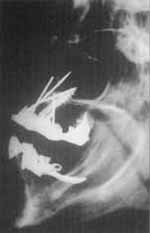
In the radiological exam of paranasal sinuses, we visualized two needled implants penetrating the nasal fossae and touching the nasal septum, and one screwed implant transfixing the palate (Figure 2).
We suggested a conservative treatment, removing the exteriorized portion of the implants in order to preserve and fix the dental prosthesis. The surgery was conducted under general anesthesia and after retraction of turbinates with vasoconstrictor, we had good visualization of implants inside the nasal fossa. They were sectioned and ground with a micromotor and diamond bur, until close to the point they emerged in the maxillary bone, preserving the mucosa of the adjacent nasal floor. We prescribed antibiotics for 10 days (cephalexine).
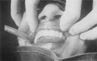
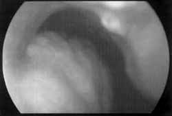
Postoperatively, the patient progressed with regression of symptomatology. There was no damage to the fixation of the implant (Figure 3), with good epithelization of nasal cavity, which covered the implants (Figure 4).
Figure 2. Paranasal sinus x-rays showing: two needled implants penetrating nasal fossae, reaching the nasal septum and one screwed implant transfixing the palate.
Figure 3. Post-operative status: we could observe there was no damage to the fixation of the implant.
Figure 4. We observed good epithelization of the nasal cavity after the removal of exteriorized portion of the implants.
DISCUSSION
The presence of an intra-nasal and/or sinusal foreign body is a reservoir of bacteria and, consequently, it favors recurrent infections. Treatment consists of its removal, generally leading to removal of implant6.
In patients who have a very thin bone layer on the upper arch, some authors have proposed the placement of bone graft under the mucoperiosteum of maxillary sinus, elevating the floor before the placement of the dental implant, in order to reduce oroantral fistulas and other sinusal complications5, 8, 9, 11.
In the case we presented, needled and screwed implants were used to fix the external prosthesis. They are normally made of tantalum and they are fixed by perforating the bone tissue in depth. Upon placement, we should be concerned about length (they are normally long), direction and angulation, which should be adequate to the area of implantation.
In order to avoid perforation of the floor of the mouth and maxillary sinus, it is required to perform CT scan to evaluate anatomy and measure bone density1.
If there are more severe complications, such as osteomyelitis and fistulas, the whole implant should be removed and the affected bone should undergo surgical debridement. In the case presented here, these complications were not identified, and therefore we indicated the section of the intranasal portion of the implant in order to preserve the fixation of the prosthesis.
Due to resistance of material, we used diamond burs to section it, and the bur was completely worn after the procedure.
The implant was ground up to the emergence of the bone, enabling regeneration of nasal mucosa, which recovered the region completely.
Some authors advocate only the removal of the exteriorized portion of the implant, preserving it in place, provided that there is perfect fixation of the prosthesis. If there is implant instability and/or major complications, complete removal is recommended1.
FINAL COMMENTS
Upon placement of dental implants, there may be exteriorization into the nasal or sinusal cavity, favoring infections. In the absence of other major complications, we could try to remove only the exteriorized portion of the implant, preserving the support of the prosthesis and preventing its removal.
REFERENCES
1. BERMAN, C. L. - Osseointegration. Complications. Prevention, recognition, treatment. Dent. Clin. North Am. 33(4): 63563, 1989.
2. BEUMER III, JOHN; LEWIS G.; STEVEN: - Sistema de Implantes Branemark: Procedimentos Clínicos de Laboratório Fatores Importantes para se Conseguir a Manter a Osseointegração, 2ª edicão, ed. Pancast, 1996, 1-13.
3. BRANEMARK, P. I.; ZARB, H. A and ALBREKTSSON, T. -Tissue Integrated Prostheses. Osseointegration in Clinical Dentistry. Quintessence, Publishing Co., Inc., Chicago, 1985.
4. De FOER, C.; FOSSION, E.; VAILLANTJ. M. - Sinus aspergillosis. J. Craniomaxillofac. Surg.; 18(1): 33-40, 1990.
5. ENGELKE, W.; DECKWER, I. - Endoscopically controlled sinus floor augmentation: A preliminary report. Clin. Oral. Implants. Res. 8(6): 527-31, 1997.
6. MIODUSKI, T. E. JR; GUINN, N. J. - Dental implants. Permanent replacement for lost teeth. A ORNJ. 51(3): 729, 731, 733-4 passim, 1990.
7. QUINEY, R. E.; BRIMBLE, E.; HODGE, M. - Maxillary sinusitis from dental osseointegrated implants. J. Laryngol. Otol., 104(4): 333-4, 1990.
8. REGEV, E. et. al. - Maxillary sinus complications related to endosseous implants. Int. J Oral. Maxillofac. Implants., 10(4): 451-61, 1995.
9. TIMMENGA, N. M. et. al. - Maxillary sinus lifts for the insertion of dental implants. J. Oral. Maxillofac. Surg., 55(9): 936-9, 1997.
10. UEDA, M.; KANEDA, T. - Maxillary sinusitis caused by dental implants: report of two cases. J. Oral. Maxillofac. Surg., 50(3): 285-7, 1992.
11. ZIMBLER, M. S. et. al. - Antral augmentation, osseointegrution, and sinusitis: the otolaryngologist's perspective. Am. J. Rhinol., 12(5): 311-6, 1998.
* Associate Professor of the Discipline of Clinical Otorhinolaryngology at Faculdade de Medicina da Universidade de São Paulo.
** Ph.D. and Professor of the Discipline of Clinical Otorhinolaryngology at Faculdade de Medicina da Universidade de São Paulo.
*** Post-Graduate studies under course at the Discipline of Clinical Otorhinolaryngology at Faculdade de Medicina da Universidade de São Paulo.
**** Otorhinolaryngologist at Clínica Otorhinus - São Paulo/ SP.
Study conducted at the Discipline of Clinical Otorhinolaryngology of Hospital das Clinicas da Faculdade de Medicina da Universidade de São Paulo and at Centro de Estudos
Alexandre Medicis da Silveira, Clinica Otorhinus. Study presented at the 11th Reunião da Sociedade Brasileira de Otologia and 2nd Encontro Brasileiro de Trabalhos Científicos em Otorrinolaringologia, on November 2-6, 1995, in Belo Horizonte/ MG.
Address for correspondence: Prof. Dr. Ricardo F. Bento - Rua Pedroso Alvarenga, 1255, Conj. 22 - 04531-012 São Paulo/ SP - Tel: (55 11) 3064-6556.
Article submitted on December 9, 1999. Article accepted on February 9, 2000.
Print: ![]()