

Year: 2000 Vol. 66 Ed. 5 - (20º)
Relato de Casos
Pages: 553 to 558
Oroantral Fistulae: Considerations Clinics and Surgical on Treatment and Report of Two Clinical Cases.
Author(s):
Leo K. Neto*,
João A. C. Navarro**,
Marcos A. Torriani***,
Paulo M. Almeida****.
Keywords: oroantral fistulae, oroantral comunication, tooth extraction
Abstract:
The present work discusses the origin, implications and treatment forms for oroantral fistulae of odontogenic origin. It is quite frequent lesion, because the dental extractions are procedures accomplished in great number in the Brazilian population. They were told two clinical cases with different surgical approachs, but that follow a same preoperative protocol, that judged indispensable for the surgical success.
![]()
INTRODUCTION
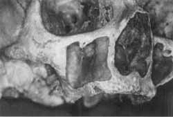
Maxillary sinuses, according to Figún and Garino, in 1989, are pneumatic cavities that are part of paranasal sinuses and are located on both sides of the face, on the maxillary bones. They are recovered by mucosa of ciliated pseudostratified columnar epithelium. Its main function is to warm inspired air and to act as a recipient of secretions originated from frontal and ethmoidal sinuses, promoting drainage of the discharges to the nasal cavity, through a very narrow canal named maxillary ostium (Figure 1). However, if drainage of maxillary sinus is inadequate, Delbalso and Hall, in 1997, reported that there could be build up of favorable bacterial means in its internal portion. Therefore, there would be less aeration of maxillary sinus, favoring growth of anaerobic pathogenic microorganisms and microaerophil bacteria.
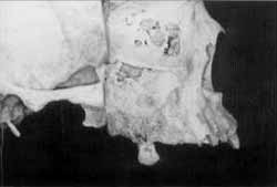
The knowledge of these details and dynamic and function of anatomical structure plays an important role in the daily practices of odontological surgeons, since Delbalso and Hall (1997) pointed out that 10 to 15% of maxillary sinuses pathologies surge from dental origin, frequently associated with upper molar and pre-molar abscesses, whose roots are intrinsically related to the floor of maxillary sinus (Figure 2). This close relation between the alveolar bone that houses the roots of molar and pre-molar teeth and maxillary sinus should be always kept in mind when performing an extraction of any dental element belonging to this group. According to the observation of Perri de Carvalho et al., in 1990, oroantral fistulae are frequently a result of post-extraction complications of upper molars, followed by surgical procedures in peri-apex of upper molars or pre-molars.
In order to define treatment, it is important to highlight that there is a difference between the terms oroantral fistula and oroantral communication. The latter, according to Laskin, in 1985, is normally small and does not cause rupture of sinusal mucosa, which is sometimes left undetected by dental surgeons. Its treatment is based on maintenance of clot inside the socket after the extraction. However, if the maxillary sinus in which there is an accidental oroantral communication has a pre-existing chronic maxillary sinusitis, there will be a high likelihood of forming an epithelized path, named oroantral fistula. From a conceptual point of view, Furtado and Guedes, in 1987, defined fistula (Latin = phistula / tube, cannula) as a narrow and deep canal, connected to a focus of suppuration or necrosis and opened to the surface by an orifice through which a purulent and sticky liquid drains. According to Gonty, in 1995, epithelization of this path takes approximately 3 weeks and no spontaneous closure will happen after this period. In this case, treatment will always be surgical; however, in order to perform a successful procedure of repairing the surgically manipulated area, the patient should not have systemic diseases and maxillary sinus should be pathology-free and decongested. It emphasizes the opinion of different authors [Cockerham et al. (1976), Gonty (1995), Hori et al. (1995), James (1980), Mion and Godoy Junior (1975), Perri de Carvalho et al. (1990), Tideman and Samman (1995) and Whitney et al. (1980)] who conduct intranasal irrigation before oroantral fistula repair surgeries.
Figure 1. View of a dry after removal of anterior wall and maxillary sinus and presence of maxillary ostium.
Figure 2. View of sagittal section in a cadaver showing the close relation of upper posterior teeth and the floor of maxillary sinus.
As to medication, Mion and Godoy Junior, in 1975, reinforced that paranasal sinuses offer more possibilities for the use of local (topic) antibiotics: First, oral and parenteral antibiotics are unlikely to reach the mucosa of sinuses at appropriate therapeutic levels; in addition, therapeutic agents applied locally at higher concentrations and for longer periods seem to be more effective. Physiology of rhinosinusal mucosa is extremely important in defense mechanism, especially ciliary beating, which has an important role in clearance of secretion. They also mentioned that regular or distilled water paralyzes immediately ciliary beating, and when sodium chloride is added to water to produce a normal saline solution, then the action of cilia is readily restored. Ciliary beating is also affected by changes in pH; therefore, to prevent rhinosinusal mediation from becoming anti-physiologic, they should have a pH between 6 and 8.3. The mediation vehicle should be aqueous, because oils interfere in cilia, forming a layer over them. Therefore, oils, should not be used because the medication dissolved in them does not reach the mucosa as desired. Association with a mucolytic agent is beneficial when intrasinusal installation with antibiotics is being done, so that viscous discharge is liquefied and viscosity is reduced, providing more penetrability of the antibiotic at the infection level. This information is the ground for pre-operative clinical management of clinical cases.
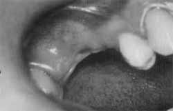
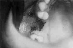
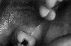
Figure 3. Intraoral aspect of gingival margin in the region of right upper first Molar and presence of oroantral fistula.
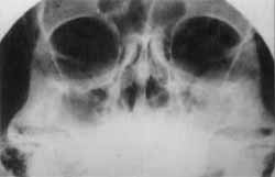
Figure 4. Waters' x-ray presenting opacification of right maxillary sinus.
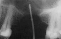
Figure 5. Periapical x-ray of gingival margin of upper molar region showing oroantral communication by means of application of contrast (gutta-percha).
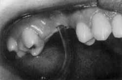
Figure 6. Insertion of catheter with urethral probe number 6, to perform intrasinusal irrigation.
After pre-operative management, it is time to choose the surgical technique. The choice is directly related to size of lesion and bone defect generated by it, according to Hanazawa et al., in 1995, and Gordon and Brown, in 1980. Therefore, surgical procedures could be performed with both soft and bone tissues, using grafts and ostectomies of alveolar margin to enhance healing of mucosa. Saad Neto and Callestini, in 1985, referred that we may apply mucoperiosteum vestibular flaps, cheek sliding flaps, palate sliding flaps, island palatine flaps, autologous bone grafts and cartilage grafts.
CASE REPORT
Case 1. Twenty-six year old female patient, with history of dental extraction in the region of left upper 1st molar for approximately 6 months, without completed healing yet (Figure 3). She also reported frequent episodes of pansinusitis, and drainage of discharge from the alveolar margin submitted to dental extraction. We conducted external x-ray with Valters' incidence (Figure 4), plus intraoral periapical exam (Figure 5) and introduction of gutta-percha cone through the residual margin at the level of left upper first molar to complement the clinical diagnosis of oroantral fistula. Next, the fistula was inserted with urethral probe number 6 (Figure 6) to enable daily irrigation of 500ml warmed sterile solution, plus final irrigation with 150mg vial of Riphamide. During irrigation, the patient was asked to place the head backwards, so that the solution was better directed through the nostrils.
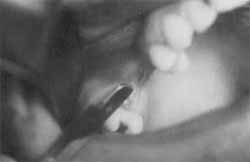
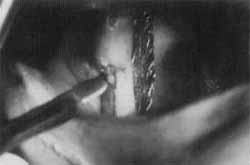
Figure 7. Surgical removal of oroantral fistula with 15 blade knife.
Figure 8. Flap of vestibular mucosa being pulled to close oroantral fistula.
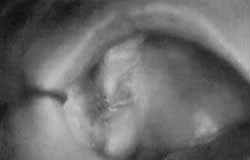
Figure 9. Post-operative results after 4 months of control.
Figure 10. Surgical removal of intraoral fistulous route, and mapping of palatine gum with sterilized laminated paper. It will be rotated to close oroantral communication.
Applications of irrigation were performed for two weeks, and in the first week a nasal decongested was added for systemic administration [Decongestant (isopropamide iodine, phenilpropanolamine chloridate, chlorpheniramine meleate) - BID) and in the second week, systemic antibiotics were used for 10 days. After clinical management with intranasal irrigation, we conducted surgical closure of oroantral fistula. The patient was submitted to posterior and middle superior alveolar nerve block, plus terminal complementation through palatine infiltration, and we used a knife 15 to remove the fistulous route (Figure 7). By means of relaxed vestibular incisions, a sliding mucosa flap from the bottom of vestibular sulcus was harvested (Figure 8) and the flap was sutured with isolate stitches and absorbable thread Vycril 4-0 to palatine mucosa, closing the fistulous route. The patient was instructed to avoid suction or yawning in order to prevent the flap from sliding away from its position. The patient was followed up weekly for 4 months after the procedure (Figure 9).
Figure 11. Three month post-op follow-up.
Case 2. Thirty-eight female patient reported dental extractions approximately 3 months before, showing incomplete healing on the posterior upper right side. The same protocol of intrasinusal irrigation and systemic medication was proposed, following the regimen described above. After the completion of clinical intervention, surgical procedure of oroantral fistula was prepared. Since it was a small sized fistula and the patient had upper total prosthesis, we decided to use a flap of keratinized mucosa from the palate. The patient was submitted to major palatine nerve block and terminal infiltration on the bottom of vestibular sulcus. We used sterilized laminated paper to map the size of the flap. Using a knife number 3 and blade number 15, we removed the fistulous route (Figure 10) and made an incision upwards towards the periosteum, based on the mapping previously done. The mucosa of periosteum was dissected, so that maxillary bone would have recover of periosteum. The mucosal flap was turned around towards the fistula, and a suture with isolate stitches was done with reabsorbable thread (Vycril 3-0). Similarly to the previous case, the same post-operative instructions were given, followed up in a weekly basis. The final visit took place 3 months later (Figure 11).
FINAL CONSIDERATIONS
Taking into account that oroantral fistulae are frequently resultant from complications of dental extractions, and that those complications impair the functioning of maxillary sinus, therefore, impacting all paranasal sinuses, dental surgeons, ENT physicians and stomatologists, should correctly diagnose the lesion and treat it in an effective fashion. Therefore there should be no presentations of maxillary sinusitis from odontogenic origin. Affected maxillary sinus should be clinically approached with irrigation that will help drainage of discharge from the sinus to nasal cavity (osteomeatal complex) and result in reduction of sinus mucosa edema. Knowledge of sinus physiology is important, since it should be associated with topic medication and systemic treatment. Surgical treatment is inevitable, since three weeks after the communication of oral and sinus cavity, there is no more spontaneous closure of the communication. The size of the lesion is one of the key factors for determining the surgical maneuver to be conducted. Postoperative instructions obediently followed by the patient contribute a lot to success rates.
REFERENCES
1. COCKERHAM, S.; WOOD, W. H.; LIND, K. Closure of a large oroantral communication by bone grafting. J. Oral Surgery, 34, DEC, p. 1098-100, 1976.
2. DELBALSO, A. M.; HALL, R. E. Diagnóstico por imagem das infecções dos espaços fasciais. In: Infecções Maxilofaciais a Orais. TOPAZIAN, R. G.; GOLDBERG, M. H., 3ª edição, São Paulo, Livraria Santos, 1997. Cap. 4, p. 112-159.
3. FIGLTN, M. E.; GARINO, R. R. Anatomia Odontológica Funcional a Aplicada. 2ª Edição, Editora Panamericana, São Paulo, 1989.
4. FURTADO, T. A.; GUEDES, A. C. M. Pele a Anexos. In: Bogliolo - Patologia. LOPES, E.R. et al. 4ª ed., Editora Guanabara, Rio de Janeiro, 1987.
5. GONTY, A. A. Application of the interseptal alveolotomy for closing the oroantral fistula - Discussion. J. Oral Maxillo fac. Surg., 53, p. 1396, 1995.
6. GORDON, N.C.; BROWN, S. L. Closure of oronasoantral defects: report of case. J. Oral. Surgery, 38, AUG, p. 600-05, 1980.
7. HANAZAWA, Y. et al. Closure of oroantral Communications Using a Pedicled Buccal Fat Pad Graft. J. Oral. Maxillofac. Surg., 53, p. 771-75, 1995.
8. HORI, M: et al. Application of the interseptal alveolotomy for closing the oroantral fistula. J. Oral Maxillofac. Surg., 53, p. 1392-96, 1995.
9. JAMES, R. B. Surgical closure of large oroantral fistulas using a palatal island flap. J. Oral Surgery, 38, AUG, p. 591-95, 1980.
10. LASKIN, D. M. Oral and Maxillo Facial Surgery. Vol Two. St. Louis, The. C. V. Mosby Company; 1985.
11. MION, D.; GODOYJUNIOR, O. M. Antibioticoterapia local, nas infecções dos seios paranasais. In: LACAZ, C.S. Antibióticos. 3ª ed. São Paulo, Editora Edgard Blucher Ltda., 1975. Cap. 26, p. 355-63.
12. PERRI DE CARVALHO, P. S. et al. Fistulas Buco-sinusais. Incidência a Tratamento. Odontológo Moderno, XVII, n° 5, MAIO, 1990.
13. SAAD NETO,M; CALLESTINI, E. A. Tratamento imediato de Comunicação Buco-Sinusal com esponja de gelatina. Revista Regional de Araçatuba A.P.CD., 6 (1), p.35-9, 1985.
14. TIDEMAN, H.; SAMMAN, N. Closure of oroantral communications using a pedicled buccal fat pad graft. Discussion. J. Oral Maxillofac. Surg., 53, p. 775-76, 1995. 15. WHITNEY, J. H. S. et al. The use of cancellous bone for closure of oroantral and oro nasal defects. J. Oral Surgery., 38, SET, p. 679-81, 1980.
* Professor of Head and Neck Anatomy at Universidade de Santa Cruz do Sul - UNISC, Santa Cruz do Sul / RS.
** Faculty Professor of Anatomy at Faculdade de Odontologia de Bauru - USP, Bauru/ SP.
*** Professor of Bucomaxillofacial Surgery and Traumatology at Universidade Federal de Pelotas - UFPel, Pelotas /RS.
**** Otorhinolarygologist.
Study presented at XXIX Jornadas Internacionales de la Asociación Odontológica Argentina.
Address for correspondence: Universidade de Santa Cruz do Sul - UNISC/ RS. Departamento de Odontologia a Enfermagem-Curso de Odontologia,
Avenida Independência, 2293 - Caixa Postal 188 a 236 - 96815 -900 Santa Cruz do Sul/ RS.
Fax (55 51) 717-1855 - E-mail: kraether@unisc.com.br
Article submitted on November 12. 1999. Article accepted on July 20. 2000.
Print: ![]()