

Year: 2000 Vol. 66 Ed. 4 - (10º)
Relato de Casos
Pages: 382 to 384
Giant Osteoma of the Maxillary Sinus. Case a Report.
Author(s):
Dayse Manrique*,
Ana C. A. Pitaluga**,
Arnaldo Guilherme***.
Keywords: nasal cavity, maxillary sinos, osteoma
Abstract:
A 14 year-old african american girl presented with a two-year history of left nasal obstruction and nasal discharge. A CT scan of the paranasal sinus revealed a 5,2 x 4 x 3,1 cm solid, lobulated tumor, extending from the left nasal cavity to the left maxillary sinus. Surgical excision by the degloving approach was dono. Histopathological examination of this lesion showed the typical features of the osteoma.
![]()
INTRODUCTION
Osteomas are the most frequent benign tumors of paranasal sinuses1,2. They are predominantly located in frontal and ethmoid sinuses; maxillary and sphenoid locations are rare. They may be diagnosed based on findings of routine radiological exams or be present with symptoms of sinusal drainage obstruction: headache, sinusitis or deformity of cranium-facial skeleton due to bone compression.
In the present case report, the authors present a case of giant osteoma in maxillary sinus, clinical diagnosis and therapeutic surgical approach.
CASE REPORT
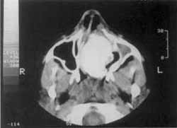
Female patient aged 14 years, presented with complaint of progressive nasal obstruction for one year, followed by left malar pain, intermittent nasal discharge on the left, without surgical or traumatic history, without complaints in other organs and/or systems. At ENT clinical exam, in anterior rhinoscopy we observed: submucous bulging, hard consistency mass, occupying the entire left nasal fossa, intact mucosa on the surface, nonpainful to the touch, deviating the nasal septum to the right and impacting the right lateral wall. At oroscopy, we observed bulging of bone palate to the left. Otoscopy without alterations. General physical exam without alterations. We ordered CT scan at axial and coronal sections and observed radiopaque and homogenous lesions, well-defined limits, occupying left maxillary sinus and left nasal fossa, with no signs of bone erosion (Figure 1):
We indicated surgical removal, carried out through transantral access (degloving), with ample exposure of lesion. Due to it consistency, it was not possible to fragment it; we chose to remove it en bloc after detachment of adjacent structures. The tumor had hard consistency, white color, measuring 5.5 x 4.0 x 3.0 cm, smooth and homogenous surface. At anatomical pathological exam we diagnosed osteoma.
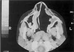
The patient progressed without complications, was discharged 24 hours after the procedure, and CT scan was carried out 2 months after the surgery (Figure 2). Eighteen-month follow-up did not show recurrence of lesion.
DISCUSSION
Osteomas are the most common benign tumors of paranasal sinuses1,2. The incidence varies from 0.01 to 0.43% of patients2,3. Despite the fact that they may be findings of radiographic exams or asymptomatic lesions, they may also progress with symptoms that depend on location. About 80% of them occur in frontal sinus, the rest present in ethmoid sinus, and rarely in maxillary or sphenoid sinus1,2,4,5,6. Maxillary sinus osteomas are rare and are present in only 5.1%0 of the cases involving paranasal sinuses2,3,5,7. Although the site of osteoma is related to the sinus, it does not grow from the sinus cavity, but rather through bone walls, occupying the cavity Afterwards1. If osteomas are multiple, they are commonly associated with intestinal polyposis, known as Fitzgerald-Gardiner3 syndrome.
Figure 1. CT scan of nasal fossa and paranasal sinuses at coronal section showing compromise of left maxillary sinus and left nasal fossa.
Figure 2. CT scan of nasal fossae and paranasal sinuses at coronal section two months after treatment, showing single cavity between left nasal fossa and left maxillary sinus.
Osteomas are benign bone proliferation masses that seldom affect maxillary sinus. They grow slowly and do not go through metastasizing, but may cause displacement and adjacent tissue erosion. They are benign bone tumors without association with malignity6. They are more frequent during the 2nd and 5nd decades of life1,2,5,6, and the incidence age is that of the period of onset of signs and symptoms1. They are more frequent in male patients in a proportion of 9:51.
As to etiology, a review of literature indicated 3 main theories for the formation of osteomas:
Embryological theory: in the development, when two tissues from different embryological origin make contact the tumors starts growing. Cranium regions in which membranous and cartilaginous elements make contact during development are: between frontal and ethmoid bones, maxillary bone comprising 2 elements, between the smaller horn of sphenoid and orbital process of frontal. According to this theory, these are the preferred sites for occurrence of osteomas.
Traumatic theory: during growth, a trauma in puberty triggered tumoral growth from a gap in the periosteum, resulting in periosteal hypereactivity3. Conversely, the low incidence of this disorder considering the large number of facial traumatism would speak against this theory.
Infectious theory: it is difficult to confirm and it is not possible to know if the infection was preset previous to the onset of osteoma or if it is secondary to ostium obstruction by the tumor. Concomitant incidence of infection and osteoma ranges from 14.5% to 30% of the cases1,6.
The most frequent symptom in patients with paranasal sinus osteoma is headache, frequently secondary to ostia obstruction or sinuses infection6.
Symptoms of maxillary sinus osteoma include pain, facial edema, rhinorrhea and sinusitis secondary to obstruction and infection2, 4, 5, 9 and deformity of craniofacial skeleton1. Maxillary sinus osteomas can rarely expand its growth into the orbit, resulting in proptosis6, diplopia, reduction of visual acuity1,9, and amaurosis4,6. They may be small and asymptomatic and be found in routine radiological exams1,2.
Radiological and CT scan diagnosis shows well-defined, radiopaque, homogenous and calcified lesion, found on the wall of the structure involved or occupying the cavity8.
When osteomas are small and asymptomatic, they may not require treatment3,5,6, and should be followed radiologically and operated if they start to expand. Symptomatic maxillary sinus osteomas should be surgically removed. The indicated access is through Caldwell-Luc9 transantral via. Recurrence was rarely reported3. If the osteoma is very large, fragmentation may be tried to avoid damage to adjacent structures4.
Histopathological diagnosis of osteoma consists of dense lamellar formation, mature bone, containing innumerous osteoblasts and little fibrous tissue 6, 8. Differential histopathological diagnosis should be made with: fibrous displasia, fibrous osteoma, characterized by the presence of connective tissue and formation of bone in a fibrous stroma3, and ossifying fibroma, constituted by the invasion of fibrous tissue into the bone, in addition to expansive characteristics and tendency to recurrence after incomplete resections10,11. Histological architecture of osteoma is not of reactive hyperplasia to trauma or bone infection3. Histopathological diagnosis should be well established, because clinical course of each pathology has already been clearly defined8.
REFERENCES
1. SAMY, L. L.; MOSTAFA, H. - Osteoma of the nose and paranasal sinuses with a report of twenty one cases. J. Laryngol. Otol., 85: 449-469, 1971.
2. SHADY, J. A.; BLAND, L. L; KAZEE, A. M.; PILCHER, W. H. Osteoma of the frontoethmoidal sinus with secondary brain abscess and intracranial mucocele: case report. Neurosurgery, 34: 920-923, 1994.
3. KARMODY, C. S. - Osteoma of the maxillary sinus. The Laryngoscope, 65: 427-434, 1968.
4. KRESPI, Y. P.; LEVINE, T. M. - Tumors of the nose and paranasal sinuses. In: PAPARELLA, M. M.; SCHUMRICK, D. A.; GLUCKMAN, J. L.; MEYERHOFF, W. L. - Otolaryngology, Philadelphia, W. B. Saunders Company, 1991, 1935-1958.
5. MILLER, N. R.; GRAY, J.; SNIP, R. - Giant, mushroom-shaped osteoma of the orbit originating from the maxillary sinus. Am. J. Ophtalmol., 83: 587-591, 1977.
6. WILKES, S.R.; TRAUMANN, J.C.; De SANTO, L. W.; CAMPBELL, R. J. - Osteoma: an usual cause of amaurosis fugaz. Mayo Clin. Proc., 54:258-260, 1979.
7. SALVOLINI, U.; CAVEZIAN, R.; PASQUET, G.; PERUGINI, S. - L'osteoma pedicule du sinus maxillaire. Une constatation radiologique rare? J. Radiol., 65: 455-461, 1984.
8. FU, Yao-shi; PERZIN, K. H. - Non-epithelial tumors of the Nasal Cavity, Paranasal Sinuses, and Nasopharynx: A clinicopathologic study. Cancer, 33: 1289-1305, 1974.
9. ZICCARD, V. B.; SMITH, J. A. - Osteoma of the maxillary antrum. Oral Surg. Oral Med. Oral Pathol., 80: 378-379, 1995.
10. BELKAHIA, A.; DIMOV, V.; GASSAB, A.; JELLOUI, B.; KEDOUS, F. - Cinq cas de fibrome ossifiant du massif facial. J. Fr. Oto-Rhino-Laryngolo. Audiophonol. Chir. Maxillo-Fac., 32: 352-356, 1983.
11. BUNDGAARD, T.; FROST JENSEN, V.; BUHL, L. - Sarcomatous change in a previously benign osteofibroma in the maxillary sinus. Arch Otorhinolaryngol. 245:22-24, 1988.
* Doctorate study under course at the Discipline of Otorhinolaryngology/ Human Communication Disorders - UNIFESP-EPM.
** Master degree in Otorhinolaryngology and Head and Neck Surgery, UNIFESP-EPM.
*** Joint Professor of the Department of Otorhinolaryngology and Human Communication Disorders.
Discipline of Otorhinolaryngology at Universidade Federal de São Paulo - Escola Paulista de Medicina.
Address for correspondence: Rua Botucatu, 740 - Vila Clementino - 04023-900 São Paulo /SP - Tel: (55 11) 573-1803 - Fax: (55 11) 570-6018.
Article submitted on June 30, 1999. Article accepted on June 9, 2000.
Print: ![]()