

Year: 1991 Vol. 57 Ed. 2 - (7º)
Artigos Originais
Pages: 81 to 84
CHRONIC SILENT OTITIS MEDIA: A CLINICAL-PATHOLOGICAL STUDY. A PRELIMINAR REPORT
Author(s):
Sady Selaimen da Costa MD. *
Michael M. Paparella MD. **
Patrícia Schachern ***
Tae H. Yoon MD. ****
Keywords: Otitis media, temporal bone
Abstract:
Chronic suppurative otitis media has been clinically defined as a chronic discharge from the middle ear in the presence of a perforation of the tympanic membrane. However, irreversible tissue pathology in the middle ear or mastoid can occur behind a non perforated tympanic membrane.
One hundred forty four human temporal bones with chronic otitis media were divided into two groups: Those with perforated (26) and those with non perforated (118) tympanic membranes. The following pathological findings were documented: granulation tissue, ossicular changes, cholesterol granuloma, cholesteatoma and tympanosclerosis. The areas in which these findings were assessed were: epitympanum, aditus ad antrum, sinus tympani, round window niche, htpotympanum and tympanic membrane.
![]()
Introduction
Chronic otitis media has traditionally been described as a chronic inflammation of the middle ear and mastoid associated with perforation of the tympanic membrane and(1-3) otorrhea. Although this definition encompasses most clinical cases, in temporal bones studies chronic otitis media is generally categorized as present, when there is histopathologically evidence ofintractably pathologic tissue within the middle ear cavity with or without perforation of the tympanic membranel(4-8).
In 1979, Paparella et al.(9) introduced the concept of silent insidius otitis media based on clinical and otopathological evidence of chronic infection in the middle ear cleft without obvious pathologic finding in the tympanic membrane.
The clinical finding and symptons of otitis media were subdue and not as apparent as in most cases of chronic otitis media. According to Paparella, chronic silent otitis media is defined as a condition in which irreversible tissue pathology is found within the middle ear cleft associated with an intact drum. It can be either undetected or undetectable depending on the clinical picture.
Although there has been evidence to support the concept that chronic otitis media can occur behind a non-perforated tympanic membrane, there has not, to our knowledge, been a study comparing the pathology of the middle ear cases of chronic otitis media with and withoud tympanic membrane perforation. Also we want to correlate the pathological finding of the temporal bones with the clinical informations extracted from the subjects clinical records.
Matherials and Methods
One hundred forty-four human temporal bones from 96 subjects were examined for the presence of intractable pathologic tissue in the middle ear. All bones belonged to the collection of the University of Minnesota.
Temporal bones which had been previously fixed, decalcified, embedded in celloidin, and cut at a thickness of 20 microns were examined using light microscopy for evidence of chronic otitis media. Chronic otitis media was defined as the presence of irreversible changes within the middle ear such as cholesteatoma, cholesterol granuloma, granulation tissue, tympanosclerosis and ossicular changes.
Temporal bones classified as having chronic otitis media were further divided into two groups, those with perforated tympanic membranes and those with non-perforated tympanic membranes, for comparison of their middle ear histopathology. The following locations were examined for the presence of histopathological findings: the epitympanum (from the tegmen tympani to the level of a line across the posterior ligament of the incus), the mesotympanum (from a line through the posterior incudal ligament to another line passing the inferior margin of the umbus of the malleus), the hypotympanum (below the Tine passing the inferior aspect of the umbo of the malleus), the aditus as antrum, the mastoid, Prussak's space, the facial recess, the oval and round window niche, the sinus tympani and the orifice of the Eustachian tube.
The clinical histories from the last admission of these subjects were abstracted for history of ear disease, audiological data, chartnotes on physical examinations of the head and neck, and biographical data such as age and sex.
Results
Temporal bone Findings
One hundred forty-four temporal bones from 96 subjects ranging in age from 10 months to 88 years were classified histologically as having chronic otitis media.
Fifty-five of the subjects were males and 41 females. Non-perforated tympanic membranes were found in 116 bones while only 28 were perforated.
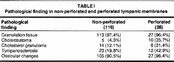
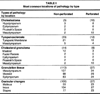
Categories of pathologic tissue are compared in non-perforated and perforated tympanic membranes in Table I. The most common locations of pathologic tissue by type are listed in Table II.
The sites in which granulation tissue was most frequently encountered in temporal bones with non-perforated tympanic membranes were the mesotympanum and mastoid and in perforated tympanic membranes in the mastoid, mesotympanum and epitympanum. Ossicular changes, ranging in severity from small erosion to complete destruction were found with similar frequency in both perforated and non-perforated tympanic membranes with the incus, stapes and malleus being affected in decreasing order.
Tympanosclerosis was observed more frequently in the group with perforated tympanic membranes with the most common site being the tympanic membrane. Cholesterol granuloma occurred with equal frequency in both groups. The common sites in the group with non-perforated tympanic membranes were the mastoid and facial recess and in the perforated group, the mastoid and epitympanum.
Acquired cholesteatomas were encountered more frequently in the group with perforated tympanic membranes, however in four of the cases with perforation, the tympanic membrane perforation was not considered to be the cause of the cholesteatoma. The common sites for cholesteatoma in the non-perforated group were the hypotympanum, epitympanum, Prussak's space and mesotympanum and in the perforated group the mesotympanum, hypotympanum and epitympanum.
Clinical Histories
Twenty-four of the 96 subjects were reported as having normal finding upon physical examination of the head and neck, Nineteen of these subjects were from the non-perforated tympanic membrane group and five from the perforated group. Interestingly, eleven of these twenty-four subjects were under the age seven (out of a total of 18 children under the age of seven). In the group with non-perforated tympanic membranes, the tympanic membrane appeared histologically normal in 22 temporal bones.
Discussion
Otitis media has a worldwide distribution and represents one of the most prevalent childhood diseases(10). The disease has been shown to follow a continuum of changes in which one type of otitis media become another(11, 14).
Although chronic suppurative otitis media has been described as a chronic discharge from the middle ear in the presence of a perforation of the tympanic membrane(1, 3), ours results lend support to the hypotesis which irreversible tissue pathology can also occur behind a non-perforated tympanic membrane. In fact this study shows that intractable tissue pathology behind a non-perforated tympanic membrane was observed to be four times more common than with a perforated tympanic membrane, further more the histopathological findings in both groups were fairly similar. Confronting the above mentioned findings with their clinical records we have found a poor correlation.
After review of chartnotes on the 96 subjects, 24 of them (25%) were found to have been reported as having normal ear nose and throat examinations.
Nineteen subjects belong to the group of intact tympanic membranes and five carne from the group with perforated tympanic membranes.
Although 24 of the 96 subjects with chronic otitis media were reported to have a normal physical examination of the head and neck at their last admission, they did not all present clinical findings compatible with chronic silent otitis media, since five of them came from the group with perforations of the tympanic membrane and were obviously misdiagnosed. Also in the group with non-perforated tympanic membranes, most of the subjects presented with gross abnomalites at the level of the tympanic membrane such as retractions, tympanosclerosis or atelectasis wich should have been observed by otoscopic examination. Eleven of the patients in this group were children so the misdiagnosis could be due to the difficulty of adequately examining small kids. In 22 subjects with non-perforated tympanic membranes, they appeared histologically normal and many of them presented with only focal pathology in the middle ear. Otoscopy examination alone in these subjects would not suggest the middle ear findings if not accompanied by a high index of suspicion.
We consider these cases as true chronic silent otitis media cases. Maybe these situations could account to explain many otological symptons once it has been demostrated that otitis media with intact drum can produce insidious slowly developing labyrinthine complications such as endolynphatic hydrops or sensorineural hearing loss, the primary portal of entry for toxin passage being the round window membrane(15). Also they could explain otherwise undiagnosed conductive deafness as shown in previous report(16).
Conclusion
The results of this study suggest that the clinical definition of chronic otitis media may not encompass the majority of patients with this condition. Further more, otoscopic examination alone was not sufficient to rule out middle ear pathology. The clinician should, therofore, be aware that a non-perforated tympanic membrane does not necessarily preclude the presence of gross pathological changes of the middle ear cleft.
Our current endeavours are towards an statistical analyzes of the kind of pathology, side by side in both groups. We are also trying to broaden pending questions regarding the pathogenesis of the otitis media, the continuum, and the interaction middle and inner ear as well as their clinical and surgical implications.
Bibliography
1. Goin DW: Chronic inflammatory diseases of the middle ear and mastoid, in English (ed): Otolaryngology: A Textbook. Hogerstown MD, Harper & Row, 1976, pp. 176-175.
2. DeWeese DD, Saunders WH: Diseases of the middle ear and mastoid, in DeWeese, Saunders: Textbook of Otolaryngology. St. Louis CV Mosby Co, 1964, pp. 378-396.
3. Kenna MA: Microbiology of chronic suppurative otitis media in children. Ann Otol Rhinol Laryngol 1988; 97 (Suppl. 131): 9-13.
4. Takahara T, Sando I: The common sites for otitis media in human temporal bones. A quantitative histopathological study. Auris Nasus Larynx 1985; 12 (Suppl. 1): 5173-5176.
5. Friedmman 1: The pathology of otitis media. J CM Pathol 1956, 9: 229-236.
6. Schuknecht HF: The Pathology of the Ear. Cambrides MA, Harvard University Press, 1974 pp 215-21.
7. Proctor B: Chronic otitis media and mastoiditis, in Paparella, Shumrick (eds): Otolaryngology 2nd ed. Philadelphia, WB Saunders Co, 1980, pp. 1455-1489.
8. Meyerhoff WL, Paparella MM, Kim CS: Pathology of chronic otitis media. Ann Otol Rhinol Laryngol 1978; 87: 749-760.
9. Paparella MM Goycoolea MV, Meyerhoff WL, et al.: Endolymphatic hydrops and otitis media. Laryngoscope 1979; 89 749-760.
10. Meyerhoff WL: Pathology of chronic suppurative otitis media. Ann Otol Rhbrol Laryngol 1988; 97 (Suppl. 131): 21-24.
11. Howie VM, Ploussard JH, Sloyer, J: The otitis prone condition. Am J Dis Child 1975; 129. 676-678.
12. Shurin, PA, Pelton SI, Donner A, et al.: Persistence of middle ear effusion in children. N Engl J Med 1979; 300: 1121-1123.
13. Juhn SK, Paparella MM, Kim CS, et al.: Pathogenesis of otitis media. Ann Otol Laryngol1977; 86: 481-492.
14. Goycoolea MV, Paparella MM, Carpenter AM, et al.: A longitudinal study of cellular changes in experimental otitis media. Otolaryngol Head Neck Surg 1979; 87: 685-700.
15. Papparella MM, Shea D Meyerhoff WL, et al.: Silente otitis media. Laryngoscope 1980; 90:1089-1098.
16. Paparella MM, Koutroupas S: Exploratory tympanotomy revisited. Laryngoscope 1986; 96(9): 978-985.
From the Otopathology Lab, University of Minnesota, MPLS, USA
* Research-Fellow, Otopathology Lab, University of Minnesota
** Chairman Emeritus, Dept. of Otolaryngology, UM, MPLS, USA
*** Researcher, Otopathology Lab, UM, MPLS, USA
**** Researcher, Otopathology Lab, UM, MPLS, USA
Address for correspondence: Sady Selaimen da Costa, rua Prudente de Morais, 432 - Ap. 64 - Ribeirão Preto, São Paulo, Brasil, 14015
Presented at the Midwinter Meeting of the Association for Research in Otolaryngology, Clearwater FL, February 1989.
This paper was supported in part by the International Hearing Foundation, Minneapolis, MN, USA.

Print: ![]()