

Year: 1996 Vol. 62 Ed. 4 - (7º)
Artigos Originais
Pages: 332 to 338
Laryngeal Framework Surgery for Voice Improvement.
Author(s):
Flávio Mignone Gripp, MD*,
Hans F Mahieu, MD, PhD*,
Maria Emilia Cardoso Gadelha, MD**,
Paulo Augusto de Lima Pontes, MD, PhD***.
Keywords: Laryngeal diseases, laryngeal cartilages, vocal cord paralysis
Abstract:
Correction of dysphonia, resulting from glottic insufficiency, can be perforrned step-wise as a single stage procedure under local anaesthesia using thyroplasty typeI, arytenoid adduction, cricothyroid approximation or combinations of these procedures. The timing and determination of the type of the procedure is mainly dependent upon: the etiology; the degree of dysphonia; the life expectancy and other patient related factors; the type and degree of glottic insufficiency; the mobility of the vocal folds; the degree of level difference between both vocal folds; the age and physical condition of the patient. In principle, it is advisable to start with a thyroplasty type I procedure and if necessary, arytenoid adduction and cricothyroid approximation can be performed additionally. Depending upon the above mentioned factors, the sequence of the procedures may be changed individually. These procedures were effective for the voice restauration, without important complications. On these study, we present our results related to the treatment of forty patients submited to these techniques, discuss the planning of the surgery and the advantages of LFS.
![]()
INTRODUCTION
The primary goal in the treatment of laryngcal disease, except in cases of laryngeal cancer and bilateral recurrent nerve paralysis, is the restoration of normal voice quality. Phonosurgery, a term first described by von Leden in thc early sixties, encompass a growing list of procedures specifically designed to voice improvement (1-3).
Many cases of organic dysphonia are caused by an incomplete glottis closure during phonation. The surgical treatment strategy to incomplete glottis closure is generally based on trying to medialize the affected vocal fold(s).
The still most frequent applied method to achieve such medialization is the endolaryngeal injection of soluble substances, such as teflon, silicone or collagen. Despite its simplicity and relative safety, in many cases intracordal injection fails to achieve the desired result.
To overcome some of the limitations and disadvantages of intra-cordal injection, laryngeal framework surgery has been developed as a viable and more versatile treatment, and more and more surgeons prefer to use the laryngeal framework type of operations as described by Isshiki (4-6), which offer a much more physiological approach to the problem of glottic insufficiency.
Laryngeal framework surgery (LFS) can be performed step-wise as a functionally monitored single stage procedure under local anaesthesia using thyroplasty type I (TTI), arytenoid adduction (AA), cricothyroid approximation (CTA) or combination of these procedures.
MATERIAL AND METHODS
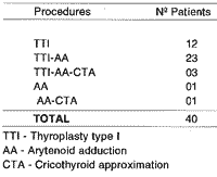
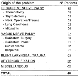
Forty patients with dysphonia resulting from glottic insufficiency were treated by laryngeal framework surgery, underwent either as a solitary procedure or as a combination of procedures (Table I). The causes for their glottic insufficiency are listed in Table II.
It involved 23 men and 17 women, with a mean age of 51 years, ranging from 20 to 73 years.
Surgical procedures:
The laryngeal framework Surgical procedures are performed under local anaesthesia following an intramuscular premedication of atropine sulfate (0,5 mg) and morfine (10 mg). The local anaesthesia is achieved with infiltration of 1% lidocaine hydrochloride with 0,001% epinephrine.
The endolaryngeal situation is monitored by fiberoptic videolaryngoscopy during the surgery. The quality of the voice is also monitored and is the most important assessment parameter during the surgery. Thus, continuous assessment of the voice and the endolaryngeal situation can be achieved by meticulous listening and flexible videomonitored laryngoscopy, to achieved the best voice result.
Prophylatic antibiotic treatment and codeine administration to prevent coughing is continued for 5 days postoperatively. Post operative voice rest ranges from 3 to 6 days, depending upon the laryngoscopy findings. Occasinally steroid administration is added in case of exceptional postoperative endolaryngeal swelling.TABLE I - Surgical procedures in patients presenting glottic insuficiency
TABLE II - Glottic insufficiency due to different etiolologies
Thyroplasty type I - The medialization achieved by this procedure is more outspoken in the anterior part of the vocal fold. The skin incision is made horizontally on the anterior portion of the neck at the lower margin of the thyroid cartilage extending from 1 em paramedially on the unaffected side to the posterior margin of the thyroid cartilage on the affected side. After exposure of the thyroid cartilage on the affected side, a rectangular cartilage "window" is incised (approximately 5x10 mm) either with a knife or in case of cartilage calcification, with a small cutting burr. Care is taken to preserve a part of the endolaryngeal perichondrium. The rectangular "window" is then depressed inwards to various depths during phonation to determine the optimal degree of depression. For fixation of the cartilaginous window in this position three basic techniques can be used: - a small wedge of silicone; - a silicone plug; - sutures through the window.
Overcorrection at the sub - or supraglottic level is recognized by a pressed and hyperkinetic, rough voice quality and by a protruding false vocal fold that can be seen at laryngoscopy, respectively.
Arytenoid addudion -The medialization with th is procedure is noticeable over the entire length of the vocal fold, but sometimes is not totally sufficient in the anterior position of the vocal fold as is often the case in vocal fold atrophy. The arytenoid adduction technique furthermore enables correction of a level difference between both vocal folds. The incision is similar to one of the type I thyroplasty, but slight longer posteriorly, extending past the posterior margin of the thyroid cartilage. For a good exposure of the arytenoid region the cricothyroid joint is dislocated. After the palpation of the muscular process of the arytenoid, the crico-arytenoid joint is located and can be opened. Care should be taken at this stage not to enter the piriform sinus which can overly the muscular process. After opening the crico-arytenoid joint, three Goretex sutures 2-0 are fixed at the muscular process and the surrounding musculature. The sutures are polled in the general direction of the lateral crico-arytenoid and thyro-arytenoid muscles, while monitoring the voice, to establish the optimal direction of traction. Overcorrection can be recognized when the voice quality presents roughness and pressure.
Cricothyroid approximation - Cricothyroid approximation mimics the funetion of the cricothyroid muscles, thus stretching the vocal folds. This procedure can also be used to raise the vocal pitch. Four (or two for unilateral cricothyroid approximation) thick (1-0 or 2-0) double non-absorbable sutures are used. The approximation is achieved with sutures that are inserted just behind the anterior part or the lower margin of the cricoid cartilage; superiorly the needles are withdrawn directed along the inner surface of the thyroid cartilage and directed outwards through the ala, slightly below the level of the vocal folds, 1.0 to 1.5 cm laterally from the midline. Care should be taken not to enter in the laryngeal lumen, which can be detected by fiberoptic control and by coughing. The voice is tested while the sutures are being pulled taughtly. Thus the cricoid and thyroid are approximated anteriorly and as a consequence the vocal folds are stretched. lf the result is satisfatory, it is advisable to use small silicone bolsters to disperse the pressure on the thyroid ala.
Pre-and postoperative evaluation consisted of :
- laryngeal videostroboscopy evaluation;
- phoniatric judgement of voice quality (phon judg.), (seven point scale: 7 = normal voice, 6 = nearly normal voice, 5 = slighty breathy voice, 4 = moderately breathy voice, 3 = severely breathy voice, 2 = aphonic moments, 1 = aphonic);
- maximal phonation time during sustained phonation of the vowel /a/ in seconds (max.phon.);
- phonetogram measured according to the recommendations of the European Union of Phoniatricians (10); from this phonetogram were derived: a) the maximal dynamic range of vocal intensity (max.dyn.), b) tire dynamic range of vocal intensity at the level of the habitual vocal pitch of thespeaking voice (dyn.MPSV), maximal vocal intensity (max.int. );
- furthermore, the patients were asked to express their opinion about the voice result achieved.
The postoperative evaluations were performed 3 and 12 months following surgery. Comparisons were made between preoperatively and 3 months postoperatively obtained data in 12 patients.
Statistical analysis of the data was performed with student's test (p<0.05 = significant).
RESULTS
Among the forty patients underwent to these procedures, from five of them no reliable postoperative evaluation was available, because they were terminally ill or they were referred to us from other countries and were not available at the appropriate time for evaluation.
All patients tolerated well the surgery under local anaesthesia conditions. The first ten patients did not receive the prophylatic antibiotic treatment and mo of these patients developed a postoperative wound infection, which healed without further complications soon after antibiotic treatment was initiated. Since the introduction of the prophylatic antibiotic treatment no wound infections were observed.
One patient developed a mild postoperative stridor which quiekly responded to corticoesteroid medication.
One older patient experienced a dyspnca d'effort and slight stridor following a combined thyroplasty type I and arytenoid adduction. Her apparently unaffected vocal fold demonstrated a reduced abductory mobility and the vocal fold itself was rather flaccid. This was the only patient who required reversal of the surgical procedure. The silicone wedge and the cartilage window of the thyroplasty type I were both removed. Six months following this procedure the voice remained good and the dyspnea had disappeared. Videostroboscopic evaluation demonstrated a markedly improved glottis closure three months postoperatively in 34 of the 35 patients. In one patient the glottis closure was judged to be the same as preoperatively, although the patient reported a subjective voice improvement. Twenty of the 35 patients were judged to have a normal or near normal voice 3 months after laryngeal framework surgery.
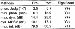
The mean pre-operative data of the 35 evaluated patients and their mean postoperative results 3 months following surgery are summarized in Table III. All differences between pre- and postoperative results were significant.
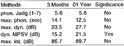
The longterm results of 12 patients one year after surgery, compared to their results 3 months after surgery are presented in table IV. Only the further improvement of the dynamic range of vocal intensity at the level of the mean pitch of the speaking voice after one year is significant.
Three months after surgery 23 of the 25 patients said to be very satisfed with the voice result and 11 patients were moderately satisfed. One patient, who was dysphonic following a blunt laryngeal trauma, was disppointed with the result. Although the voice had been improved with the vocal fold medialization, scarred vocal fold mucosa still interfered with normal phonation.
DISCUSSION
Laryngeal framework surgery was frst described by Payr in 1915 (7). Vocal fold medialization was achieved by inward depression of an anteriorly based thyroid cartilage flap. Meurman (8) described laryngeal implantation of cadaveric and autologous costal cartilage. He implanted the cartilage through a midline thyrotomy, between the innerthyroid perichondrium and the thyroid ala. Despite good results, in some patients injection, cartilage extrusion, and massive edema (requiring tracheotomy in one patient), made further investigations necessary. Afterwards, laryngeal diseases that involved unilateral recurrent nerve paralysis started to be treated with injection of soluble substances, such as teflon, silicone and collagen.
Isshiki et al (4, 9) improved on Payr's technique by medializing a rectangular thyroid cartilage island attached to the inner thyroid perichondrium at the level of the true vocal cord (Isshiki thyroplast type I). Since, laryngeal framework surgery has been proved to be a successful technique for glottic insufficiency. To overcome some of the limitations and disadvantages of intracordal injection, laryngeal framework surgery has been developed as a viabie and more versatile treatment.
The timing and planning of laryngeal framework surgery is an important issue which requires adjustment to each individual case, and is largely dependent upon:
- the etiology of the incomplete glottis closure;
- the degree of dysphonia;
- the time that has passed since the onset of dysphonia;
- the appearence and mobility of the vocal folds;
- the position of the affected vocal fold;
- the degree of compensation of the contralateral vocal fold;
- the life expectancy and physical condition of the patient;
- other patients related factors (profession, age, etc);
- other complaints related to laryngeal dysfunction (aspiration, hyperventilation, etc);
- last but not least, the experience of the surgeon.
TIMING - Etiology is a very important factor in timing especially if the dysphonia is due to a recurrent laryngeal or vagus nerve paralysis.TABLE III Comparision mean pre- and post-operative (3 months) voice results (N=35)
TABLE IV Compararison mean short and longterm post-operative voice results (N=12)
If, in case of a paralysis, the continuity of the nerve is intact (e.g. idiopathic paralysis) or believed to be intact, spontaneous recovery of function may be expected up a year following the onset of the paralysis. Therefore it is advisable to plan a surgical treatment no sooner than a year following onset, unless other factors prevail.
If the paralysis is the result of a severed nerve without reconstruction (e.g. ressection of vagus nerve tumor, ressection of the recurrent nerve due to thyroid malignant tumor, ete), no spontaneous recovery of function can be expected. In these circumstances, laryngeal framework surgery can be taken into consideration sooner. If, in such cases, the glottic insufficiency is relatively small, it is worthwhile to wait for compensation of the unaffected vocal fold to occur. Voice therapy can be advised during this period, but in our opinion will not stimulate compensation. If compensation has not been achieved sufficiently within 6 months, surgical treatment can be planned.
If the dysphonia is a result of trauma, it is advisable to wait at least 6 months before framework surgery is contemplated. By that time, scar tissue will have settled down.
If the glottic insufficiency is the result of a fixed cricoarytenoid joint, there is no reason to postpone surgical treatment. Laryngeal electromiography is very helpful to differentiate between laryngeal paralysis and fixation of thc crico-arytenoid joint.
In patients with a recurrent laryngeal nerve paralysis as a result of an intra-thoracic malignancy, laryngeal framework surgery is usually performed as soon as is convenient for the patient, because of the short life expectancy of these patients. If the mobility of both vocal folds is normal, although a dysphonia due to glottic insuffciency is present, laryngeal framework surgery can be planned if voice therapy remains unsuccessful.
The degree of dysphonia can influence the timing of laryngeal framework surgery as well. If the degree of dysphonia is very expressive, we tend to operate sooner than one year following the onset.
Sometimes patients have other complaints related to the laryngeal dysfunction such as aspiration or hyperventilation as a result of the large airflow during the phonation. These facts themselves can be a reason to plan surgery sooner than one year following the onset.
In selected cases professional and social factors may urge us to improve the voice sooner as well.
PLANNING - Our personal strategy in planning medialization, laryngeal framework surgery is performed as follows, unless specific factors, as mentioned below, incite us to change our policy: in cases of dysphonia with a slight to moderate degree of glottic insufficiency, we start with a thyroplasty type I. If, during the surgery, it is found to result in an insufficient improvement of the voice, an arytenoid adduction is performed additionally.
Often, however, a good voice can be obtained by means of a thyroplasty type I only. Prior to finish the surgery, the effect of a manual crico-thyroid approximation is tested as a rule. If this procedure results in a better voice, the cricoid and thyroid cartilages are approximated permanently with sutures.
The reasons why we prefer this sequence of procedures are:
1) Thyroplasty type I is a much more simple and less time consuming procedure than an arytenoid adduction.
2) Usually no endolaryngeal edema is observed during a thyroplasty type I, whereas arytenoid adduction results in a not insignificant edema of the arytenoid region. This may interfere with the voice monitoring during surgery. Even in those cases were a combination of thyroplasty type I and arytenoid adduction is considered likely to be required, it seems wise to start the thyroplasty type I. Presence of endolaryngeal edema at the time of per-operative voice monitoring should be ruled out by fiberoptic examination of the larynx during the surgery. lf significant edema or even hematoma is present, overcorrection is required to get a good post-operative voice result.
3) Thyroplasty type I is a procedure which can more easily be reversed than an arytenoid adduction.
4) Swallowing complaints and pain during the first postoperative days are much more often observed following arytenoid adduction than following thyroplasty type I.
Exceptions of the policy described above can occur:
1) In cases with an large glottic insufficiency and/or a level difference between both vocal folds. In such cases an arytenoid adduction alone can be sufficient and we therefore start with this procedure. If, during the surgery, it is found to result in an insufticient improvement of the voice, a thyroplasty type I is performed additionally.
2) In selected cases of isolated fixation of the crico-arytenoid joint, an effort can be made to explore the crico-arytenoid joint and fix the arytenoid in a more adducted position. However, if the fixation of this joint is only part of a more extensive fibrosis, it is much safer to limit the surgery to simpler procedures like thyroplasty type I and cricothyroid approximation.
3) In cases of normal laryngeal mobility, if medialization is required, it should be achieved without arytenoid adduction.
4) In cases with bowed or flacid vocal folds, tension correction by means of cricothyroid approximation seems to be the first choice, if necessary bilateral thyroplasty type I can be added.
5) In older patients the medialization required for the best voice result may compromise the airway as a result of a diminished tension of the unaffected vocal fold, associated with older age. This flaccid but otherwise unaffected vocal fold can be adducted passively by the Bernoulli effect during inspiration resulting in a relative airway obstruction. Therefore, it is wise not to be overzealous in correcting thc dysphonia of older patients. The some holds true for patients with a poor physical condition, for whom operating time and peroperative stress should be reduced to the minimum. In both patient groups, we tend to limit the surgical procedure to a thyroplasty type I and sometimcs a cricothyroid approximation.
6) If the surgeon is not yet experienced in laryngeal framework surgery, it is advisable to limit the procedures to the thyroplasty type I and cricothyroid approximation initially, because these procedures are relatively simple. This may not lead to the best obtainable voice result in all cases, but it will definitely improve the voice without much risks involved.
CONCLUSION
1) Laryngeal framework surgical procedures performed under local anaesthesia with functional vocal control constitute a safe and reliable method to treat dysphonia resultíng from glottic insufficiency.
2) Laryngeal framework procedure is an effective way to restore the voice and no serious complications were observed.
3) The voice tunning during the procedure is very important to obtain a good result.
BIBLIOGRAPHY
1. VON LEDEN, H. - Phonosurgery. Acta Otorhinolaryngol Iber Am, 22: 291-9, 1971.
2. ISSHIKI, N. - Recent Advances in Phonosurgery. Folia Phoniatr, 31: 119-54, 1980.
3. HilRANO, M. - Surgical Alterations of Voice Quality. In: Otolaryngology-Head and Neck Surgery: Update I. C. Cumings et al (Eds). Mosby, St Louis, pp. 249-62, 1989.
4. ISSHIKI, N.; OKAMURA, H.; ISHIKAWA, T. -Thyroplasty type I (lateral compression for dysphonia due to vocal cord paralysis or atrophy). Acta Otolaryngol, 80: 465-73, 1975.
5. ISSHIKI, N.; TANABE, M.; SAWADA, M. - Arytenoid adduction for vocal fold paralysis. Acta Otoiyngol, 140: 555-8, 1978.
6. ISSHIKI, N. - Phonosurgery: Theory and Practice. Springer Verlag, Tokyo, Berlin, New York, 1989.
7. PAYR, A. - Plastik am Schildknorpel zur Behebung der Folgen Einseitiger Stimmbandlahmung. Dtsch Med Wochenschr, 43: 1256-70, 1915.
8. MEURMAN, Y. -Operative Mediofixation of the vocal cord in complete unilateral paralysis. Arch Otolaryngol, 55: 544-53, 1952.
9. ISSHIKI, N.; TAIRA, T.; TANABE, M. -Surgical alterations of the vocal pitch. J Otolaryngol, 12:335-40, 1983.
10. SCHUTTE, H. K.; SEIDER, W. - Recommendation by the Union of European Phoniatricians: Standardizing Voice Arca Measurement -Phonetography. Folia phoniatr, 35: 28688, 1983.
* Department of Otorhinolaryngology and Head and Neck Surgery Free University Hospital, Amsterdam, The Netherlands.
** Post-graduation course in Otorhinolaryngology and Head and Neck Surgery, Universidade Federal de São Paulo - Escola Paulista de Medicina; staff at Instituto da Laringe, São Paulo, Brazil.
* ** Department of Otorhinolaryngology and Human Communication Disorders, Universidade Federal de São Paulo - Escola Paulista de Medicina, director of Instituto da Laringe, São Pauto, Brazil.
This study was realized at the Department of Otorhinolaryngology and Head and Neck Surgery, Free University Hospital, Amsterdam, The Netherlands De Boelelaan, 1117- 108 1 HV
Amsterdam, The Netherlands Phone: 31 (20) 548-6688.
Artigo recebido em 03 de abril de 1996.
Artigo aceito em 15 de maio de 1996.
Print: ![]()