

Year: 1993 Vol. 59 Ed. 1 - (3º)
Artigos Originais
Pages: 17 to 24
Permeability of the mucoperiosteum in otitis media. An experimental study in the cat.
Author(s):
Marcelo M. Hueb, M.D., M.S.*,
Marcos V Goycoolca, M.D., M.S., Ph.D.**,
Carlos B. Ruah, M.D.***,
David Muchow****
Keywords: otitis media, middle ear
Abstract:
This experimental study investigated the dynamics of tracers (latex microspheres) of different sues in the inflamed middle ear mucosa of the Eustachian tube-obstructed cot. Latex (polystyrene) microspheres caused no inflammation of the mucoperiosteum. The permeability of the middle ear mucosa (including the round window membrane) appeared to be dependent on size. Microspheres tended to be phagocytized by macrophages in early stages of the inflammatory process. At later stages they tended to traverse the epithelial layer and were observed with macrophages or within lymphatics in the connective tissue layer. Small and medium sized microspheres were occasionally observed in the perilymphatic space, medial to the round window membrane.
The importance of this study in regards to the permeability of the mucoperiosteum in otitis media and its relationship to middle/inner ear interaction will be discussed The possible use of different drugs linked to different sized and charged microspheres to selectively treat middle ear or inner ear pathologies is a/so discussed.
![]()
INTRODUCTION
The mucociliary system seems to be the fundamental mechanism for eliminating foreign matter from the middle ear1-3. The capability of absorption by the normal and pathological middle ear mucoperiosteum has been demonstrated recently in different animal models with a variety of tracers4-10. Under inflammatory conditions with increased vascularity and cellular activity, the pathological mucoperiosteum seems to absorb tracers faster than the normal mucoperiostcum.
Experimental studies have demonstrated that the round window encumbrance leas the same inflammatory changes observed in the mucoperiosteum of otitis media-induced animalis11. Various experimental studies have shown that the round window membrane is permeable to antibiotics, local antisepticst5, Horseradish peroxides, toxin's-ZU, latex microspherest; and to macromolecules such as triturated album. The round window membrane leas also beck shown to be permeable to cationic ferreting and impermeable to anionic ferreting, establishing that its permeability is also charge dependant,24.
These studies have undoubtedly unveiled the semi permeable characteristics of the middle ear mucoperiosteum and round window membrane, suggesting a middle ear 25 and a round window membrane/inner ear defense system26. My objectives of this study work to evaluated the predictability of the inflamed/infected mucoperiosteum to microspheres of different sizes and its importance in the interaction between the middle and inner ears.
MATERIAL AND METHODS
A group of 12 adult cays wilting an average of 3.5 kg were submitted to bilateral Eustachian tube obstruction under carmine hydrochloride anesthesia (30 mg/kg), sterile technique was used throughout. Through a soft palate incision fine-grade silicone sponges work introduced bilaterally in the tuba orifices to occlude it completely. At the procedure, latex microsphercs (Seradyn Inc.) of negative surface charge and of different sizes (diameters 0.85, 3.08, and 9.64 micrometers; 0.25 ml for each size) were injected in the left middle ear cavity through a posterior bulla approach. My right medic cars work used as otitis media controls. The soft palate incision and the incision used for the posterior bulla approach work closed using absorbable sutures.
Animals work then subdivided into 4 groups of 3 each and sacrifices by decapitation under deep anesthesia at 1, 2, 4 and 12 weeks after the procedure. The temporal bones work removed, immediately placed in a fixative solution (10% buffered funneling), dccalci6ed in 5% trichloroacetic aid, dehydrated through graded series of alcohols, and processed using cello din mucous. From superior to inferior 20u sections work cut in a sliding microtome. Each tenth section was then stained with hematoxylin and coca. Sections work studied under light microscopy at various magnifications. The presence of latex (polystyrene) microspheres was evaluated in the effusion, mucoperiosteum, Eustachian tub, round window membrane, and inner ear (perilymph). My cells involved in the cellular response to their presence work also evaluated. All his parameters work contact at each different stage to Asses the dynamics of this microspheres.
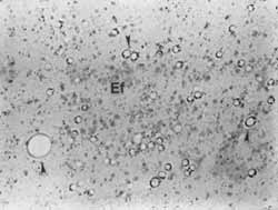
FIGURE 1 - Microspheres (arrow heads are seen in the effusion (Ef) at 1 week after Eustachian tube obstruction. Original x420.
RESULTS
The histopathological aspects of the middle ears with the microspheres contact to those of the muddle cars without them, revealed no significant differences. Furthermore, microspheres of any size were not noticed in the blood or lymphatic vessels at the contralateral ears at any of the 4 stages studied.
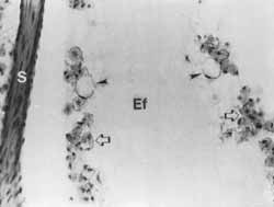
FIGURE 2 - Medium sized microspheres are observed within macrophage (arrow heads). Observe numerous macrophages without microspheres (open arrows). S. (stapes), Ef(effusion). Original x420.
In the study ears, large quantification of micro spheres of ail three sizes were observed in the effusion at the first two stages (1 and 2 weeks), decreasing in number with increasing survival time (Figure I). Occasionally, small (O.RS micron) and medium (3.08 micron) sized micro spheres wreck observed surrounded by polymorphonuclears or phagocytes by macrophages at the 1 week stag animals. With increasing survival time, micro spheres were much more frequently noticed inside macrophages (Figure 2). At the 2 weeks stage small and medium sized micro spheres were observes traversing the epithelial layer (Figure 3). At this stage, the presence of small and medium sized micro spheres was occasionally noticed in the perylimphatic space or traversing the runic window membrane (Figures 4 and 5).
At the next stage (I month after Eustachian tub obstruction) micro spheres wreck observed leas cottony in the middle ear cavity, being mistily observed in the mucoperiosteum. It was not guncotton to observe the presence of small micro spheres inside macrophages or lymphatic of the sub epithelial layer at talus stage (Figure 6). At the last stage studied (3 months survival time) the Comings were similar to those found for the 1 month stage, however, the presence of micro spheres were noticed to decrease significantly when compared to the previous 3 stages.
Mononuclear cans were not observed nearby the micro spheres at any of the 4 stages studied. The larger (9.ó4 micron) micro spheres were not observed inside macrophages, mucopcriosteum, lymphatic or blood vesicles, or perylimphatic space in any of the 4 stages after Eustachian tube obstruction.
DISCUSSION
Experimental Eustachian tube obstruction has back shown to cause punt lent otitis media in the cat, which progresses toward horrification with irreversible tissue changes 11,25,27,29. My result found for the middle ears of the animals in our stocky corroborate these previously reported experimental. Our findings that the middle ears showed similar result in both groups of animals suggest dial the presence of micro spheres was not an additional factor for the inflammatory process secondary to the Eustachian tube obstruction. The findings that the micro spheres did not reach contra lateral control ears could be explained by the fact that the absorption of this tracers was latitude to the lymphatic system of the studied ears and/or the amount aft micro spheres introduced in these ears was not sufficient to go through the synthetic circulation and reach the contra lateral ears.
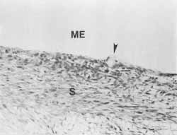
FIGURE 3 - A medium sized microsphere (arrow head) is seen traversing the epithelial layer at the promontory level, S (subepithelial layer), ME (middle ear). Original x420.
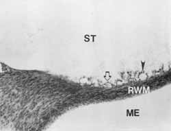
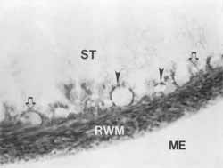
FIGURE 4 - Micro spheres are observed in the perilymphatic space (arrow head) or traversing the rotted window membrane (open arrow). ST (scala tympani), ME (middle ear), RWM (round window rnembrane). Original x210
FIGURE 5 - Higher imagination of figure 4. Micro spheres are observed in the per lymphatic space (arrow heads) or traversing the roundup window membrane (open arrows). ST (seal hvnhuni), ME (middle ear), RWM (rondo window membrane). Original x420
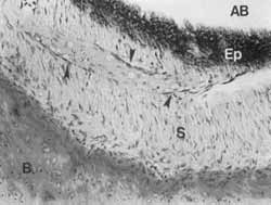
FIGURE 6 - Small sized micro spheres are observed inside a lymphatic used (arrow heads) in the sub epithelial layer (S) of the anterior bridal (A B). Up (epithelial layer), B (bore). Original x420
Our findings are suggestive drat macrophages are Quiet cells involved in the reaction to the micro spheres. It is also suggestive that these cans are responsible for the initial clearance of submicron (0.85) and up to 3 micron foreign bodies from the middle ear effusion. As small (0.85) microspheres were observed inside lymphatic of the muck eriostemon in later stages, it is suggestive that talc lymphatic system in the cat is associated with a later clearance of sub micron particles forte the twiddle car cavity. Larger (9.64 micron) ecospheres were not observed inside macroclimates or lymphatic vessels, suggestitivg that the twiddle ear mucoperiosteum of the car is not printable to particles of this size.
Our results are also suggestive, as previously reported, that the basic mechanism of particle absorption seems to involve pinocytosis7-9-14. It also corroborates previous suggestions that once die substance reaches the connective tissue layer it is processed by macrophages or traverses to lymphatic and/or blood vessels7-9-14.
it is widely known that on evasions, pungent otitis media can resulting inner car complications (e.g. labyrinthitis). Bacteria, toxins, and a variety of other substances could enter the inner ear through a diversity of natural orifices, blood and lymph vessels, micro fissures, and oval and round windows, etc. The round window is the only "soft tissue barrier between the middle and inner ears and it leas been shown to be pintable to a wide range of substances 9' 1 0, 1223. Despite being pair of the mueuperiosteuln of the trickle car, the round window membrane leas been de nonstarter to have ills permeability decreased once the inflammatory changes become establisliedt4. Furthermore, the round window membrane leas also been shown to have attic permeability related to the size and surface charge of the substance in question14,24. This semi permeable characteristic of the round window membrane would be an supporting element involved in treatment of inner car pathologies using middle car drug delivery system. Different conges linked to different sized and charged microspheres could selectively tract middle car or inner car disorders. Micro spheres are cornnte3ccially available linked to alacrity of drugs and substances
In conclusion, our results are suggestive dial latex mi~ exospheres are inert, acne that die permeability of the middle ear mucus (including the round window temperance) is selective for size and electric charge as previously demonstrated 14-24. the clearance of substances frock the nitride ear cavity sets to involve macrophages, and at later stages, the lymphatic system. The use of potentially zootoxic antibiotics linked to large latex (polystyrene) microspheres could become another effective method of safely treating choric middle ears disorders.
ACKNOWLEDGMENTS
The authors would like to thank Ms. Noriko Morizono and Sherry Lamey for processing the temporal bone, and Ms. Carolyn Sheridan for photography and technical support. My support of the FUNEPU, Uberaba, Brazil is also acknowledges.
REFERENCES
1. HOLMGREN, G.: Recherché experimental les sure les functions de IA tromped d'Eustache. Acta Otolaryngol (Stockh) 20: 381-387, 1934.
2. SADÉ, J. AND ELIEZER, N.: Secretory otitis media and the nature of the mucociliary. Acta Otolaryngol (Stockh) 70: 351-357, 1970.
3. LIM, D.J.: Functional morphology of the mucosa of the middle ear and Eustachian tube. Ann Otol Rhinol, 85 (supplem. 25): 26-43, 1976.
4. BORTNIK, E. AND PROUD, C.: Experimental absorption of fluids from the middle car. Arch Otolaryngol 81: 237-242, 1965.
5. TERRAHE, K. AND WESTPHAL, U.: Electron microscopy resorption studies of healthy and inflamed middle ear mucosa. Arch Etp Oltrert Nasen K 199:458-461, 1971.
6. HAYE, R.: Resorption of hotseradish peroxidase from the middle car cavity. Z Zellforsch 123: 209-214, 1972.
7. LIM, D.J. AND HUSSL, B.: Macromolecular transport by the middle ear and its lymphatic system. Acta Otolaryngol (Stockh) 80: 19-31, 1975.
8. SAKAGAMI, M.; HARADA, T.; JUHN, S.K.; DUVALL, A.J. AND MATSUNAGA, T.: Uptake of horseradish peroxidase by the middle car mucosa in experimentally induced otitis media. Eur Arch Otorhinolarvrrgol248: 113-118, 1990.
9. HUEB, M.M.; GOYCOOLEA, M.V.; RUAH, C.B. AND MUCHOW, D.C.: Dynamics of microspheres in the inflamed middle ear mucosa of the cat. Presented at the Fifih International Symposium on Recent Advances in Otitis Media. Ft. Lauderdale, Florida, May 20-24, 1991. In press, proceedings.
10. GOYCOOLEA, M.V.; MUCHOW, D.C.; HUEB, M.M; AND RUAH, C.B.: In Seach of Missing Lins in Otology. IV. Permeability of the Round Window Membrane to Microspheres. An Electron Microscopy Study in Chinchillas. In press, Laryngoscope.
t 1. GOYCOOLEA, M.V.; PAPARELLA, M.M.; JUHN, S.K. ET AL: Oval and Round Window Changes in Otitis Media: Potential Between Middle Earand temer Ear. Laryngoscope 90: 1387-1391, 1980,4.
12. SMITH, B.M. AND MYERS, M.G.: the penetration of gentamycin and neonty(in into perilymph across the round window membrane. Otolaryngol Head Neck Surg 87: 888-891, 1979.
13. OKUNO, T. AND NAMURA, Y.: Permeability of the round window membrane. Arch Otorhinolaryngol 240: 103-106, 1984.
14. GOYCOOLEA, M.V.; MUCHOW, D. AND SCHACHERN, P.: Experimental Studies on Round Window Structure: Function and Permeability. Laryngoscope, 98 (supplem. 44): 1-20, 1988a.
15. MORIZONO, T. AND SIKORA, M.: Compound action potencial input-output decntitment effect of tropically applied antiseptics. Arch Otolaryngol Head Neck Sug 109:677-681, 1983.
16. SADO, S. AND KIMURA, R.S.: Distribution of HRP in the inner car after injection in to the middle ear cavity. Acta Otolaryngol 97.593-610,1984.
ACKNOWLEDGEMENTS
17. SCHACHERN, P.A.; PAPARELLA, M.M.; GOYCOOLEA, M.V. ET AL: The Permeability of the Round Window Membrane Duting Otitis Media. Arch Otolaryngology Head Neck Surg 113. 625-629, 1987.
I8. GOYCOOLEA, M.V.; PAPARELLA, M.M; GOLDBERG, B. ET AL: Permeability of the Round Window Membrane to Staphylococcal Pyrogenic Exotoxin. IntJ Pediatr Otol 1: 301-308, 19806.
19. BRADY, D.R.; PEARCE, J.P. AND JUHN, S.K.: permeability of the Round Window Membrane to 22Na or RISA. Arch Otolaryngol214: 183-184,1976.
20. SCHACHERN, P.A.; PAPARELLA, M.M; GOYCOOLEA, M.V. ET AL: Mie Round Window Membrane Following the Application of Staphylocoecal Exotoxin: An Electron Microscopie Study. Laryttgoseope, 91:2007-2016,1981.
21. GOYCOOLEA, M.V.: Inner Ear Complications of Otitis Media: Passage of Macromolecules and Pyrogenic Staphylococeal Exotoxin from Middle to Inner Ear. A thesis submitted to the faculty of the Graduate School of the University of Minnesota (Master Thesis) December, 1979a.
22. GOLDBERG, B.; GOYCOOLEA, M.V.; SCHILIEVERT, P. ET AL: Passage of Albumin from Middle to Inner Ear in Otitis Media in the Chinchilla. Ant J Otol2(3): 210-214,198 1.
23. LUNDMAN, L.A.; HOLMQUIST, L. AND BAGGER-SJOBACK:Round Window Membrane Permeability. An In Vitro Model. Acta Otolaryngol (Stockh) 103: l-9, 1987.
24. GOYCOOLEA, M.V.; MUCHOW, D.; MARTINEZ, G.C.; AGUILA, P.B.; GOYCOOLEA, H.G.; GOYCOOLEA, C.V.; SCHACHERN, P. AND KNIGHT, W.: Permeability of the Human Round-Window Membrane to Cationic Ferritin. Arch Otol Rhinol Laryngol 114.1247-1251,1988b.
25. GOYCOOLEA, M.V.; PAPARELLA, M.M; JUHN, S.K. AND CARPENTER, A.M.: The cells involved in the middle ear defense system. Ann Otol Rhinol Laryngol 89 (supplem 68): 121-128,1980c.
26. GOYCOOLEA, M. V.; PAPARELLA, M.M.; GOLDBERG, B. £T AL: Permeability of the Round Window Membrane in Otitis Media. Arch. Otolaryngol. 106:430-433, 1980d.
27. GOYCOOLEA, M.V.; Pathogenesis of Otitis Media: An Experimental Study in the Cat. A thesis submitted to the faculty of the Graduate School of the University of Minnesota (Ph.D. Thesis), June, 1978.
28. GOYCOOLEA, M.V.; PAPARELLA, M.M.; CARPENTER, A.M. AND JUHN, S.K.: A Longitudinal Study of Cellular Changes in Experimental Otitis Media. Otolaryngol Head Neck Surg 87.- 685-700, 1979b.
29. GOYCOOLEA, M.V.; PAPARELLA, M.M.; JUHN, S.K. AND CARPENTER, A. M.: Otitis media with perfuration of the tympanic membrane: A longitudinal experimental study. Laryngoscope 90: 2037-2045, 1980e.
30. SERADYN INC.: Particle Techonology Division, Form# 1661-84, 1984.
O Dr. Hueb é Professor Assistente da FMTM, e staff; Hospital Santa Llícia, Uberaba, MG.; Mestre em Otorrinolaringologia e Pós-Graduando (Doutoramento), FMRP-USP, S.P.; e "instructior" no Departamento de Otorrinolaringologia da Universidade de Minnesota, Minneapolis, MN, Estados Unidas
Este estudo foi apoiado em parte pela 3M Minnesota, pela verba 8P50-DC-00133 "National Institute of Deaffness and Other Communicative Disorders", Estados Unidos, e Audia Chile, Santiago, Chile.
Endereço para correspondência: Dr. Marcelo M. Hueb - Hospital Santa Lúcia - Av. Santos Dumont, 409 - 38010 - Uberaba - M.G. - Brasil.
* Departamento de Orolaringologia da Universidade de Minnesota, Minneapolis, MN, Estados Unidas; Departamento de Otorrinolaringologia, Faculdade de Medicina do Triângulo Mineiro e Hospital Santa Lúcia, Uberaba, M. G., Brasil.
** Departamento de Orolaringologia da Universidade de Minnesora, Minneapolis, MN, Estados Unidos; Audia Chile e Clínica Las Condes, Santiago, Chile.
*** Departamento de Orolaringologia da Universidade de Mitutesota, Minneapolis, MN, Estados Unidos e Clínica Drs. Ruah, Lisboa, Portugal.
**** Departamento de Orolaringologia da Universidade de Minnesota, Minneapolis, MN, Estados Unidos e Audia Chile.
Print: ![]()