|

| Código de la Imagen : 3684 | |
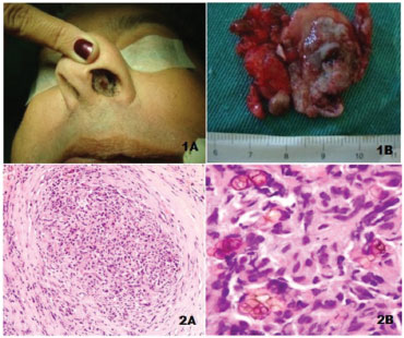
| Figure 1 Clinical image of the septal lesion (1A). | |
Imagen publicada en: |
|
|
|
| 1 | |
| Descripción: | |
|
|
| Autor (es) del artículo de origen: | |
| Lídio Granato1; Ísis Rocha Dias Gonçalves1; Tomás Zecchini Barrese2; Carlos Kayoshi Takara1 | |
| Título y link del artículo: | |
| Primary chromohifomycosis of the nasal septum | |
| oldfiles.bjorl.org/conteudo/acervo/acervo_english.asp?id=4555 |
All rights reserved - 1933 /
2026
© - Associação Brasileira de Otorrinolaringologia e Cirurgia Cérvico Facial